Devastating destruction of the surface of the human eye can occur as a result of a myriad of conditions, leading to significant loss of vision and associated suffering that can be very difficult to treat. Successful transplantation of ex-vivo expanded ocular surface stem cells (SCs) marks an exciting advance in application of engineered tissue for rehabilitation of severely damaged ocular surfaces co-incident with ocular SC failure. Over the last 15 years, these techniques have evolved and expanded to embrace various tissue types and now include ex-vivo expansion and transplantation of corneal limbal, conjunctival and oral mucosal epithelium. The growth of this field and resultant explosion of associated terminology has led the Cornea Society to recommend a standardised nomenclature based on anatomical type (limbal, conjunctival and other mucosal grafts), source of tissue (autologous, allogeneic-cadaveric or living related donor) and technique of tissue transplantation (Ref. Reference Daya, Chan and Holland1). The purpose of this review is to summarise various techniques and advances made specifically in the field of ex-vivo SC expansion and tissue transplantation, to report on the clinical results from these techniques, and to evaluate future barriers and challenges to the success and application of these techniques in patients.
The ocular surface
The ‘ocular surface’, a term first coined by Thoft and Friend, encompasses the outermost surface of the human eye and inner surface of the eyelid consisting of corneal and conjunctival epithelia (Ref. Reference Thoft and Friend2). The ocular surface is a functional unit that possesses unique biological, immunological and antimicrobial functions that serve to protect the eye, and which together with the tear film at the cornea, provide a smooth optical surface for vision (Refs Reference Thoft and Friend2, Reference Kinoshita3). Two closely related but phenotypically distinct populations of cells derived from conjunctiva and cornea constitute the epithelia of the ocular surface (Ref. Reference Schermer, Galvin and Sun4).
The conjunctiva composed of stratified columnar epithelium, is a vascularised membrane that covers the anterior scleral surface of the eye (bulbar conjunctiva) and inner surface of the eyelid (tarsal conjunctiva). The junction between the tarsal and bulbar conjunctiva is an area called the fornix, a loose and flexible area of conjunctiva allowing easy movement of the lids and eyeball (forniceal conjunctiva). In contrast, the corneal epithelium composed of stratified squamous epithelium, is avascular (an essential feature, as the cornea is the transparent window of the eye and its main optical element) and is limited to the anterior surface of the clear cornea. The demarcating border of these distinct populations of epithelial cells is the thin circumferential corneal limbus, presumed home to the majority of corneal epithelial SCs and a functional barrier that prevents conjunctival invasion of the cornea.
SCs of the ocular surface
Evidence for SCs, specialised populations of adult cells, first emerged from pioneering work on bone marrow derived cells in the 1960s (Ref. Reference Till, McCulloch and Siminovitch5). SCs are self-renewing, slow cycling (label retaining in proliferation studies), can proliferate indefinitely and give rise to daughter cells of variable proliferative capacity that in turn repair and replace cells lost through natural turn over and injury (Ref. Reference Barrandon and Green6). SCs and their progeny are divided according to their proliferative capacity in vitro into holoclones, meroclones and paraclones. Among these, the holoclone has the greatest proliferative and colony forming capacity and is considered the true SC. Meroclones are reservoirs of transient amplifying cells, whereas the paraclone is a transient amplifying cell which has a limited capacity for division and generates aborted colonies containing only terminally differentiated cells (Ref. Reference Barrandon and Green6). The transition from holoclone to meroclone to paraclone is unidirectional. Evidence for a population of slow cycling corneal epithelial SCs located at the limbal area of the cornea emerged in 1989, explaining the long-standing observation that new corneal epithelial cells migrated centripetally (from limbus to central cornea) to heal areas of injured epithelium (Refs Reference Cotsarelis7, Reference Thoft and Friend8).
SC populations throughout the body reside in protected niche environments that shield and nurture their ‘stemness’. As by definition a SC can give rise to any type of cell in the body (pluripotency), its progeny appear dictated by the specific micro environment of the niche in which the SC population exists (Ref. Reference Li9). Thus, limbal SCs give rise to corneal epithelial cells, whereas SCs in the bulge area of the hair follicle give rise to dermal constituent cells (Ref. Reference Pasolli10). At the limbus, corneal epithelial SCs appear to reside within ‘limbal crypts’ located between the palisades of Vogt in the superior and inferior limbus (Ref. Reference Shortt11). However, a different anatomical feature termed ‘limbal epithelial crypts’ have also been suggested to be a putative SC niche (Refs Reference Shanmuganathan12, Reference Dua13). The authors describe the limbal epithelial crypts as ‘distinct anatomical extensions from the peripheral aspect of the limbal palisades, consisting of a solid cord of cells extending peripherally or circumferentially’. It is not yet clear how these putative limbal SC niches may function together and whether one is more active than the other in homoeostasis versus repair. Despite the majority of corneal epithelial SCs being located at the limbus, scattered ectopic populations of corneal epithelial SCs have been identified within the bulbar conjunctiva (Ref. Reference Kawasaki14). Recent evidence has shown that in mouse and pig corneas cells with stem like properties of self-renewal and proliferation exist in the central cornea of these animals (Ref. Reference Majo15). The exact location and distribution of corneal epithelial SC's are yet to be definitively resolved.
Epithelial SCs of the conjunctiva occur more abundantly and have been demonstrated throughout the fornix and bulbar conjunctiva (Ref. Reference Pellegrini16). Clonal analysis of conjunctival SC's has shown them to be bi-potent; being able to give rise to both conjunctival epithelium and specialised mucin producing goblet cells (Ref. Reference Pellegrini16).
Devastating ocular surface injury leading to SC deficiency
Renewal of the ocular surface is dependent on a precious reserve of resident SCs that repairs and replenishes surface epithelium throughout an individual's lifetime. This process is critical for maintaining the uniquely transparent structure of the cornea, the main optical component of the eye. Limbal stem cell deficiency (LSCD), through genetic abnormality, inflammation, infection or trauma results in a chronically unstable corneal surface associated with significant morbidity from visual loss, pain, infection and scarring (Fig. 1). It is beyond the scope of this review to provide a comprehensive list of all conditions leading to LSC damage, however common conditions include aniridia (an inherited abnormality of the PAX 6 gene leading to anterior segment dysgenesis and LSCD), thermal and chemical injury (causing localised and global ocular surface damage), immuno-inflammatory conditions such as Steven–Johnson's syndrome (SJS) and ocular cicatricial pemphigoid (causing varying degrees of localised and global ocular surface failure), contact lens wear and iatrogenic injury to the ocular surface (for instance through the use of mitomycin C). These conditions can selectively cause localised corneal or conjunctival SC failure or globalised ocular surface damage (Refs Reference Dua17, Reference Ahmad18). The severity of the disease in these cases is dependent on whether partial or total SC deficiency has occurred.
Colour photograph of eye with chronic limbal stem cell failure. There is direct invasion of the corneal surface by conjunctival tissue due to loss of the limbal barrier. The corneal surface is vascularised and unstable. Internal structures of the eye are normal.

The ocular surface is unable to repair and regenerate itself in the face of SC dysfunction. As the two main functions of the LSCs are to replenish the corneal epithelium and form a barrier to conjunctival epithelial invasion onto the cornea, LSCD results in chronic breakdown and ulceration of the corneal epithelial surface and conjunctivalisation and vascularisation of the cornea. These sequelae lead to chronic pain, discomfort, increased risk of infection, scarring, opacification and loss of vision. Conjunctival cell dysfunction (CCD) leads to loss of mucin producing goblet cells and hence lubrication of the ocular surface (particularly as lacrimal gland dysfunction often occurs in tandem with ocular surface destruction). Loss of ocular mobility as a result of scarring and formation of adhesions within the eyelids, and ultimately, keratinisation of the ocular surface can turn the ocular surface skin like. The combination of severe LSCD and CCD renders the ocular surface a hostile, unstable environment that is incompatible with visual function. Although LSCD may occur independently together with relatively normal conjunctival function (as in the case of aniridia), CCD often leads to or exacerbates LSCD. Ocular SCD has an overall estimated incidence of 240 new cases per year in the UK, and in severe cases is a devastating disease state with significant patient morbidity (Ref. Reference Shortt, Tuft and Daniels19). The true global burden of SCD is difficult to ascertain. Ironically, in many of these cases, beneath a significantly dysfunctional ocular surface, the remaining structures and visual potential of the eye remain preserved. Hence, ocular surface restoration not only provides relief from pain and disfigurement but also the chance for vision in many of these patients.
Options for SC transplantation
Kenyon and Tseng in 1989 showed that the transplantation of autologous limbal tissue restored a normal epithelial phenotype in the grafted eye, demonstrating for the first time the feasibility of LSC transplantation (Ref. Reference Kenyon and Tseng20). Although autologous limbal tissue transplantation is still a cost effective, successful option with no risk of immune rejection for cases of unilateral LSCD, many cases of SC deficiency may have limited availability of autologous tissue, particularly where disease processes are bilateral, and also risks inducing SC failure in the donor eye (Ref. Reference Jenkins21). When allogeneic tissue is utilised (either from a living related donor or cadaver) there is a significant risk of immune rejection and eventual SC failure. When availability of healthy limbus is limited, ex-vivo cultured limbal epithelial transplantation and other ex-vivo techniques offer opportunity for SC expansion from a small autologous biopsy specimen with much lower risk to the patient.
Critical to the successful treatment of these devastating ocular surface diseases has been the paradigm shift in our appreciation of the important role of biological, immunological and microbiological factors and their interdependence and influence on the likely survival of transplanted SCs on the ocular surface, and the need for an integrated approach to ocular SC therapies that addresses all factors underlying ocular surface failure on a case by case basis (Ref. Reference Kinoshita3).
For the reader unfamiliar with this field, it is worth noting that the main focus of ex-vivo expansion techniques has been towards restoration of the cornea and treatment of LSCD. This emphasis has been partly because of the importance in terms of comfort and vision that the corneal surface be made stable, but also because attempts at restoring the cornea in severe cases of cicatricial ocular surface failure coincident with a dry non-lubricated chronically inflamed surface have universally resulted in failure. We must thus assume that the case series in the literature must all include wet eyes even in cases of total LSCD. Conjunctival restoration is in comparison in its infancy.
Ex-vivo corneal limbal SC expansion techniques
Pellegrini et al., in a seminal paper reported the successful treatment of 2 patients with LSCD treated with ex-vivo expanded limbal SC sheets in 1997 (Ref. Reference Pellegrini22). Many groups have since shown that limbal epithelial stem cell (LESC) culture is both feasible and repeatable, and have reported several series of patients treated with ex-vivo expanded cell sheets. Although a number of variations in technique of ex-vivo LESC expansion have been described, all share common fundamental features that follow an initial biopsy of a small piece of tissue from the limbal region of the healthier eye:
1. The limbal epithelial cells are isolated from the biopsy sample.
2. These cells are cultured on a substrate under appropriate conditions.
3. Once a confluent sheet of cells is achieved the cell layer may be stratified before it is transplanted together with its substrate onto the recipient eye.
There are considerable variations in the technique at each of these stages and little agreement about an optimal methodology. Each stage is considered in greater detail herein.
Separation of limbal epithelium from the underlying stroma
A number of protocols for preparation of the biopsy specimen prior to culture have been described and broadly yield either a cell suspension culture or explant culture system. No evidence in terms of superiority of either system has yet been demonstrated. Many groups use trypsin/EDTA digestion to yield cell suspensions for subsequent culture (Refs Reference Pellegrini22, Reference Schwab, Reyes and Isseroff23). Digestion with dispase yields separated intact cell sheets, with some evidence that the use of dispase preserves clonogenicity, and p63 positive progenitors better than trypsin/EDTA (Refs Reference Tsai, Li and Chen24, Reference Nakamura, Inatomi, Sotozono, Koizumi and Kinoshita25, Reference Kawakita26). Other groups have opted for culturing the biopsy specimen in-toto or following maceration prior to culture (Refs Reference Sangwan27, Reference Shimazaki28, Reference Kawashima29). This approach has the theoretical advantage of retaining the LESC's within their anatomical niche microenvironment at the time of culture, which may preserve clonogenecity and colony forming efficiency. Recent evidence has however demonstrated that such explant cultures grown on amniotic membrane, a commonly used substrate, yield epithelial cells that become less SC like with distance of migration away from the explant 0 (Ref. Reference Kolli30). LESCs may also invade the underlying stroma within the explant undergoing epithelial-mesenchymal transition with declining growth potential over time (Ref. Reference Li31). Proximity of LESCs to their niche may therefore be vital to maintaining their undifferentiated state during explant culture in the absence of a 3T3 feeder layer. Despite the theoretical risk of xenogeneic transfer from 3T3 murine feeder cells, it is worth stating that there is a long history of using irradiated 3T3 feeder cells without any recorded complications from their use. There may be an increased risk with mitomycin C versus radiation as a method of growth arrest.
Culture conditions and media
Corneal LSC's were first serially co-cultured with a feeder layer of growth arrested murine 3T3 cells which were shown to maintain the cells in an undifferentiated state (Refs Reference Barrandon and Green6, Reference Pellegrini16, Reference Rheinwald, Harris, Trump and Stoner32). These growth arrested mesenchymal cells may act as a surrogate niche environment for the LESCs. Cultures have been obtained in systems where there is no contact with the co-cultured feeder cell layer, and also in the absence of a feeder layer although there is no definite evidence that these systems actually support holoclones. Clonogenicity and lifetime in culture are increased by adding foetal calf serum, hydrocortisone, epithelial growth factor, cholera toxin and other factors to aid adhesion and stratification (Refs Reference Pellegrini16, Reference Lindberg33, Reference Rheinwald and Green34).
The use of animal derived components in these culture systems and evidence of xenogeneic RNA in cultured cells have raised concerns regarding the possibility of zoonosis or xenogeneic immune responses after transplantation, and has driven the development of xenofree culture systems that continue to maintain cells in an undifferentiated state (Ref. Reference Johnen35). Bovine derived serum has been replaced with autologous and cord blood derived serum (Refs Reference Ang36, Reference Nakamura37, Reference Nakamura38). Factors such as cholera toxin and murine 3T3 feeder cells have also been removed from culture systems in an attempt to exclude xeno-derived culture components (Refs Reference Shahdadfar39, Reference Bergström40). An absence of firm evidence that LESCs are maintained in these systems, which would require lengthy single cell cloning experiments, mean that we cannot yet discard the 3T3 method. Human amnion derived epithelial cells have also been used as an allogeneic feeder cell layer as have human bone marrow derived mesenchymal cells (Refs Reference Chen41, Reference Omoto42). Several groups have reported clinical success in patients with LSCD following LESC transplantation from xenofree systems (Refs Reference Nakamura37, Reference Sangwan43, Reference Zakaria44, Reference Meller45, Reference Kolli46, Reference Di Girolamo47, Reference Shimazaki48).
Choice of substrate
The first clinical application of cultured LESCs could not be replicated on a larger scale because of fragility of the cultured sheet of epithelium (Ref. Reference Pellegrini22). A significant improvement was the use of supporting materials for cell culture, transportation and transplantation onto patients. Most common among these has been the use of human amniotic membrane (HAM) derived from the placenta, which has previously been described as a treatment in itself to restore the damaged ocular surface and limbal stroma (Refs Reference Chen41, Reference Omoto42, Reference Sangwan43, Reference Zakaria44, Reference Meller45, Reference Kolli46, Reference Di Girolamo47, Reference Shimazaki48, Reference Kim and Tseng49, Reference Anderson50, Reference Gomes51). Prior to usage for LSC culture, amniotic membrane is processed and preserved by various methods, and can be treated with NH4OH and/or EDTA, with either removal (denuded) or retention (intact) of the epithelium of the HAM (Ref. Reference Shortt52). The jury remains undecided regarding the use of intact or denuded HAM in terms of which better maintains holoclones and the LSC phenotype in culture, and protocols vary between groups (Refs Reference Grueterich, Espana and Tseng53, Reference Hernandez Galindo54, Reference Koizumi55). Native, intact amniotic membrane (retaining the amniotic epithelium) contains high levels of Epidermal growth factor (EGF), Keratinocyte growth factor (KGF), Hepatocyte growth factor (HGF) and basic Fibroblast growth factor (bFGF), which are potentially involved in epithelial proliferation and epithelium–stromal interactions (Ref. Reference Kenyon56).
Fibrin matrices have also been used as substrates for cell culture and shown to maintain holoclones and the LSC phenotype in culture (Refs Reference Meyer-Blazejewska57, Reference Higa58). Owing to the adhesive nature of fibrin, transplanted epithelial sheets may be attached without sutures, following which the fibrin matrix is rapidly degraded over 24–48 h (Refs Reference Colabelli Gisoldi59, Reference Rama60, Reference Rama61). Synthetic polymers have also been used as temperature responsive substrates that can be detached from culture plates by altering ambient temperature and have been used effectively for treatment of LSCD in patients (Refs Reference Sitalakshmi62, Reference Nishida63). Other novel approaches have included LSC culture on the surface of contact lenses and the use of scaffolds derived from the cocoons of silk worms and plastic compressed collagen (Refs Reference Di Girolamo64, Reference Chirila65, Reference Levis, Brown and Daniels66).
Cell stratification
Following culture of a monolayer of limbal epithelial cells, cell stratification is typically achieved by air lifting the cells such that proliferation takes place at an air fluid interface (Ref. Reference Pruniéras, Régnier and Woodley67). However, there is some evidence that this process may induce squamous metaplasia of cells 0 (Ref. Reference Li68).
Alternative cell sources in cases of bilateral LSCD
Autologous ex-vivo LESC culture is contingent on the availability of even a small area of healthy limbus from which a biopsy can be obtained. In cases of severe disease, where this may not be possible, the options that remain for ex-vivo ocular surface reconstruction are either the allogeneic expansion of limbal epithelial cells from cadaveric or living related donor tissue, or the use of autologous buccal mucosal epithelial cell cultures.
Despite ready culture of allogeneic tissue using the techniques described, clinical results with allogeneic tissue have been variable and complicated by the need for systemic immunosuppression to avoid tissue rejection (Ref. Reference Pauklin69). In a study of 10 patients with variable causes of LSCD, Daya et al reported improvement in symptoms in 7 patients at final follow up (Ref. Reference Daya70). Interestingly, DNA analysis at 9 months showed no ex-vivo donor SC DNA, bringing into question the actual source of renewed epithelium. It is possible that the transplanted cell sheet acts as a biological cellular bandage that aids healing or restitution of an SC niche, however in truth these observations really highlight gaps in our knowledge regarding epithelial homoeostasis and biology.
In recent years, the use of autologous buccal mucosal tissue as an alternate SC source for ex-vivo expansion of epithelial cultures has been shown to be useful in ocular surface reconstruction in cases of severe bilateral LSCD (Refs Reference Nishida63, Reference Ang71, Reference Burillon72, Reference Priya73). Evidence suggests that these autologous oral epithelial cells survive on the ocular surface up to 22 months after transplantation and perhaps longer, and do not adopt a corneal phenotype (Ref. Reference Chen74). Cultured oral mucosal epithelium also expresses mucins and cytokeratin 3 that may aid in their role in ocular surface reconstruction (Refs Reference Hori75, Reference Juhl, Reibel and Stoltze76).
Several authors have demonstrated the use of ex-vivo cultured conjunctival tissue for treatment of LSCD in animal models and have shown both histologically and immunohistochemically that the subsequent epithelium in these cases shared morphology and characteristics of corneal epithelium (Refs Reference Tanioka77, Reference Meller, Dabul and Tseng78, Reference Ono79, Reference Ang80, Reference Scuderi81, Reference Tan, Ang and Beuerman82).
Clinical results of ex-vivo SC expansion therapies for corneal surface reconstruction
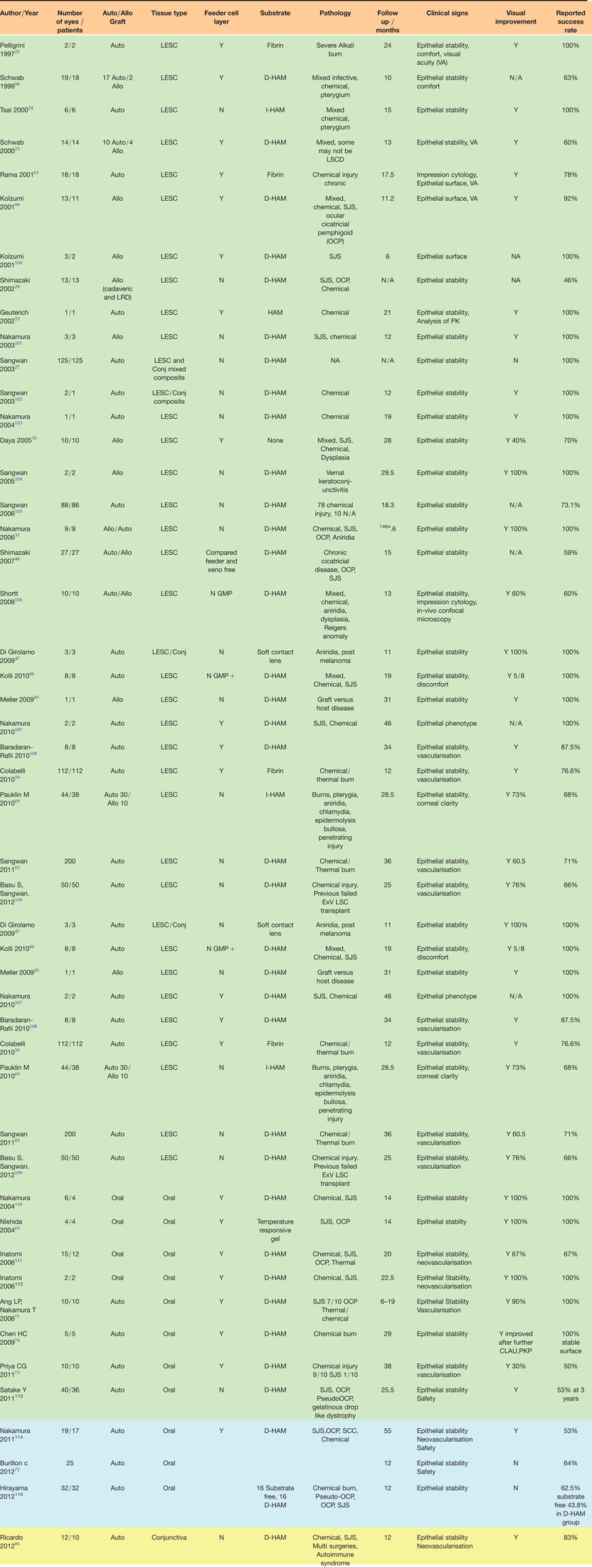
Ex-vivo epithelial tissue engineering techniques offer hope to many patients afflicted with total corneal LSCD (Fig. 2). Since Pellegrini's landmark study in 1997, there have been many different groups that have reported their results of ex-vivo expanded LESC culture and oral mucosal SC culture and transplantation in treatment of such patients. The details of these studies in terms of laboratory methodology, range of treated conditions and clinical results are summarised in Table 1.
(a) Colour preoperative photograph of the eye of a 34 year old patient with PAX6 haplo-insufficiency with limbal epithelial stem cell failure resulting in corneal vascularisation and conjunctivalisation. The cornea is opaque and details of intraocular structures such as the iris are difficult to discern. Vision was hand movements only. (b) The same cornea 2 years after ex-vivo expanded limbal epithelial stem cell transplantation. Note that the cornea appears clearer and that intraocular structures are now clearly visible. The corneal epithelial surface is also now stable. Vision improved to 6/36, further improvement was limited by foveal hypoplasia.
Tabulated summary of published studies using ex-vivo expanded engineered tissue for the treatment of ocular surface disease. Green rows are limbal epithelial stem cell cultures (LESC), blue rows are oral mucosal stem cell cultures. The yellow row is conjunctival epithelial stem cell cultures. Allo = Allograft. Auto = Autograft. D-HAM= denuded human amniotic membrane, I-HAM = intact human amniotic membrane. SJS = Steven Johnson Syndrome. OCP = Ocular cicatricial pemphigoid. PK = penetrating keratoplasty
There is considerable variation in success rate between groups in these reported clinical series, ranging from 46% to 100% for cultured LESC transplantation, and 43–100% for cultured oral mucosal transplantation. Sangwan et al (2011) reported success rates of 71% in 200 patients that were followed up for a period of 36 months after cultured LESC transplantation using a xeno-free culture technique (Ref. Reference Sangwan43). Clinical parameters of success in this study included long-term corneal epithelial stability and improvement in vision. Notably, all patients in this study had LSCD because of chemical or thermal injury, signifying an acute injury rather than chronic disease process. In contrast, Shimazaki et al (2007) treated 27 patients with a diverse range of diseases including ocular cicatricial pemphigoid, SJS and chemical injury, reporting a success rate, defined by corneal epithelial stability, of 59% at 15 months (Ref. Reference Shimazaki48). In terms of cultured oral mucosal transplantation, Satake (2011) report a success rate of 53% at 3 years in 40 patients with a diverse range of pathologies (Ref. Reference Satake97).
Ricardo et al. (2012) have recently described the use of cultured conjunctival epithelium for restoration of the corneal surface, reporting success rates of 83% in terms of epithelial stability at 12 months after surgery (Ref. Reference Ricardo100). Confocal microscopy and impression cytology of the corneas of these patients postoperatively showed corneal epithelial phenotype in a few patients (16.6%) and mixed corneal epithelial/conjunctival phenotype in others (66%). No corneal epithelial cells were found in the 2 cases that failed. In 3 patients who underwent subsequent penetrating keratoplasty, analysis of excised corneal button demonstrated a stratified epithelium, which stained positively for cytokeratin (CK3) and cytokeratin (CK19) suggestive of a mixed cell phenotype.
What is apparent when we consider these studies is the variation in technique and methodology used in the ex-vivo expansion and culture of both LESC's and oral mucosal epithelium. This variability represents a biotechnology that is still evolving and seeking optimisation. Despite the significant variation in success rates in patients treated with cultured epithelial cell sheets, these results must be viewed in the context of patients with devastating ocular surface failure where there is no other option for treatment. A reasonable proportion of these patients may require further surgery to improve vision, including keratoplasty once a stable ocular surface has been achieved.
Factors determining success after ex-vivo expanded cell transplantation
The clinical success following ex-vivo expanded cell therapies must be interpreted in the context of the underlying cause of ocular surface failure. Outcomes following a severe but self-limiting episode such as a chemical injury are likely to be different to that following an ongoing inflammatory state such as mucus membrane pemphigoid (MMP) or SJS, where the environment for cell survival remains hostile.
Evidence of transplanted limbal SC survival is mixed. DNA analysis after allogeneic cell expansion and transplantation was unable to detect donor DNA 9 months after transplantation (Ref. Reference Daya70). In all these cases, the previously unstable ocular surface remained healthy and intact. Analysis of the excised recipient cornea in cases where keratoplasty was performed some time after ex-vivo tissue transplantation offers a unique insight into the state of the corneal surface, and has shown an intact multi-layered corneal epithelium in cases of previously confirmed LSCD and ocular surface breakdown. Although this does not definitely confirm de novo SC transfer to the recipient ocular surface, it does show that the transplantation of autologous ex-vivo engineered tissue encourages regeneration of the ocular surface. This may occur through de-novo SC transfer, or through a biological therapeutic effect that remains poorly understood. The ability to temporally track transplanted cells in-vivo in an animal model will allow greater insights into ocular surface biology in these patients in future (Ref. Reference Mort101).
Successful limbal SC transplantation onto a hostile ocular surface requires a heightened awareness of other ongoing factors such as host immune response to the transplant, ongoing inflammation and the role of the microbiological milieu in terms of chronic inflammation and infection. Addressing all these elements in an integrated approach to treating these devastating ocular surface disorders will probably lead to improved treatment strategies and outcomes in this clinically challenging group of patients (Ref. Reference Kinoshita3).
Clinical results of ex-vivo SC expansion therapies for conjunctival reconstruction
Ocular surface diseases such as SJS, MMP, epidermolysis bullosa, pterygium excision as well as chemical and thermal burn injury can lead to severe scarring and loss of conjunctival tissue. This leads to a cascade of cicatricial sequelae such as forniceal shortening, entropian, trichiasis, symblepharon, ankyloblepharon and dysfunctional blinking, which together with dry eye disease arising from goblet cell loss and keratinisation, renders the entire ocular surface hostile and prone to breakdown and infection (Ref. Reference Schrader102). Ex-vivo transplantation of cultured tissue to the corneal surface is often likely to fail in this environment and hence is rarely undertaken. Osteo-odonto keratoprosthesis remains the final option in these severely scarred dry eyes (Ref. Reference Tan103). Conjunctival restoration, forniceal reconstruction and lid surgery may thus be required if successful corneal restoration is to be achieved.
Kim et al described the use of autologous nasal mucosa for conjunctival restoration in combination with oral mucosa of the cornea in 6 patients with severe chemical injury and ocular surface cicatrisation (Ref. Reference Kim104). All patients improved and goblet cells were observed on the ocular surface. The technique is uncomfortable for the patient and the ease and number of times that tissue can be harvested may be limited. A number of groups have described the use of ex-vivo cultured conjunctival epithelium for restoration of conjunctiva in a number of diverse clinical cases 0 (Refs Reference Tanioka77, Reference Meller, Dabul and Tseng78, Reference Ono79, Reference Ang80, Reference Scuderi81, Reference Tan, Ang and Beuerman82). Forniceal conjunctival biopsies appear to grow most effectively in culture and can maintain SC properties even after cryo-preservation (Refs Reference Eidet105, Reference Schrader106). Despite the apparent success of conjunctival culture and transplantation to areas of localised ocular surface damage, there have thus far been no reports of the use of tissue engineered conjunctiva for ocular surface restoration in eyes with severe conjunctival cicatrisation arising from diseases such as MMP and SJS.
Hurdles from bench to bedside
An analysis of the cost of producing an ex-vivo expanded LSC sheet for transplantation in the UK in 2010 was estimated as being over £10 000 per patient. Cost has hence been a major barrier to widespread implementation of ex-vivo cultured tissue in Europe (Ref. Reference Miri, Al-Deiri and Dua107).
Stringent regulatory rules designed to safeguard patients now apply to all tissue banks providing cellular therapies including LESCs. These good manufacturing practice (GMP) measures require that ex-vivo expanded tissue be produced in accordance with the Advanced Therapy Medicinal Products (ATMP) EU Directive. In the UK, the Medicine and Healthcare products Regulatory Agency (MHRA) is responsible for ensuring that these stringent standards are met. In the United States (US), the Food and Drug Administration (FDA) and the American Association of Tissue Banks oversees accreditation and compliance with regulations by tissue banks. These strict regulatory considerations in the Europe and the US increase the cost and limit the number of tissue banks producing engineered tissue in these regions.
A number of groups from India have demonstrated that ex-vivo SC programmes can be established and implemented cost effectively and on a scale that has eluded the West. Reliance Life Sciences (Mumbai, India) now produces and delivers substrates and SCs within India and further afield. Such a centralised model of SC culture and delivery may be an efficient and economical model for SC therapies in future.
Conclusions
Epithelial tissue engineered by ex-vivo expansion of the patient's own epithelial SCs is a significant advance in restoration of the ocular surface in patients with severe ocular surface disease as a consequence of ocular surface SC deficiencies. These tissue engineering techniques continue to evolve and improve and are still variable in their efficacy. As our understanding of the biology of SCs and the ocular surface continues to grow, the clinical success rates of these techniques is also likely to improve. It is widely accepted that the engineered tissue derived from autologous tissue sources has significant advantage in terms of survival and immune tolerance. However, autologous tissue may not always be available in cases of severe bilateral ocular disease, and although allogeneic tissue together with immunosuppression are possible, recent discoveries herald the possibility of exciting future therapies derived from autologous mesenchymal and induced pluripotent stem cells (IPSCs) that could potentially yield a multitude of engineered tissue types (Refs Reference Rashid and Vallier108, Reference Branch109). Although IPSCs are yet to be used for ocular surface reconstruction, a recent study has shown successful corneal epithelial cell generation from dermal fibroblast IPSCs. Further refinement of these techniques will be required before this potentially unlimited source of corneal epithelial SCs reaches clinical application (Ref. Reference Hayashi110).
Conflicts of interest
None of the authors have any financial interests in the subject matter of this review.
Acknowledgements
The authors thank Dr Alex Shortt of UCL Institute of Ophthalmology, 11–43 Bath Street, London, EC1 V 9EL, UK, for kind contribution of the clinical photographs that compose Figure 2.