Adolescence is an important developmental period during which mental health can shape the rest of the life span. The incidence of depression increases during adolescence (Cole et al., Reference Cole, Tram, Martin, Hoffman, Ruiz, Jacquez and Maschman2002), and adolescent-onset depressive disorders increase the likelihood of later recurrences (Pine, Cohen, Gurley, Brook, & Ma, Reference Pine, Cohen, Gurley, Brook and Ma1998). Other indices of positive mental health (or psychological well-being), such as self-esteem, decline across the adolescent period (Robins, Trzesniewski, Tracy, Gosling, & Potter, Reference Robins, Trzesniewski, Tracy, Gosling and Potter2002). Existing research has explored how both environmental and biological factors may contribute to adolescent mental health outcomes. Family environmental factors, such as positive and negative parenting behaviors and styles, have been found to be particularly important predictors of both depression (McLeod, Weisz, & Wood, Reference McLeod, Weisz and Wood2007; Yap, Pilkington, Ryan, Kelly, & Jorm, Reference Yap, Pilkington, Ryan, Kelly and Jorm2014) and well-being (Lekes, Gingras, Philippe, Koestner, & Fang, Reference Lekes, Gingras, Philippe, Koestner and Fang2010). Furthermore, biological factors such as alterations in brain structure have also been hypothesized to underlie the development of depressive symptoms and disorder, with limbic and prefrontal regions most commonly implicated (e.g., Hulvershorn, Cullen, & Anand, Reference Hulvershorn, Cullen and Anand2011; Vijayakumar et al., Reference Vijayakumar, Allen, Dennison, Byrne, Simmons and Whittle2017; Whittle et al., Reference Whittle, Lichter, Dennison, Vijayakumar, Schwartz, Byrne and Alle2014). Less work has been done linking brain structure with measures of positive mental health in adolescents, although significant associations have been reported between brain structure (limbic and prefrontal regions) and well-being (Ent et al., Reference Ent, Braber, Baselmans, Brouwer, Dolan, Hulshoff Pol and Bartels2017; Kong, Ding, et al., Reference Kong, Ding, Yang, Dang, Hu, Song and Liu2015; Kong, Wang, Hu, & Liu, Reference Kong, Wang, Hu and Liu2015; Lewis, Kanai, Rees, & Bates, Reference Lewis, Kanai, Rees and Bates2014; Takeuchi et al., Reference Takeuchi, Taki, Nouchi, Hashizume, Sassa, Sekiguchi and Kawashima2014) and other constructs linked to positive mental health, such as mindfulness (Friedel et al., Reference Friedel, Whittle, Vijayakumar, Simmons, Byrne, Schwartz and Allen2015) and cognitive reappraisal of emotions (Vijayakumar et al., Reference Vijayakumar, Whittle, Yücel, Dennison, Simmons and Allen2014).
While there is support for parenting behaviors and brain structure as independent predictors of adolescent mental health outcomes, less is known about how these environmental and biological factors work together to influence such outcomes. Although some evidence indicates that parenting behaviors may influence brain structure during adolescence (Kok et al., Reference Kok, Thijssen, Bakermans-Kranenburg, Jaddoe, Verhulst, White and Tiemeier2015; Luby et al., Reference Luby, Barch, Belden, Gaffrey, Tillman, Babb and Botteron2012; Whittle et al., Reference Whittle, Vijayakumar, Dennison, Schwartz, Simmons, Sheeber and Allen2016), and that this is one plausible mechanism by which parenting may influence risk for depression, it is also important to consider that these environmental and biological factors may act as independent factors that interact with each other to predict outcomes (Schriber & Guyer, Reference Schriber and Guyer2016). That is, parenting may differentially predict adolescent depressive symptoms depending on an individual's brain development. Such Biology × Environment interactions may explain in part why some individuals who experience poor parenting do not develop depression (Dennison et al., Reference Dennison, Sheridan, Busso, Jenness, Peverill, Rosen and McLaughlin2016). Only three studies, to our knowledge, have investigated such a model, all using cross-sectional brain imaging data, and all focusing on depression outcomes. In earlier work with the cohort examined in the current study, we found support for the hypothesis that brain structure is an independent risk factor that interacts with aggressive maternal behavior to predict adolescent depressive symptoms (Whittle et al., Reference Whittle, Yap, Sheeber, Dudgeon, Yücel, Pantelis and Allen2011; Yap et al., Reference Yap, Whittle, Yücel, Sheeber, Pantelis, Simmons and Allen2008). Results suggested that females with larger hippocampi were more sensitive to the depressogenic effects of aggressive parenting (Whittle et al., Reference Whittle, Yap, Sheeber, Dudgeon, Yücel, Pantelis and Allen2011). Yap et al. (Reference Yap, Whittle, Yücel, Sheeber, Pantelis, Simmons and Allen2008) reported that boys with a smaller left paralimbic anterior cingulate, larger right amygdala, and girls with smaller amygdalae were more sensitive to parental aggression in prediction of depression. More recently, Schriber et al. (Reference Schriber, Anbari, Robins, Conger, Hastings and Guyer2017) reported that adolescent girls with relatively large left hippocampal volumes demonstrated increased sensitivity to low levels of family connectedness and high levels of community crime exposure in the prediction of depression.
In the current study, we aimed to advance our understanding of these issues in several ways. First, and as discussed further below, we investigated the role of structural brain development (i.e., change across time) as a factor that interacts with parenting to predict adolescent depressive symptoms. Second, in order to comprehensively investigate both negative and positive mental health outcomes, we investigated depressive symptoms in addition to psychological well-being. Third, prior research has generally interpreted Biology × Environment interactions qualitatively by visual inspection of simple slope plots; our goal was to more objectively interpret interactions, making inferences about biological sensitivity that might be indicative of vulnerability versus susceptibility.
Diathesis-Stress and Differential Susceptibility: Biology × Environment
Diathesis-stress and differential susceptibility are two conceptual frameworks that outline distinct approaches to understanding how Biology × Environment interactions predict developmental outcomes. Diathesis-stress is a developmental theory, which implies an optimal trajectory of human development that can be derailed by adversity. It describes the relationship between individual vulnerability to psychopathology and environmental stressors; more stress relates to poorer outcomes (Monroe & Simons, Reference Monroe and Simons1991; Monroe, Slavich, & Gotlib, Reference Monroe, Slavich and Gotlib2014). Differential susceptibility (Belsky, Reference Belsky1997), and the related biological sensitivity to context (Ellis, Essex, & Boyce, Reference Ellis, Essex and Boyce2005), are two evolutionary-developmental theories hereafter referred to as “differential susceptibility,” given the considerable similarities between the two models (Belsky & Pluess, Reference Belsky and Pluess2009; Ellis, Boyce, Belsky, Bakermans-Kranenburg, & van IJzendoorn, Reference Ellis, Boyce, Belsky, Bakermans-Kranenburg and van Ijzendoorn2011). Differential susceptibility theory argues that multiple developmental trajectories result from human adaptive capabilities. It assumes we have evolved to adapt to environments that vary in both adversity and support. The model hypothesizes that susceptibility to environmental influence (positive and negative) is instantiated biologically, and that people vary in this susceptibility. Highly susceptible individuals “suffer” more in adverse circumstances than individuals with low susceptibility, but also thrive more in supportive environments. This theory argues that if interpreted in a diathesis-stress framework, one might mislabel high susceptibility as “vulnerability” because development is only examined in a negative context. Therefore, both positive and negative contexts should be examined in order to detect differential susceptibility, in which highly susceptible individuals (compared to less susceptible) would be predicted to show a stronger response to positive and negative environmental stimuli for better and worse, respectively. However, few studies investigating Brain × Environment predictors of adolescent mental health have explicitly utilized such a design (including Whittle et al., Reference Whittle, Yap, Sheeber, Dudgeon, Yücel, Pantelis and Allen2011, and Yap et al., Reference Yap, Whittle, Yücel, Sheeber, Pantelis, Simmons and Allen2008, described above). To our knowledge, Schriber et al. (Reference Schriber, Anbari, Robins, Conger, Hastings and Guyer2017) have published the only study to include both positive and negative environmental factors in the examination of Brain × Environment predictors of depression. Positive mental health outcomes, however, were not examined. Given that differential susceptibility focuses on sensitivity to environmental influences for better and worse, we argue that investigating positive outcomes (and not just the absence of negative outcomes) is important to comprehensively understand the relevance of Biology × Environment interactions for mental health outcomes.
Biology × Environment research that supports diathesis-stress or differential-susceptibility has predominantly focused on Gene × Environment interactions (Bakermans-Kranenburg & van IJzendoorn, Reference Bakermans-Kranenburg and van IJzendoorn2015; Belsky et al., Reference Belsky, Jonassaint, Pluess, Stanton, Brummett and Williams2009). However, interest is growing in examining Brain × Environment interactions, whereby brain structure is explored as a marker of biological susceptibility or vulnerability (Schriber et al., Reference Schriber, Anbari, Robins, Conger, Hastings and Guyer2017; Schriber & Guyer, Reference Schriber and Guyer2016; Whittle et al., Reference Whittle, Yap, Sheeber, Dudgeon, Yücel, Pantelis and Allen2011; Yap et al., Reference Yap, Whittle, Yücel, Sheeber, Pantelis, Simmons and Allen2008). Investigating structural brain development as a vulnerability or susceptibility factor may be particularly informative, given that measures of brain development (especially during sensitive periods, such as childhood and adolescence) have been suggested as potentially more important in predicting outcomes (such as depression) as compared to structure at any one particular time point (Ducharme et al., Reference Ducharme, Albaugh, Hudziak, Botteron, Nguyen, Truong and Karama2014; Vijayakumar et al., Reference Vijayakumar, Allen, Youssef, Dennison, Yücel, Simmons and Whittle2016; Whittle et al., Reference Whittle, Lichter, Dennison, Vijayakumar, Schwartz, Byrne and Alle2014).
Distinguishing Between Differential Susceptibility and Diathesis-Stress Effects
Researchers have often distinguished between diathesis-stress and differential susceptibility effects by visually inspecting interaction plots. That is, after identifying a significant interaction between an independent variable and a moderator, simple slope plots are then compared to prototypical differential susceptibility and diathesis-stress effects. Such effects can be visually approximated, but recently there has been a push in the literature to use statistical guidelines to more systematically distinguish between the frameworks (Belsky & Widaman, Reference Belsky and Widaman2018; Roisman et al., Reference Roisman, Newman, Fraley, Haltigan, Groh and Haydon2012; Widaman et al., Reference Widaman, Helm, Castro-Schilo, Pluess, Stallings and Belsky2012). These guidelines are increasingly being used in the literature (e.g., Belsky et al., Reference Belsky, Newman, Widaman, Rodkin, Pluess, Fraley and Roisman2014; Rioux et al., Reference Rioux, Castellanos-Ryan, Parent, Vitaro, Tremblay and Séguin2015). To clarify, a differential susceptibility effect is indicated by a disordinal interaction (regression lines cross), meaning that a moderator has a different effect in two conditions of an independent variable in the prediction of an outcome variable. A diathesis-stress effect is indicated by an ordinal interaction (regression lines will not be parallel and will not cross), indicating that a moderator has more effect under one level of an independent variable (Kochanska, Kim, Barry, & Philibert, Reference Kochanska, Kim, Barry and Philibert2011; Roisman et al., Reference Roisman, Newman, Fraley, Haltigan, Groh and Haydon2012; Widaman et al., Reference Widaman, Helm, Castro-Schilo, Pluess, Stallings and Belsky2012).
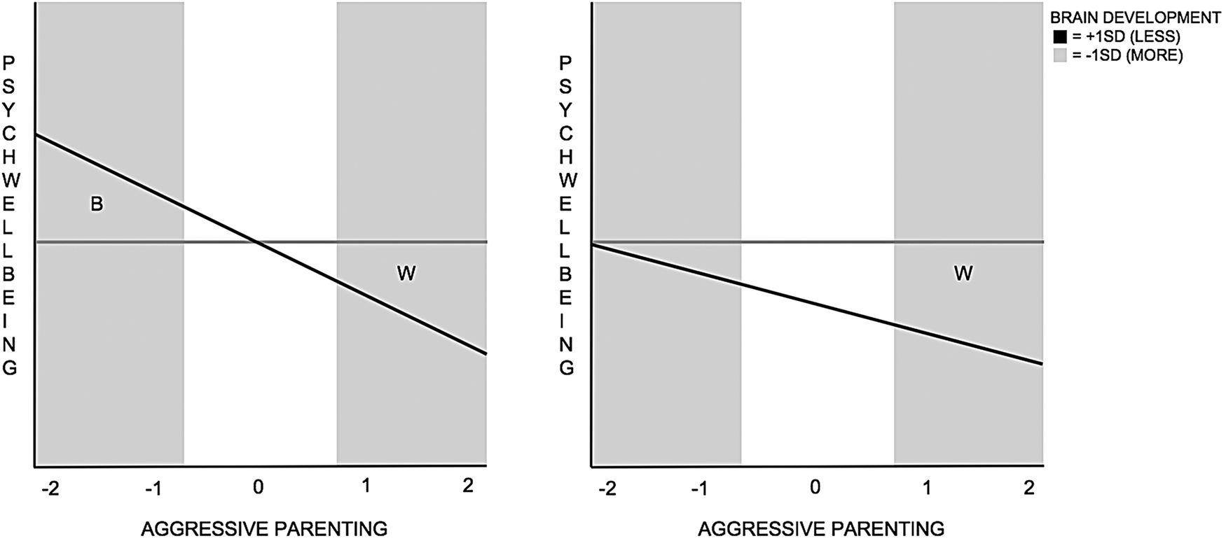
Roisman et al. (Reference Roisman, Newman, Fraley, Haltigan, Groh and Haydon2012) developed indices to distinguish between diathesis-stress and differential susceptibility. The Roisman approach includes significance testing and the implementation of three indices to assess whether interaction effects support either framework. These indices are outlined in the Method section and Figure 1. Briefly, a test of the regions of significance (RoS) examines the values of the independent variable for which the moderator and outcome variable are significantly associated (Kochanska et al., Reference Kochanska, Kim, Barry and Philibert2011; Roisman et al., Reference Roisman, Newman, Fraley, Haltigan, Groh and Haydon2012). The proportion of the interaction (PoI) and the proportion affected (PA) supplement RoS on X. The PoI captures the proportion of the entire interaction that reflects a better rather than a worse outcome for participants. Similar to the PoI, the PA captures the proportion of individuals affected positively by the interaction between the independent variable and the moderator. An alternative method of distinguishing between models has also been developed by Widaman and colleagues (Belsky & Widaman, Reference Belsky and Widaman2018; Widaman et al., Reference Widaman, Helm, Castro-Schilo, Pluess, Stallings and Belsky2012). Widaman and colleagues suggest that, when considering evidence in support of two competing models outlined a priori, significance testing is not required. Rather, the F ratio is assessed and then the point at which the regression lines cross is calculated to detect an ordinal or disordinal interaction (Widaman et al., Reference Widaman, Helm, Castro-Schilo, Pluess, Stallings and Belsky2012). If the F ratio is not close to zero (roughly greater than 1) and the crossover point is at the boundary or outside the observed values of the independent variable, then diathesis-stress is supported, and if it is within the observed range of values, then differential susceptibility is supported; however, specific F ratio and crossover values are not specified.
Prototypical differential susceptibility (left) and diathesis-stress (right) effects. The gray line indicates more brain change, and the black line less brain change. Roisman indices include the regions of significance on X (RoS on X), which examines values of the independent variable (X) for which the moderator (Z) and outcome variable (Y) are significantly associated. Significant associations must fall within +/–2 SD of the mean of X, as highlighted by the shaded areas. Interactions are broken down into two parts: better (B) and worse (W) outcomes predicted by the interaction. (B is uniquely predicted by differential susceptibility and a distinguishing factor between model effects.) B is expressed by two further indices: proportion of the interaction that reflects better outcomes (PoI; B/[B+W]) and proportion of individuals affected positively by X (PA). Differential susceptibility is inferred if Z and Y are significantly associated at both high and low values of X (W and B areas), the PoI = 40%–60%, and the PA ≥16% (scores closer to 50% provide stronger evidence). Diathesis-stress is inferred if Z and Y are significantly associated at high values of X (W area), the PoI equals <20%, and the PA equals <16% (scores closer to 0 provide stronger evidence).

The Current Study
The current study examined whether maternal parenting behaviors and adolescent structural brain development interacted to predict positive and negative mental health outcomes during late adolescence. We specifically assessed whether positive and aggressive maternal parenting behavior and structural brain development (indexed by change in cortical thickness/subcortical volume from early to late adolescence) interacted to predict late-adolescent depressive symptoms and psychological well-being. Frontal and subcortical structures were analyzed as regions of interest given prior evidence for their considerable structural development during adolescence, and associations with depressive symptoms (Forbes et al., Reference Forbes, Ryan, Phillips, Manuck, Worthman, Moyles and Dahl2010; Hulvershorn et al., Reference Hulvershorn, Cullen and Anand2011; Vijayakumar et al., Reference Vijayakumar, Allen, Dennison, Byrne, Simmons and Whittle2017; Whittle et al., Reference Whittle, Lichter, Dennison, Vijayakumar, Schwartz, Byrne and Alle2014) as well as well-being (Ent et al., Reference Ent, Braber, Baselmans, Brouwer, Dolan, Hulshoff Pol and Bartels2017; Kong, Ding, et al., Reference Kong, Ding, Yang, Dang, Hu, Song and Liu2015). Furthermore, it has been suggested that structural change in the frontal cortex during adolescence may underpin sensitivity to the social environment (Fuhrmann, Knoll, & Blakemore, Reference Fuhrmann, Knoll and Blakemore2015; Gogtay et al., Reference Gogtay, Giedd, Lusk, Hayashi, Greenstein, Vaituzis and Thompson2004; Nelson, Leibenluft, McClure, & Pine, Reference Nelson, Leibenluft, McClure and Pine2005).
For significant interactions, we investigated whether diathesis-stress or differential susceptibility effects were supported using the Roisman indices. We considered that the Roisman (rather than Widaman) approach was more appropriate given that, even though our aim from the outset was to distinguish between differential susceptibility and diathesis-stress effects, we wanted to test for localization of effects. That is, our aim was to identify which brain regions (within frontal cortex and subcortex) interacted with parenting to predict adolescent depressive symptoms and well-being. As such, it was deemed important to employ a more conservative approach, with significance testing, to first identify regions that interacted with parenting to predict outcomes.
Evidence in support of the diathesis-stress hypothesis would be demonstrated if a given pattern of brain development was associated with more depressive symptoms and/or less well-being in the context of more aggressive or less positive maternal parenting. In comparison, differential susceptibility would be supported if, in addition to the above hypothesized effect, this same pattern of brain development was also associated with “better” mental health outcomes (i.e., fewer depressive symptoms and/or increased psychological well-being) in the context of more positive maternal parenting or less aggressive maternal parenting.
The existing research (Schriber & Guyer, Reference Schriber and Guyer2016; Whittle et al., Reference Whittle, Yap, Sheeber, Dudgeon, Yücel, Pantelis and Allen2011) led us to hypothesize that we would find evidence for differential susceptibility, by which structural brain development might alter individual sensitivity to maternal parenting in the prediction of adolescent outcomes for better and worse. However, there is minimal prior research to guide hypotheses about whether more or less change in brain structure might confer sensitivity to environmental factors. Findings that relate to brain development and adolescent depression indicate a slower rate of cortical thinning in the prefrontal cortex (i.e., less thinning) in those with relatively higher depressive symptoms (Ducharme et al., Reference Ducharme, Albaugh, Hudziak, Botteron, Nguyen, Truong and Karama2014). However, reduced (or less) thinning of other regions involved in emotion processing has also been associated with positive traits in adolescents (e.g., increased mindfulness; Friedel et al., Reference Friedel, Whittle, Vijayakumar, Simmons, Byrne, Schwartz and Allen2015) and reduced risk for psychopathology also (Bos, Peters, Van de Kamp, Crone, & Tamnes, Reference Bos, Peters, Van de Kamp, Crone and Tamnes2018; Cannon et al., Reference Cannon, Chung, He, Sun, Jacobson, Van Erp and Heinssen2015). Thus, while evidence exists that more and less cortical thinning across adolescence might be “adaptive,” it is unclear which pattern of development might indicate vulnerability or susceptibility to the environment. Research informing hypotheses about subcortical structural development is even less clear.
Method
Participants
Adolescents were recruited as part of the Orygen Adolescent Development Study (ADS) study. A cohort of 2,453 final-year primary school children were recruited from schools in metropolitan Melbourne and completed the Early Adolescent Temperament Questionnaire—Revised (EATQ-R; Capaldi & Rothbart, Reference Capaldi and Rothbart1992; Ellis & Rothbart, Reference Ellis and Rothbart2001). Of the individuals recruited, 415 were invited to take part in the study based on their scores on the effortful control and negative emotionality dimensions of the EATQ-R (subset previously detailed by Yap et al., Reference Yap, Allen, Shea, Parsia, Simmons and Sheeber2011). Those selected were oversampled from the extreme ends of the distribution for these temperamental factors. This sought to increase interindividual differences in psychological well-being within the sample. An equal number of adolescents were drawn from standard deviation ranges above and below the mean: (a) 0–1, (b) 1–2, (c) 2–2.5, and (d) greater than 2.5, thus emphasizing high and low scores.
Of this subset, 245 adolescents consented to participate further. Participants were invited to complete magnetic resonance imaging (MRI) brain scans at three time points, aged approximately 13 years at Time 1 (T1), 17 years at Time 2 (T2), and 19 years at Time 3 (T3). A total of 177 participants completed brain scans at one or more time points. A researcher trained in neuroanatomy examined the quality of raw and processed MRI images. Data was excluded for 9 participants due to poor image quality and reconstruction. Two participants were also excluded due to having a full-scale intelligence quotient score below 70, as assessed by the Wechsler Intelligence Scale for Children at baseline (Wechsler, Reference Wechsler2003). After exclusions, 166 participants remained for analysis (N = 86 males), and of these, 73 participants underwent three scans, 55 had two, and 38 had only one. This subset of 166 participants is described elsewhere by Vijayakumar et al. (Reference Vijayakumar, Allen, Youssef, Dennison, Yücel, Simmons and Whittle2016).
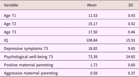
For the present study, only participants who completed outcome measures at T3 were included in analyses. Accordingly, the final data set used for analyses included 118 participants for depressive symptoms and 119 adolescents for psychological well-being. Sample characteristics are listed in Table 1. Shapiro–Wilk normality tests were implemented to assess the distributions of the EATQ-R dimensions for the analyzed sample. Effortful control was normally distributed p > .05, and though negative emotionality was not normally distributed p = .03, but was closer to normal than the selected sample of 415 individuals. Distributions are illustrated in online-only supplementary Figure S.2.
Sample characteristics

Note: T1, Time 1. T2, Time 2. T3, Time 3.
Measures
MRI image acquisition
Scans were completed at T1 on a 3Tesla GE scanner at the Brain Research Institute, Austin and Repatriation Medical Centre, Melbourne, Australia. The following parameters were used: repetition time = 36 ms; echo time = 9 ms; flip angle = 35°, field of view = 20 cm, 124 T1-weighted contiguous slices (voxel dimensions = 0.4883 × 0.4883 × 1.5 mm). For T2 and T3, scans were completed on a 3Tesla Siemens scanner at the Royal Children's Hospital, Melbourne, Australia. Parameters included the following: repetition time = 1900 ms; echo time = 2.24 ms; flip angle = 9°, field of view = 23 cm; 176 T1-weighted contiguous slices (voxel dimensions = 0.9 mm3).
MRI image processing
Images were processed at Melbourne Neuropsychiatry Centre, Melbourne, Australia and were transferred to an SGI/Linux workstation for analysis. The FreeSurfer image analysis suite was used for cortical and subcortical reconstruction (http://surfer.nmr.mgh.harvard.edu). FreeSurfer enables topologically correct and geometrically accurate surface models of inner and outer cortical boundaries and subcortical volume models. To address issues such as geometric distortion and voxel dimension drift, which can compromise longitudinal data collected at multiple sites, images were processed through the longitudinal stream of FreeSurfer 5.3 (Reuter, Schmansky, Rosas, & Fiscil, Reference Reuter, Schmansky, Rosas and Fischl2012). This created a within-subject unbiased template space and average image across time points using robust, inverse consistent registration. Such a template is used in segmentation processes for each time point. It provides common information about anatomical structures and significantly increases reliability and statistical power (Reuter & Fischl, Reference Reuter and Fischl2011; Reuter, Rosas, & Fischel, Reference Reuter, Rosas and Fischl2010). Images were inspected for reconstruction quality, and manual edits were made where necessary.
Furthermore, reliability analysis was carried out given that different scanners were used at T1 and T2/T3, ensuring that changes in cortical thickness and subcortical volume over time were not due to measurement bias related to the scanner platforms and acquisition parameters. An independent sample of adults underwent scans at both sites, and a reliability analysis indicated that the change in scanners between T1 and T2 did not produce any bias in number of cortical and subcortical regions. The majority of regions analyzed in this study were examined in the interscanner reliability study: those that were checked were regarded as reliable. Further detail has been outlined in previous papers analyzing data from the same cohort (Dennison et al., Reference Dennison, Sheridan, Busso, Jenness, Peverill, Rosen and McLaughlin2016; Vijayakumar et al., Reference Vijayakumar, Whittle, Yücel, Dennison, Simmons and Allen2014, Reference Vijayakumar, Allen, Youssef, Dennison, Yücel, Simmons and Whittle2016, Reference Vijayakumar, Allen, Dennison, Byrne, Simmons and Whittle2017).
Calculation of brain development scores
A central aim of this study was investigating individual variability in developmental change as a marker of differential susceptibility or vulnerability. Brain development was operationalized here by measuring structural brain development: specifically, change in cortical thickness and subcortical volume across time. Brain development scores were calculated to capture trajectories of change in thickness for 11 cortical and 6 subcortical regions (outlined in online-only Supplemental Table S.1). Traditional change scores calculate the difference between two measurements, such as scores at baseline and follow-up. However, this approach was not employed because the data set was unbalanced as individuals underwent different numbers of scans (1–3) across time points. Two participants had data for T1 only, three had data for T2 only, and one participant had data for T3 only. To make use of all available data across the three time points, we calculated a change score for each individual reflected by the random age slope from linear mixed models. These analyses were implemented using the Linear and Nonlinear Mixed Effects Models (nlme) package in R 3.3.3 (Pinheiro, Bates, DebRoy, & Sarkar, Reference Pinheiro, Bates, DebRoy and Sarkar2018; R Development Core Team, 2008). Random slopes from linear mixed models are based on all available data for each individual, regardless of whether they underwent one, two, or all three scans. These variables were calculated by identifying models of best fit for each brain region. Models were compared using various combinations of age and sex to predict structural brain development. Prior research indicates that sex is an important predictor of brain development (Herting et al., Reference Herting, Johnson, Mills, Vijayakumar, Dennison, Liu and Tamnes2018; Wierenga et al., Reference Wierenga, Bos, Schreuders, Peper, Tamnes and Crone2018). As such, we chose to employ a strategy that was consistent with a number of prior studies on normative brain development (Ducharme et al., Reference Ducharme, Albaugh, Hudziak, Botteron, Nguyen, Truong and Karama2014; Mills, Goddings, Clasen, Giedd, & Blakemore, Reference Mills, Goddings, Clasen, Giedd and Blakemore2014; Mutlu et al., Reference Mutlu, Schneider, Debbané, Badoud, Eliez and Schaer2013; Raznahan et al., Reference Raznahan, Shaw, Lerch, Clasen, Greenstein, Berman and Giedd2014), which was to identify best fitting developmental models based on age and sex. For each model, the random effect of the intercept and age-slope for each i th subject were specified by the d i and d(age)i terms while e ik represented the residual error term. To identify the best fitting development model we examined the following: a null model with no fixed effect predictors (Y = Intercept + d i + d[age]i + e ik); a linear model including age as the only fixed effect predictor (Y = Intercept + d i + d[age]i + β1[age] + e ik); a model including a quadratic effect for age (Y = Intercept + d i + d[age]i + β1[age] + β2[age2] e ik); a model including a main effect of sex (Y = Intercept + d i + d[age]i + β1[age] + β2[sex] + e ik), and another model with an Age × Sex interaction term (Y = Intercept + d i + d[age]i + β1[age] + β2[sex] + β3[Age × Sex] + e ik). More complex models with additional fixed effects were selected only if p < .05 for the added parameter and the Bayesian information criterion was reduced by at least two indicating a better fit. Random slopes for age were extracted from the best fitting model for each brain region. These values are used to indicate brain development over time. Positive and negative random slope scores indicate an increase and decrease, respectively, in the change of an individual's structure over time relative to the average group-level change. For instance, the orbitofrontal cortex has been found to thin across adolescence on average for this sample (Vijayakumar et al., Reference Vijayakumar, Allen, Youssef, Dennison, Yücel, Simmons and Whittle2016), so a negative score would indicate relatively greater thinning while a positive score would represent relatively reduced thinning. For the sample used in this study, random slopes and traditional difference scores (calculated by subtracting T3 from T1 scores) were highly correlated (reported in Table S.2 of online-only supplementary material) indicating that random slopes appropriately capture observed change in brain development.
Depressive symptoms
Adolescent depressive symptoms were measured at T1 and T3 using a self-report measure, the Center for Epidemiological Studies-Depression Scale (CES-D; Radloff, Reference Radloff1977), which includes 20 items that assess mood, somatic complaints, relations with others, and motor function over the past week. The CES-D has good validity and reliability for adolescents and is widely used with this population (Beck, Brown, Epstein, & Steer, Reference Beck, Brown, Epstein and Steer1988). Cronbach's α for the current sample was 0.89. Depressive symptom data was not imputed for either time point.
Psychological well-being
At T3 participants completed the self-report Young Adult Quality of Life questionnaire (YAQOL; Chen et al., Reference Chen, Cohen, Kasen, Gordan, Dufur and Smailes2004). This measure includes a scale called positive outlook (derived from five items about feeling optimistic and satisfied) designed to measure psychological well-being. We operationalized psychological well-being using this positive outlook scale. Higher scores indicate better well-being with a minimum score of zero and a maximum of 100. The YAQOL is designed specifically for young adults between the ages of 18 and 25 and has good validity and reliability as reported by Chen et al. (Reference Chen, Cohen, Kasen, Gordan, Dufur and Smailes2004). Cronbach's α for the current sample was 0.87.
Mother–child interactions
At T1, 160 mother–child dyads (out of the broader 245 participants) completed two family interaction tasks. Dyads completed an Event Planning Interaction (EPI) during which they planned fun activities together. These activities were chosen from the Pleasant Events Checklist, a modified version of the Pleasant Event Schedule (Macphillamy & Lewinsohn, Reference Macphillamy and Lewinsohn1982). Dyads also completed a Problem-Solving Interaction (PSI) during which participants discussed and resolved areas of conflict chosen from the Issues Checklist, such as “talking back to parents” (Prinz, Foster, Kent, & O'Leary, Reference Prinz, Foster, Kent and O'Leary1979). Each task lasted 20 min and was audio-visually recorded. Of the final sample (N = 119), 98 mother–child dyads had complete parenting data. Missing positive and aggressive maternal behavior data were imputed using the Expectation–Maximization function in SPSS version 22. Little's missing completely at random test indicated that data were missing completely at random (χ2 = 0.09, df = 1, p = .77), suggesting the appropriateness of a single imputation method.
Coding of parent–child observational data
Affective and verbal content from the parent–child recordings was coded using the Living in Family Environments coding system, an observational coding system in which a new code is entered each time the verbal or affective content changes within an interaction (Hops, Davis, & Longoria, Reference Hops, Davis and Longoria1995). The system includes 10 codes for affective content (e.g., happy and angry) and 27 codes for verbal content (e.g., validation and affection). A positive parenting behavior construct was created including affective behavior that was happy or caring, along with approving, validating, affectionate, or humorous comments made with neutral affect. A negative (aggressive) parenting behavior construct included codes with contemptuous, angry, or belligerent affect, and also included cruel, provocative, annoying/disruptive, or argumentative verbal statements made with neutral affect. For both constructs, the frequency of behavior was recorded, that is, the average number of times per minute that a mother expressed aggressive/positive behavior. Prior research conducted with the data set used for the present study indicates that context influences the impact of parental behavior on adolescent outcomes (Schwartz et al., Reference Schwartz, Dudgeon, Sheeber, Yap, Simmons and Allen2011). That is, parental aggression expressed during a task that is intended to be pleasant (i.e., EPI) or parental positive affect conveyed during problem solving (i.e., PSI), when conflict might otherwise easily arise, have been shown to predict adolescent outcomes such as depression (Schwartz et al., Reference Schwartz, Byrne, Simmons, Whittle, Dudgeon, Yap and Allen2013). Similar findings have been reported in marital research; for example, positive affect during marital conflict improves marital satisfaction (Gottman, Coan, Carrere, & Swanson, Reference Gottman, Coan, Carrere and Swanson1998). Therefore, for this study, frequency of positive maternal behavior during the PSI, and frequency of aggressive parental behavior during the EPI, were used in analyses. The scores for positive and aggressive maternal behavior were significantly negatively correlated r = –.41, p < .001. Researchers with extensive training who were blinded to participant details and study hypotheses coded the parent–child interaction data. A separate researcher coded approximately 20% of recordings to estimate interscorer agreement: κ coefficients (a conservative index that controls for chance agreement) for aversive and positive parenting constructs were 0.70 and 0.86 (Hops et al., Reference Hops, Davis and Longoria1995). The Living in Family Environments coding system has good validity as a measure of family processes and has been used previously in studies of adolescent depression (Katz & Hunter, Reference Katz and Hunter2007).
Statistical analysis
Linear regressions were performed using R to examine whether interactions between maternal behaviors and brain development (change in cortical thickness/subcortical volume) from early to late adolescence predicted adolescent depressive symptoms and psychological well-being at T3. CES-D scores at T3 and psychological well-being scores from T3 were used as dependent variables. All independent variables were centered for analysis, and sex was entered as a covariate. CES-D at T1 was included as a covariate for depression models (we could not control for baseline well-being as we did not administer the YAQOL at T1). Analyses were performed for hypothesis-driven regions in the frontal lobe and subcortex, with separate models run for each left and right hemispheric regions (acquired from FreeSurfer 5.3's Desikan-Killiany atlas and listed in online-only Supplemental Table S.1). Inclusion of temperament dimensions effortful control and negative emotionality as covariates did not alter significant results. Three-way interaction terms (Parenting × Brain × Temperament) were not significant for either outcome variable.
For each dependent variable, statistical analysis was run for 11 cortical and 6 subcortical regions and two parenting variables (positive and aggressive maternal behavior), totaling 68 comparisons. As such, the p value was set at .01028 using a modified version of the Benjamini and Yekutieli method to correct for multiple comparisons utilizing a false discovery rate of 5% (Narum, Reference Narum2006). Interaction effects were the focus of this study; therefore, only models with significant Brain × Parenting effects were further interpreted. Other nonsignificant models are reported in online-only Supplemental Table S.3.
According to the Shapiro–Wilk normality test, in the final sample, depressive symptoms were positively skewed (p < .001), while psychological well-being was negatively skewed (p < .001), consistent with prior research in community samples (Chen et al., Reference Chen, Cohen, Kasen, Gordan, Dufur and Smailes2004). Kurtosis was present for depressive symptoms (z = 2.11, p < .05), but not for psychological well-being (z = 1.65, p > .05). For all significant models (outlined in the Results section), regression residuals were examined using the Shapiro–Wilk normality test. Distributions were not normal, suggesting that normality assumptions for parametric testing were violated. Bootstrapping was implemented using R, resampling many smaller samples from the sample. Statistics were then calculated for these “bootstrap” samples allowing the sampling distribution to be estimated. The standard deviation of the estimated sampling distribution was used to calculate the standard error of the statistic, which was then used to calculate significance tests (Field, Reference Field2009).
Roisman indices
Roisman indices and model effects are illustrated in Figure 1. Roisman et al. suggest several indices to distinguish between diathesis-stress (ordinal) and differential susceptibility (disordinal) effects for continuous variables: the regions of significance (RoS) on X, the proportion of the interaction (PoI), and the proportion affected (PA). A test of the RoS on X examines the values of the independent variable (i.e., parenting, X) for which the moderator (i.e., brain development, Z) and the outcome variable (i.e., depression/well-being, Y) are significantly associated (Kochanska et al., Reference Kochanska, Kim, Barry and Philibert2011; Roisman et al., Reference Roisman, Newman, Fraley, Haltigan, Groh and Haydon2012). This provides a formal test to assess whether the association between Z and Y occurs at values of X that fall within a particular range of interest (+/–2 SD of the mean of X, or 95% of a normally distributed sample). For the RoS on X, evidence for a diathesis-stress effect (ordinal interaction) is observed if a significant association between Z and Y occurs at the values of X that indicate greater adversity, that is, low levels of X for positive parenting, or high levels of X for aggressive parenting. Evidence for a differential susceptibility effect (disordinal interaction) is observed if a significant association between Z and Y occurs at both low and high levels of X (positive/aggressive parenting).
The PoI and the PA are indices that supplement the RoS on X and, unlike the RoS on X, which relies on significance testing, the PoI and PA are unaffected by sample size (Roisman et al., Reference Roisman, Newman, Fraley, Haltigan, Groh and Haydon2012). The PoI captures the proportion of the interaction that reflects a better (B) rather than worse (W) outcome for participants (B/[B+W]). Prototypically, differential susceptibility is inferred when B = 50% though Roisman et al. indicate that values of 40%–60% are highly consistent with differential susceptibility (other researchers have suggested that this range could be further widened; Del Giudice, Reference Del Giudice2017). For diathesis-stress, however, the Roisman guidelines stipulate that a PoI score of 0% lends support to diathesis-stress; they do not stipulate a range of scores that could provide evidence for this model (no 20% margin). Therefore, the stipulated range for the PoI index is not comparable for both diathesis-stress and differential susceptibility. To address this, we applied a comparable margin of 20%, such that a PoI score of 0%–20% provides support for diathesis-stress. We considered that, without such a margin, the criteria that indicated support for differential susceptibility was more easily satisfied compared to the criteria for diathesis-stress. Calculation of the PoI requires a crossover value, the value of X at which the regression lines cross over in a disordinal interaction. For a negative outcome variable (depression), better outcomes fall below this crossover point, and for positive outcome variables (well-being), better outcomes fall above the crossover point. Similar to the PoI, the PA captures the proportion of individuals affected positively by the interaction between X and Z. The PA is a pragmatic evaluation of a differential susceptibility effect: if the RoS on X index is satisfied but the PA indicates that only 2% of individuals are benefited, then arguably the model is not very useful (Roisman et al., Reference Roisman, Newman, Fraley, Haltigan, Groh and Haydon2012). Prototypically, a value of 50% for the PA index lends support to differential susceptibility; 0% supports diathesis-stress. A value of 16% or less indicates questionable support for differential susceptibility, while less than 2% indicates clear lack of support (these percentages fall 1 and 2 SD above the mean of a normal distribution). Finally, Roisman et al. also recommend that quadratic terms be entered for X (X 2) and the interaction term XZ (ZX 2) to eliminate the possibility of erroneously detecting a differential susceptibility effect by imposing a linear model on quadratic data. Thus, significant quadratic terms for the independent variable were also tested and controlled for where necessary. Interaction plots and calculation of the Roisman indices were carried out using an online instrument developed by Chris Fraley (www.yourpersonality.net/interaction). Independent variables were standardized for plotting.
Results
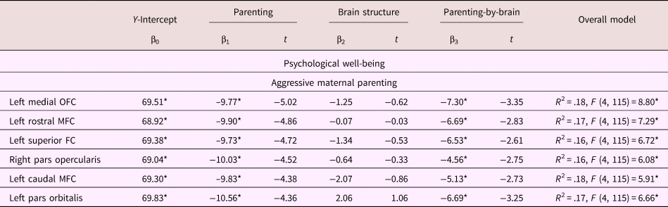
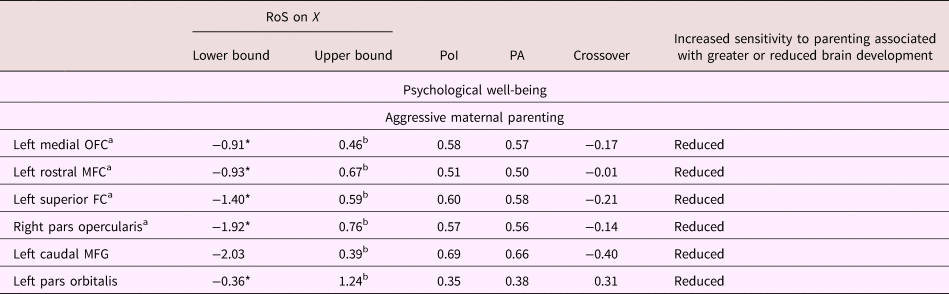
Significant results for regression main effects and interactions are reported in Table 2 and results for Roisman indices are reported in Table 3. Significant interactions (that survived correction for multiple comparisons) occurred in the prediction of psychological well-being only.
Results for regression models, which included significant Parenting × Brain interactions

Note: The regression equation: Y(depression/well-being) = β0(Intercept) + β1X(parenting) + β2Z(brain) + β3XZ(Parenting × Brain). Statistical significance was determined from models using mean-centered independent variables. Regression coefficients (i.e., standardized beta weights; β) were derived from regressions using standardized independent variables (z scores); interaction terms were also calculated from z scores. According to Friedrich (Reference Friedrich1982), β coefficients of interaction terms obtained from unstandardized coefficients are incorrect (i.e., the z scores of product terms is not equal to the product of z scores). Depressive symptoms and well-being were significantly negatively correlated, r (119) = –.52, p < .001. Corrected α level for 68 comparisons (false discovery rate = .05) using Benjamini and Yekutieli method (Narum, Reference Narum2006). OFC, orbitofrontal cortex. MFC, middle frontal cortex. FC, frontal cortex. *p < .01028.
Roisman indices reported for regression models, which included significant Parenting × Brain interactions

Note: aModel meets criteria for differential susceptibility according to Roisman et al. (Reference Roisman, Newman, Fraley, Haltigan, Groh and Haydon2012). Parenting behaviors were not significantly correlated with thickness of any of the brain regions in these models. bOutcomes for which the lower and/or upper RoS for parenting (RoS X) fall within +/–2 SD of the parenting mean. RoS, regions of significance. PoI, proportion of the interaction. PA, proportion affected. OFC, orbitofrontal cortex. MFC, middle frontal cortex. FC, frontal cortex. *p < .01028.
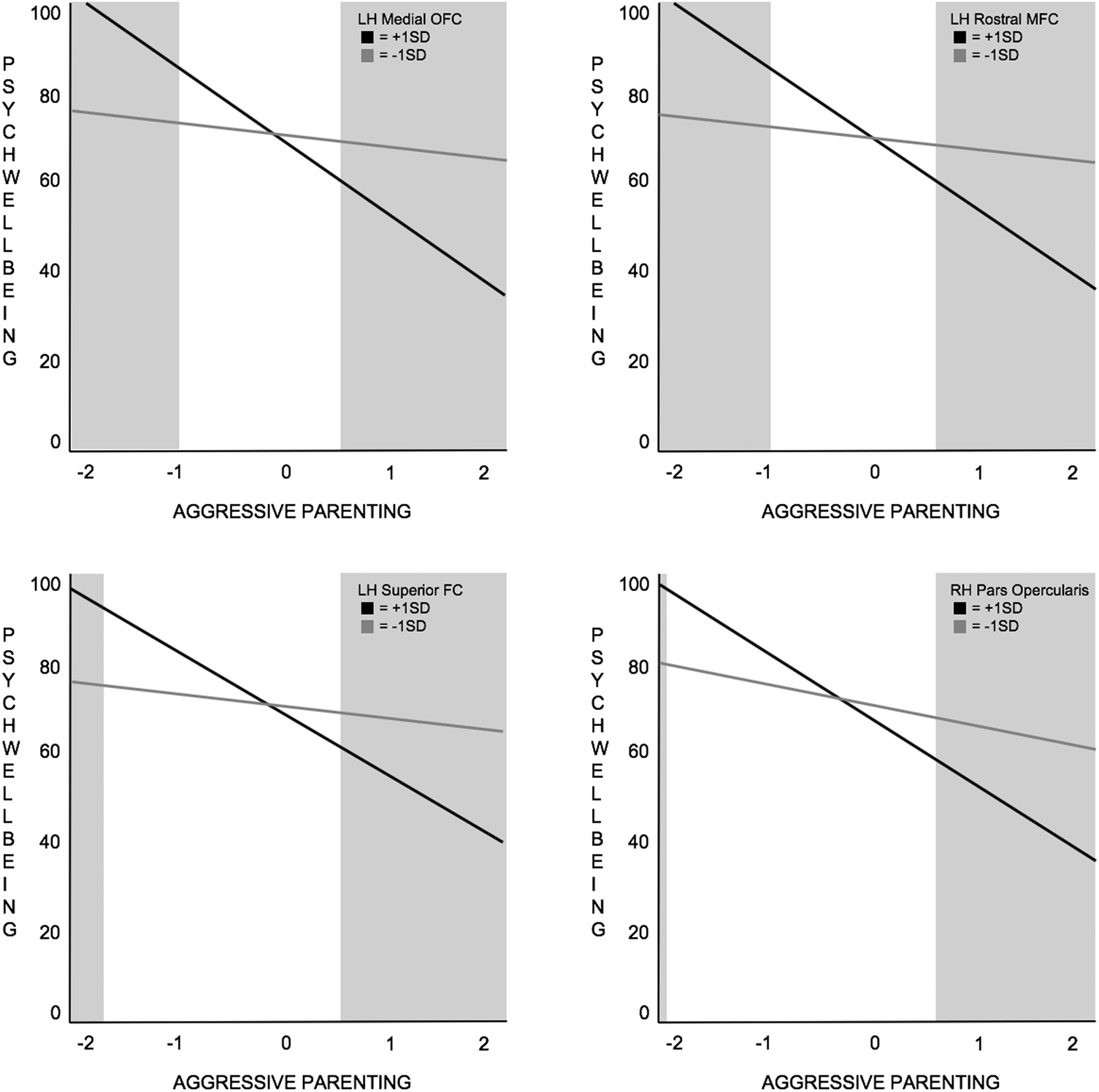
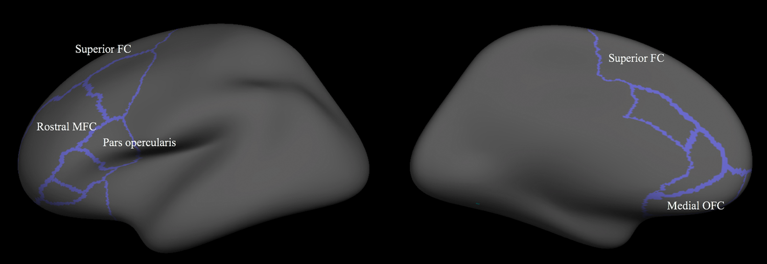
Aggressive maternal behavior interacted with development of the left medial orbitofrontal cortex, left caudal and rostral middle frontal cortices, left superior frontal cortex, left pars orbitalis, and right pars opercularis in the prediction of adolescent psychological well-being. Only models that included the left medial orbitofrontal, rostral middle frontal and superior frontal cortices, and right pars opercularis satisfied Roisman indices as differential susceptibility effects (see Table 2 and 3 for model results). For all regions, reduced thinning was associated with increased susceptibility to aggressive maternal behavior, as Figure 2 illustrates. Furthermore, as shown in online-only Supplemental Figure S.1, models that did not meet Roisman criteria showed similar effects to those that did. Positive maternal behavior did not significantly interact with brain development in any region to predict well-being. Subcortical regions were not implicated in any model in the prediction of psychological well-being (see Supplemental Table S.3 for all results). Brain regions highlighted in differential susceptibility effects are outlined in Figure 3. We considered it possible that including baseline depressive symptoms may have contributed to a lack of significant findings for depression models. To rule out this possibility, depression models were rerun excluding CES-D T1 scores. Excluding the CES-D T1 covariate did not change results, as shown in Table S.4 of the online-only supplement, suggesting that controlling for baseline depression was not driving the results.
Interaction plots illustrating differential susceptibility effects. Adolescent psychological well-being predicted by aggressive maternal parenting and structural brain development. From left to right (upper figures): left medial orbitofrontal cortex and left rostral middle frontal cortex; (lower figures): left superior frontal cortex and right pars opercularis. All figures illustrate differential susceptibility effects for which reduced cortical thinning is associated with higher susceptibility to high and low levels of aggressive parenting. RH, right hemisphere. LH, left hemisphere. MFC, middle frontal cortex. FC, frontal cortex. OFC, orbitofrontal cortex.

Illustration of regions of interest (outlined). Labeled regions are those that were implicated in significant models.

Discussion
The aim of this study was to examine whether structural brain development interacted with positive and/or aggressive maternal parenting behavior to predict positive and/or negative late adolescent outcomes, specifically depressive symptoms and psychological well-being. Furthermore, we examined whether results were consistent with diathesis-stress or differential susceptibility according to indices by Roisman et al. (Reference Roisman, Newman, Fraley, Haltigan, Groh and Haydon2012). This is the first study to investigate brain development in the context of these Biology × Environment interaction models, and to investigate both positive and negative environmental contexts and outcomes. Although structural development in both frontal cortical and subcortical regions were analyzed, significant interactions were only identified with frontal structures, and results were consistent with differential susceptibility.
Our findings indicated that development of the left medial orbitofrontal cortex, rostral middle frontal and superior frontal cortices, and right pars opercularis interacted with aggressive maternal behavior to predict adolescent psychological well-being, illustrating differential susceptibility effects. Individuals with reduced thinning in these regions demonstrated high susceptibility to aggressive maternal behavior. That is, they reported higher psychological well-being in the context of reduced aggressive maternal behavior and lower psychological well-being when subjected to more aggressive behavior. The orbitofrontal region is involved in processing socioemotional contextual information (Casey, Getz, & Galvan, Reference Casey, Getz and Galvan2008; Nelson et al., Reference Nelson, Leibenluft, McClure and Pine2005; Schriber & Guyer, Reference Schriber and Guyer2016). More specifically, the medial orbitofrontal cortex is thought to play a role in processing the reward value of reinforcing stimuli (Kringelbach, Reference Kringelbach2005). We speculate that it is perhaps as a consequence of these socioemotional functions that medial orbitofrontal cortex development is particularly sensitive to high and low levels of aggressive parenting behaviors in the prediction of well-being. Development of the superior frontal and rostral middle frontal cortices are implicated in emotion regulation, and specifically, these regions have been linked to the reappraisal of negative emotion (Goldin, McRae, Ramel, & Gross, Reference Goldin, McRae, Ramel and Gross2008). Moreover, reduced cortical thinning of these regions has been linked to poorer adolescent emotion regulation (Vijayakumar et al., Reference Vijayakumar, Whittle, Yücel, Dennison, Simmons and Allen2014). It is possible that these regulatory functions relate to the increased sensitivity to high and low levels of aggressive parenting. Our findings also identified the pars opercularis, a region that is part of the inferior frontal cortex and supports multiple functions involved in interpersonal interactions and communication, including processing of language, facial emotion recognition, and empathy (Liakakis, Nickel, & Seitz, Reference Liakakis, Nickel and Seitz2011; Skuse & Gallagher, Reference Skuse and Gallagher2009). The pars opercularis itself is involved in speech, language comprehension, and cognitive control (Clos, Amunts, Laird, Fox, & Eickhoff, Reference Clos, Amunts, Laird, Fox and Eickhoff2013). Cortical thickness in this region has been linked to depression (Qiu et al., Reference Qiu, Lui, Kuang, Huang, Li, Li and Gong2014); however, our findings suggest that negative environments may also interact with development of the right pars opercularis, heightening sensitivity to high and low levels of negative contexts in the prediction of positive developmental outcomes.
Past research has found associations between greater cortical thinning in prefrontal regions (overlapping with those implicated here) and better emotional outcomes related to emotion regulatory abilities and lowered depressive symptomatology (Ducharme et al., Reference Ducharme, Albaugh, Hudziak, Botteron, Nguyen, Truong and Karama2014; Vijayakumar et al., Reference Vijayakumar, Whittle, Yücel, Dennison, Simmons and Allen2014, Reference Vijayakumar, Allen, Dennison, Byrne, Simmons and Whittle2017). Conversely, some evidence suggests that reduced thinning in frontal regions is associated with positive psychological traits and reduced vulnerability to psychopathology (Bos et al., Reference Bos, Peters, Van de Kamp, Crone and Tamnes2018; Cannon et al., Reference Cannon, Chung, He, Sun, Jacobson, Van Erp and Heinssen2015; Friedel et al., Reference Friedel, Whittle, Vijayakumar, Simmons, Byrne, Schwartz and Allen2015). Our findings might help to explain these inconsistent results by suggesting that more or less thinning is neither “good” nor “bad,” and that associations between structural brain development and psychological functioning are better explained by considering environmental experience. That is, brain development may influence functional outcomes in conjunction with environmental adversity and support.
Of note, only models whereby brain development interacted with aggressive parenting to predict psychological well-being were significant and supported differential susceptibility. It is unclear why individual differences in brain development were not associated with sensitivity to positive maternal behaviors, or why brain development and parenting behaviors did not interact to predict adolescent depression. It is possible that other aspects of neurobiology not captured here (e.g., brain function) may better capture sensitivity to positive maternal behavior. We have previously found that reduced frequency of positive maternal behaviors is associated with risk for clinical depression (i.e., major depressive disorder; Schwartz et al., Reference Schwartz, Simmons, Whittle, Byrne, Yap, Sheeber and Allen2017). Thus, it is possible that brain development may interact with positive maternal behaviors to predict more extreme or simply other mental health outcomes not examined here. Furthermore, it is plausible that for positive parenting the “dose” required for a significant interaction effect is higher than that for aggressive parenting. That is, low-level aggression over a short period might trigger a significant interaction effect, but for positive parenting perhaps a larger dose over a longer period of time is required for a similar effect. An additional possibility might relate to the complex interplay of environmental and biological factors that contribute to the development of depression and well-being. It is possible that the inclusion of additional environmental variables, such as community and peer-related factors, may better explain individual variability in adolescent outcomes particularly in relation to depression (Cicchetti & Natsuaki, Reference Cicchetti and Natsuaki2014; Sellström & Bremberg, Reference Sellström and Bremberg2006).
While our study is the first to investigate longitudinal brain development as a moderator of environmental influences on adolescent mental health, there are also limitations to consider. First, we limited our study to investigating frontal cortical thickness and subcortical volume development (rather than other properties of brain structure). While this was consistent with prior research on parenting and depression (Schriber et al., Reference Schriber, Anbari, Robins, Conger, Hastings and Guyer2017; Whittle et al., Reference Whittle, Yap, Sheeber, Dudgeon, Yücel, Pantelis and Allen2011), future research could examine whole-brain approaches, using other measures of structural change such as surface area, as well as measures of functional development. Second, we examined psychological well-being as a positive outcome variable, so as to examine positive adolescent development explicitly rather than just the absence of negative development (i.e., low depressive symptoms). While our operationalization of psychological well-being was limited to positive outlook (which captures positive mindsets), positive development could be captured in many other ways depending on how it is defined (Chen et al., Reference Chen, Cohen, Kasen, Gordan, Dufur and Smailes2004). Future research should also include other measures of positive adjustment, an aspect of development that is infrequently, and often less clearly, defined in the literature compared to poor development. Third, our research design was limited to one aspect of environment, namely, parenting observed in a lab setting. During adolescence individuals seek connections beyond the family and may therefore be particularly susceptible to influence by a range of environmental factors. Although our findings indicate that parenting remains an important and continuing contributor to outcomes during adolescence, future research might examine broader environmental factors such as socioeconomic status, neighborhood disadvantage, and peer relationships. This could offer a more multilevel approach to Biology × Environment interactions. Fourth and finally, in order to test for differential susceptibility, we investigated both positive and negative environmental exposure, and positive and negative outcomes. However, using single variables that range from positive on one end of the continuum to negative on the other would be a more parsimonious approach to investigating both sides of the “for-better-and-for-worse” hypothesis. Therefore, future research should investigate the use of such variables.
The current investigation is the first to employ systematic indices to explore whether brain development might be considered an indicator of differential susceptibility or vulnerability to positive and aggressive parenting in the prediction of positive and negative mental health outcomes in adolescence. Our findings highlight that reduced thinning of the left medial orbitofrontal cortex, middle frontal and superior frontal cortices, and right pars opercularis predicts high susceptibility to the effects of aggressive parenting in the prediction of psychological well-being. These findings suggest that, rather than interpreting reduced thinning of these regions as either “good” or “bad,” it may be more meaningful to consider that less structural development renders individuals more sensitive to the detrimental and beneficial impact of more or less adverse/supportive environmental influence. Our results contribute to a more nuanced picture of pathways to positive mental health. Furthermore, these findings have implications for adolescent mental health interventions, as the consideration of individual differences in neurobiological susceptibility may improve our understanding of responsiveness to treatment.
Financial support
This study was funded by the Australian Research Council (Discovery Project ID: DP130103551). Sarah Whittle was supported by an NHMRC Career Development Fellowship (ID: 1007716). Christos Pantelis was supported by a NHMRC Senior Principal Research Fellowship (628386 and 1105825).
Supplementary material
The supplementary material for this article can be found at https://doi.org/10.1017/S0954579418001475.