Introduction
Secondary or referred otalgia can represent a diagnostic challenge to the otolaryngologist. The differential diagnosis is long, and although a thorough history and examination may establish the diagnosis, dental causes of referred otalgia may be overlooked. Indeed, secondary otalgia due to dental disorders may account for up to 50 per cent of all cases referred to the ENT clinic.Reference Yanagisawa and Kveton1 In part, this may be due to deficiency in dental diagnostic skills. Full examination of the oral cavity, and in particular the dentition, is a skill acquired through formal training, as gained through the dental degree course, and is taught to a much lesser extent in formal medical training.
In patients presenting with otalgia without an obvious cause, further investigations (such as computed tomography and magnetic resonance imaging) are often performed in order to exclude other causes, most notably central nervous system lesions. With hindsight, many of these patients have a dental cause, and, had the diagnosis been made at the initial examination, costly investigations could have been prevented. Diagnosis or exclusion of temporomandibular joint (TMJ) dysfunction syndrome and common dental disorders can be performed rapidly and reliably in the ENT clinic, using standard examination instruments.
This article aims to give a succinct overview of common dental causes of otalgia, highlighting the most characteristic symptoms and signs in order to aid differential diagnosis. We also provide a guideline to enable ENT clinicians to perform a rapid and simple dental and TMJ examination, which will reliably diagnose or exclude dental otalgia.
Dental otalgia (i.e. otalgia secondary to dental disease) is the most common cause of secondary otalgia,Reference Kreisberg and Turner2 and TMJ dysfunction syndrome represents the most common dental cause.Reference Wazen3 Decay and pulpal inflammation of posterior teeth can also frequently present as otalgia. Other dental causes of otalgia include the pain of tooth eruption (common in infants), tooth fracture or a dry socket following extraction.Reference Kreisberg and Turner2, Reference Harvey4
Referred otalgia secondary to dental and TMJ disorders can be explained by the intimately related sensory innervation of the mouth and ear. The embryological development of the head and neck region, in particular the first branchial arch, explains the common innervation of the ear, jaw and dentition. The external cleft of the first branchial arch later forms the external auditory meatus. The internal pharyngeal pouch of the first arch forms the eustachian tube, the middle-ear cavity and the mastoid antrum. The cartilage of the first arch forms the incus and the malleus; the mesoderm forms the muscles of mastication. The nerve of the first branchial arch is the Vth cranial nerve, which primarily supplies the structures mentioned.
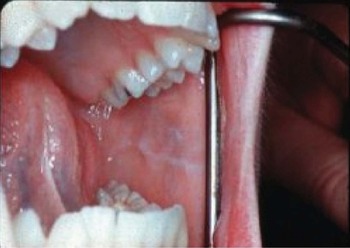
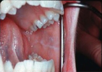
Hypertrophic linear lesion (‘linea alba’) on the buccal mucosa at the occlusal plane, consistent with bruxism.
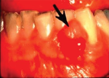
Discharging sinus due to chronic apical periodontal abscess or chronic apical periodontitis.

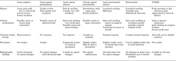
Characteristic features of common dental conditions presenting as referred otalgia

TMJDS = temporomandibular joint dysfunction syndrome
The ear has multiple innervations, including the trigeminal (Vth), facial (VIIth), glossopharyngeal (IXth) and vagus (Xth) nerves, as well as superficial sensory branches from the cervical plexus (i.e. lesser occipital (C2) and greater auricular (C2 and C3) nerves). Similarly, the TMJ is innervated by the Vth (auriculotemporal branch) and VIIth cranial nerves. The maxillary branch of the Vth nerve supplies the mucous membranes of the upper mouth and the maxillary gums and teeth. The mandibular branch of the Vth nerve also supplies the mucous membranes of the cheek, the anterior two-thirds of the tongue, the floor of the mouth and the lower teeth and gums.
Common causes of dental otalgia
We may subdivide dental causes of otalgia as being related to disorders of the oral mucosa, the teeth and the TMJ.
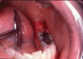
Typical appearances of pericoronitis of lower wisdom tooth.

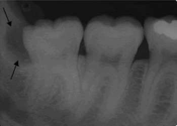
Dental X-ray showing radiolucency distal and posterior to lower wisdom tooth, commonly seen due to formation of a paradental cyst secondary to chronic pericoronitis.

Oral mucosal inflammation
Given that the trigeminal nerve (Vth) supplies the sensation of both the ear and the mucous membranes of the oral cavity, infection and inflammatory disorders of the oral cavity (e.g. aphthous ulceration) may produce otalgia.
Referred otalgia secondary to tooth disorders
Dental pain originating from the teeth (i.e. pulpitis) and periodontal tissues (periodontitis) poses a significant diagnostic problem to the trained dentist. It is therefore not surprising that these same problems pose a diagnostic challenge when presenting to the ENT clinic as secondary otalgia.
Pulpitis
The tooth is composed of an outer calcified shell (the enamel and dentine) and inner soft tissue (the dental pulp). The dental pulp contains nerves, blood vessels and connective tissue, which maintain the outer, inanimate, calcified matter. The blood supply and innervation of the dental pulp enters the tooth through small foraminae at its root apex. Pulpal pain arises secondary to inflammation of the dental pulp, and may arise as a result of thermal, chemical or infective insults. The most common cause is dental caries. As sensory innervation of the pulp comprises mainly non-myelinated, slow conducting, type C pain fibres, the pain of pulpitis is typically poorly localised.Reference Ten Cate5 Often, the patient is unable to tell whether the source is in the upper or lower jaw. The pain does not cross the midline, is characterised by aggravation with hot or cold stimuli, and persists without stimulus in more advanced stages of disease. The tooth is usually not tender to percussion. This is an important diagnostic sign which may differentiate pulpitis from acute apical periodontitis. There may be some evidence of the cause of pulpitis, such as caries, heavy restoration or loss of tooth structure. Depending upon its severity and cause, the pain can be sharp and brief, throbbing, or dull and lasting several hours to days.
Chronic pulpitis causes mild, poorly localised pain that often comes and goes over a period of weeks or months. With poor localisation and lack of positive signs, chronic pulpitis represents one of the most difficult conditions to recognise.
Apical periodontitis
Each tooth is anchored to the jaw, within its socket, by its surrounding periodontal ligament. The periodontium is well vascularised and richly supplied by somatic sensory nerve fibres. Apical periodontitis results from irreversible pulpitis and pulp death, and the subsequent extrusion of the necrotic and inflammatory material into the apical periodontal tissues, through the apical foraminae. The periodontium is well innervated with myelinated, rapid conducting, Aδ nerve fibres; therefore, this condition typically presents as a severe, throbbing pain well localised to the affected tooth.Reference Ten Cate5 Inflammation and oedema between the two hard tissues of the bone and the tooth root result in extrusion of the affected tooth. This may be evident to the patient at rest or on biting. The tooth is usually very tender to percussion. These two clinical findings are important diagnostic signs that differentiate apical periodontitis from pulpitis. There may also be bony tenderness over the apex of the tooth within the adjacent alveolar bone.
However, chronic and mild periodontal infections can present as a poorly localised, persistent, dull ache that may last hours to days. These patients are more likely to present to the ENT clinic with diffuse earache.
Acute apical abscess
Acute apical periodontitis may proceed to abscess formation. Symptoms associated with apical periodontitis may worsen. Apical tenderness is increasingly evident, and a tender swelling (‘gum boil’) may present where the abscess erodes through the alveolar bone (Figure 2). Local and systemic signs of an abscess become apparent, and the diagnosis can be made. However, patients may present early, before obvious jaw swelling, or after spontaneous drainage. The affected tooth may be raised (affecting the patient's occlusion) and tender to touch.
‘Impacted’ third molars
Partially erupted or impacted ‘wisdom teeth’ are common and do not cause pain in themselves. However, food and bacterial plaque can easily accumulate in the space between the crown of the partially exposed tooth and the overlying flap of soft-tissue (operculum), predisposing to inflammation and infection of the associated soft tissue. This is called pericoronitis (Figure 3). The swollen and oedematous operculum can be subject to occlusal trauma from the opposing teeth, which further exacerbates the inflammation. Acute pericoronitis typically presents with erythema, swelling, pain, tenderness of the gum flap and an offensive taste due to persistent oozing of pus from beneath the flap. Trismus may also be present. In more severe cases, an acute periodontal abscess may form, with facial cellulitis and spread of infection into adjacent surgical spaces. Chronic or subacute pericoronitis can frequently present as poorly localised facial and jaw pain radiating into the ear region, and therefore presenting to the unsuspecting ENT surgeon as referred otalgia (Figure 4).
Temporomandibular joint dysfunction syndrome
The TMJ is a synovial joint and is susceptible to the various arthritic disorders. The most common TMJ disorder is known by a variety of descriptive terms, including TMJ dysfunction syndrome, facial or fascial arthromyalgia, and myofacial pain dysfunction syndrome. Although the reported prevalence of signs and symptoms of TMJ dysfunction syndrome varies considerably between studies because of lack of uniform diagnostic criteria, it has been estimated that between 20–40 per cent of the population may suffer TMJ dysfunction syndrome at some time in their life.Reference De Kanter, Truin, Burgersdijk, Van't Hof, Battistuzzi and Kalsbeek6–Reference Ellis, Hupp and Tucker9 The aetiology is unknown, although many associated and predisposing factors have been described, including stress and bruxism (i.e. teeth clenching or grinding), trauma, and arthritis.Reference Mitchell10, Reference Suvinen, Reade, Kemppainen, Kononen and Dworkin11 The three cardinal features of the condition are pain (of the joint or the masticatory muscles), joint noise, and restricted movement of the jaw (trismus). It is believed that the principal factor responsible for the symptoms of pain and trismus is masticatory muscle spasm.Reference Mitchell10
Tuz et al. reported that 64 per cent of patients suffering TMJ dysfunction syndrome complained of otalgia (in comparison with only 8 per cent of the control group), and that 78 per cent of TMJ dysfunction patients had at least one otological symptom.Reference Tuz, Onder and Kisnisci12 Furthermore, it is well recognised that other otological symptoms such as tinnitus and vertigo are commonly associated with TMJ dysfunction syndrome, and that treatment results in amelioration of otological symptoms.Reference Wright, Syms and Bifano13, Reference dos Reis, Hotta, Ferreira-Jeronymo, de Felicio and Ribeiro14 Indeed, the relationship between TMJ disorders and ear problems was first documented by Wright in 1920, in a description of deafness due to the position of the mandible and TMJ.Reference Curtis15 In 1934, Costen described a syndrome of ear and sinus symptoms related to disturbed function of the TMJ.Reference Costen16
Pain is the most common and prominent complaint of patients with TMJ dysfunction syndrome.Reference Cooper and Cooper17 Classically, it is described as diffuse pain in or around the jaw joint, often radiating up to the temple and down into the neck. However, common variations in pain include deep, dull earache associated with a feeling of fullness in the ear, headaches and referred facial pains. The pain intensity may vary during the day, often being more severe at the start or end of the day. Patients with a night time habit of clenching or grinding the teeth (bruxism) often awake with joint pain which gradually abates during the day. However, in those who clench or grind during the working day, often due to stress, the symptoms are worst in the evenings.
Patients may also report clicking or clunking of the joint and a history of jaw locking. Occasionally, these noises are described as sounds deep within the ear, and it is likely that these patients will be seen initially in the ENT clinic. Joint noises are common in the asymptomatic population and are of little importance in the absence of pain or mechanical symptoms.
Examination usually elicits tenderness of the masticatory muscles on palpation when clenching, especially at the insertion of the masseter muscle along the underside of the zygomatic arch. Joint noises on auscultation (with or without a stethoscope), trismus and abnormal lateral movements of the lower jaw on opening and closing may be present. Restricted jaw movement or trismus may be due to either inflammatory pain or mechanical restriction; the latter often due to antero-medial displacement of the joint meniscus. However, in patients presenting initially to the ENT surgeons, this feature will usually be absent or minimal. Excessive lateral deviations of the mandible upon jaw opening, and staggered movements, suggest abnormal TMJ function and internal derangement.
There are no diagnostic radiological changes unless a degenerative arthropathy or lytic lesion exists. Tenderness of the TMJ and the masticatory muscles represents the most useful and important diagnostic sign. Cooper and Cooper conducted a large study investigating the incidence of TMJ and associated symptoms and signs in over 2700 patients presenting to a specialist TMJ clinic, and found acute TMJ tenderness in 67 per cent of patients.Reference Cooper and Cooper17 Muscle tenderness was elicited in the majority of patients; 85 per cent in the lateral pterygoid, 72 per cent in the medial pterygoid, 52 per cent in the masseters and 55 per cent in the temporalis muscle. Temporomandibular joint sounds were heard by stethoscope auscultation in 38 per cent of patients, and abnormal (i.e. jerky or lateral) mandibular movements were demonstrated in 47 per cent. Features consistent with bruxism include worn incisal edges, flattened occlusal surfaces of the molar teeth, scalloping of the lateral borders of the tongue and ridging of the buccal mucosa at the occlusal line. Bruxism is a major aetiological factor in TMJ dysfunction syndrome, and has been noted to be present in more than 50 per cent of cases.Reference Cooper and Cooper17
From Cooper and Cooper's study (and other sources),Reference Celic, Jerolimov and Panduric8, Reference Ellis, Hupp and Tucker9 it appears that tenderness of the masticatory muscles represents the most reliable and sensitive diagnostic sign of TMJ dysfunction syndrome, whereas tenderness of the TMJ itself is less consistent, being absent in almost half the patients studied.
Most patients are managed satisfactorily with reassurance, soft diet, local heat to the area, avoidance of clenching or grinding, and simple analgesics. However, a minority may require further investigation and treatment by oral and maxillofacial surgeons, using occlusal splints and/or anti-depressant medication in the first instance.
Basic dental examination to exclude common dental causes of otalgia
It is assumed that ear examination is normal.
Intra-oral
Muscle and soft tissue examination
The entire oral mucosa can be readily examined for signs of inflammation or infection, including ulcerations, as part of the general ENT examination. Bruxism is a common predisposing and exacerbating factor of TMJ dysfunction syndrome.Reference Ellis, Hupp and Tucker9 One of the signs of this habit is white, linear scarring (due to chronic traumatic irritation) of the buccal mucosa (the ‘linea alba’) and along the lateral border of the tongue, both adjacent to the occusal plane of the dentition (Figure 1).
The lateral and medial pterygoid muscles are gently palpated intra-orally to elicit masticatory muscle tenderness. Palpation in the upper rear of the mouth, behind and above the last molars, will detect lateral pterygoid muscle tenderness. In the lower part of the back of the mouth, on the inner aspect of the lowest corner of the jaw, are the medial pterygoid muscles. Tenderness of the pterygoid muscles is the most consistent finding in TMJ dysfunction syndrome.Reference Cooper and Cooper17
Examination of dentition
The general condition of the gingivae and teeth provides a rapid assessment of the patient's general oral health and the likelihood of significant oral or dental problems. Gross discrepancies in the relationship between the upper and lower jaws are suggested by: severe overjet (i.e. the lower incisors placed too far behind (>3 mm) the upper incisors (i.e. class II occlusion)); reverse incisal overlap (i.e. the lower incisors overlapping in front of the upper incisors (i.e. class III occlusion)); and excessive overbite (i.e. severe overlap (>3 mm) between the incisor teeth). Some have suggested that these malocclusions may cause significant stress on the TMJ and therefore may predispose to TMJ dysfunction and pain.Reference Pullinger, Seligman and Gornbein18–Reference Vanderas20 Excessive tooth wear (most readily seen on the canine and incisal edges) is a good sign of bruxism, a major contributory factor in TMJ dysfunction syndrome.
One may then focus on the molar teeth and their periodontal tissues in order to exclude obvious infective pathology. Infection of the periodontal tissues will present as localised erythema and swelling. Occasionally, a chronic, discharging sinus can be seen along the gingivae adjacent to the affected tooth. Palpation along both the lingual and buccal aspects of the gingivae and alveolar ridge will elicit exquisite tenderness adjacent to the affected tooth. The most common infective condition is pericoronitis of an impacted lower wisdom tooth.
Those teeth that been heavily restored are at greater risk of infection. Despite the lack of signs to suggest acute infection of a specific tooth, heavily restored teeth should be examined individually by percussion (with an oral mirror) and cold stimulation (by a cotton pledget soaked with ethyl chloride), to detect underlying inflammation. If a tooth is shown to be acutely tender upon tactile or thermal stimulation, the patient should be referred to their dentist for further investigation and management.
Extra-oral examination & TMJ
Firstly, one should observe for facial swelling suggestive of local dental infection (pericorontitis or dental abscess), masseter hypertrophy (bruxism) or inflammation of the TMJ. Mouth opening should be assessed for jerky and/or excessive lateral movements of the lower jaw or subluxation of the TMJ.
The TMJ should be palpated for tenderness, clicking or jerky movements upon jaw opening and closing. Auscultation with a stethoscope may detect mild clicking or crepitus.
The temporalis and masseter muscles should be gently palpated for tenderness. Mouth opening should be measured in order to detect trismus (defined as inter-incisal opening of <3.5 cm), suggestive of either mechanical block or, more commonly, inflammatory restriction due to pain or spasm.
Summary
Dental causes of secondary otalgia are very common. Knowledge of the key features of common dental pathologies can help differentiate and diagnose these conditions, precluding any unnecessary radiographic investigations. This article aims to provide a succinct review of the common dental disorders that frequently cause patients to present to the ENT clinic with (secondary) otalgia, and a brief guidance on simple dental examination for the ENT surgeon to assist in the diagnosis of these dental problems (see Table 1).