


Introduction
Knowledge of the anatomy of the nasal cavity and the paranasal sinus has become more important with the recent popularity of functional endoscopic sinus surgery. The nasal turbinates are important anatomical structures located on the lateral nasal wall. In each nasal cavity, there are usually three nasal turbinates. The inferior turbinate is a separate bone, while the middle and superior turbinates are part of the ethmoid bone.
The nasal turbinates show some anatomical variation, most frequently the concha bullosa variant.Reference Ozcan, Selcuk, Ozcan, Akdogan and Dere1 However, agenesis of the nasal turbinates is extremely rare. Only three cases of inferior concha agenesis have previously been reported.Reference Caylakli, Yilmaz, Hürcan, Ozer and Ozlüoğlu2–Reference Arslan, Aydinlioglu, Bozkurt and Egeli4 To the best of our knowledge, in the English language literature, unilateral agenesis of the middle nasal turbinate has not previously been reported. In this article, we report the first such case.
Case report
A 57-year-old man presented to our clinic with a five-month history of headache, facial pain, postnasal discharge and nasal obstruction. He had previously been prescribed antibiotics, but these had been unhelpful. His past medical history was unremarkable except for gout and kyphosis. He denied any history of nasal surgery or nasal trauma.
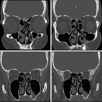
On anterior rhinoscopic examination, a nasal septal deviation towards the left side was detected, with accompanying right inferior turbinate hypertrophy. Endoscopic nasal examination revealed bilateral mucopurulent nasal discharge and absence of the left middle nasal turbinate. A coronal computed tomography scan of the paranasal sinuses indicated diffuse rhinosinusitis, and confirmed the absence of the left middle nasal turbinate (Figure 1).
Coronal computed tomography scan showing the absence of the left middle nasal turbinate.
The patient underwent bilateral functional endoscopic sinus surgery, septoplasty and radiofrequency reduction of the right inferior nasal turbinate.
The patient's post-operative course was uneventful, and his symptoms improved immediately.
Discussion
The nasal turbinates are composed of bone, soft tissue and overlying mucosa, and extend from the lateral nasal wall into the nasal cavity. On each side, there are usually three turbinates: the inferior, middle and superior. In some individuals, a fourth turbinate, the supreme nasal concha, may be observed above the superior turbinate.Reference Terrier, Weber, Ruefenacht and Porcellini5
Embryologically, the ethmoid bone and the inferior nasal turbinate originate from the prechordal plate.Reference Gumusalan3 The lateral nasal wall is smooth during the early developmental period, but over time three ridges (termed the rugae turbinate) develop, lying in an antero-posterior direction. The inferior, middle and superior nasal turbinates develop from these ridges.
• The lateral nasal wall has complex anatomy, and several anatomical variations have been reported to date
• The most common anatomical variation of the middle nasal turbinate is concha bullosa
• Unilateral agenesis of the middle nasal turbinate has not previously been reported; here, we report the first case
During this developmental process, many anatomical variations may arise. Pneumatisation of the middle nasal turbinate, termed concha bullosa, is the commonest, and may be seen in nearly half the population.Reference Ozcan, Selcuk, Ozcan, Akdogan and Dere1 Pneumatisation of the superior and inferior nasal conchae is relatively rare.Reference Ozcan, Selcuk, Ozcan, Akdogan and Dere1 Other reported anatomical variations of the nasal turbinates include the paradoxical middle turbinate, secondary middle turbinate, bifid middle turbinate, trifurcate middle turbinate, bifid inferior turbinate, middle turbinate hypogenesis, inferior turbinate hypogenesis and inferior turbinate agenesis.Reference Caylakli, Yilmaz, Hürcan, Ozer and Ozlüoğlu2–Reference Arslan, Aydinlioglu, Bozkurt and Egeli4, Reference Derosas, Marioni, Brescia, Florio, Staffieri and Staffieri6–Reference Eweiss, Khatwa and Zeitoun9
The structure and dimensions of the nasal turbinates are crucial to the maintenance of normal nasal physiology. Secretions produced by the nasal and paranasal sinus mucosal lining are transported posteriorly along the surfaces of the middle and inferior turbinates to reach the nasopharynx. Anatomical variation of the turbinates may disrupt this process of mucosal secretion drainage. In cases of hypogenetic or agenetic turbinates, the inhaled air cannot be trapped in the nasal cavity, and passes through the nasal cavity rapidly without adequate humidity and warming.
In addition to their physiological significance, the middle and inferior turbinates are often used as reference points during endoscopic sinus surgery. The middle nasal turbinate hangs down anteriorly from the ethmoidal roof and lies both medially and inferiorly to the anterior ethmoidal cells. Posteriorly, however, its insertion turns laterally behind the ethmoid bulla and forms a thick horizontal bony plate, the roof of the middle meatus.Reference Terrier, Weber, Ruefenacht and Porcellini5
Only three cases of inferior nasal turbinate agenesis have previously been reported in the English language literature.Reference Caylakli, Yilmaz, Hürcan, Ozer and Ozlüoğlu2–Reference Arslan, Aydinlioglu, Bozkurt and Egeli4 In one of these, bilateral middle turbinate hypogenesis was also present.Reference Gumusalan3 However, no case of middle turbinate agenesis has previously been reported. In our case, left-sided middle turbinate agenesis was present, with a septal spur appearing at the anatomical location of the absent middle turbinate. We surmise that temporary interruption of the development of the prechordal plate, during early embryogenesis, may have caused this rare variation. Formation of the septal spur probably occurred secondary to middle turbinate agenesis, with the septal spur developing gradually over time to fill the space left by the absent middle turbinate.



