Introduction
During fertilization the mammalian oocyte activates mechanisms that prevent polyspermy (for review see Yanagimachi, Reference Yanagimachi, Knobil and Neill1994). In the mouse the activation-dependent, cortical-granule-mediated modification of zona pellucida is the major mechanism implicated in preventing fusion of the oocyte with multiple spermatozoa (Wassarman, Reference Wassarman1990; Yanagimachi, Reference Yanagimachi, Knobil and Neill1994). The perivitelline space may be another site of polyspermy block that develops within minutes after penetration of the first spermatozoon (Sato, Reference Sato1979). This block requires the presence of zona pellucida and is absent in fertilized zona-free oocytes (Maluchnik & Borsuk, Reference Maluchnik and Borsuk1994). It is possible that this second block, which may depend on cortical granule-derived material (Szollosi, Reference Szollosi1962; Maluchnik & Borsuk, Reference Maluchnik and Borsuk1994), is responsible for preventing the fusion of a subsequent spermatozoon, which managed to enter the perivitelline space before the development of full zona block. Such multiple zona penetration is a relatively rare event in the mouse, but occurs on a regular basis during fertilization in the rabbit (Yanagimachi, Reference Yanagimachi, Knobil and Neill1994).
Additional block to sperm penetration acts at the level of the oolemma. It has been shown that fertilized, zona-free mouse oocytes can be effectively reinseminated only within a 1-h window (Wolf, Reference Wolf1978; Maluchnik & Borsuk, Reference Maluchnik and Borsuk1994). Several studies suggest that this block against sperm penetration is induced by sperm–oocyte fusion, and does not occur if the oocyte is activated by artificial agents (Komar, Reference Komar1982; Borsuk & Tarkowski, Reference Borsuk and Tarkowski1989; Maleszewski, Reference Maleszewski1992; Horvath et al., Reference Horvath, Kellom, Caulfield and Boldt1993; Maleszewski & Bielak, Reference Maleszewski and Bielak1993) or if spermatozoa are injected directly into the cytoplasm (Maleszewski et al., Reference Maleszewski, Kimura and Yanagimachi1996; Wortzman-Show et al., Reference Wortzman-Show, Kurokawa, Fissore and Evans2007).
Although the exact molecular mechanism of the sperm–egg fusion remains unclear, several studies showed that the integral oolemma protein CD9 plays a crucial role in gamete interaction during fertilization (Chen et al., Reference Chen, Tung, Coonrod, Takahashi, Bigler, Chang, Yamashita, Kincade, Herr and White1999; Kaji et al., Reference Kaji, Oda, Shikano, Ohnuki, Uematsu, Sakagami, Tada, Miyazaki and Kudo2000; Le Naour et al., Reference Le Naour, Rubinstein, Jasmin, Prenant and Boucheix2000; Miyado et al., Reference Miyado, Yamada, Yamada, Hasuwa, Nakamura, Ryu, Suzuki, Kosai, Inoue, Ogura, Okabe and Mekada2000). Even less is known about the molecular mechanisms of oolemma block to the sperm penetration. In our earlier study, using in situ immunofluorescence and western blot techniques, we demonstrated that in mouse zygotes the loss of oolemma fusibility with spermatozoa is not linked to the decrease of CD9 protein in the oolemma (Komorowski et al., Reference Komorowski, Szczepanska and Maleszewski2003). On the other hand, the ability of growing oocytes to fuse with spermatozoa seems to be related directly to the appearance of CD9 protein on the oocytes surface (Komorowski et al., Reference Komorowski, Baranowska and Maleszewski2006). It was recently demonstrated using transmission electron microscopy (TEM) that CD9 protein is localized in the oocyte microvillar membrane and may be involved in the control of microvillar morphology (Runge et al., Reference Runge, Evans, He, Gupta, McDonald, Stahlberg, Primakoff and Myles2007). In the present study we examined at the ultrastructural level, if any changes in the organization of oolemma and in the level and localization of CD9 occur in mouse oocytes during fertilization. We found that the morphology of oolemma changes after fertilization. Moreover, in the oolemma of the zygote, there is significantly less CD9 and microvilli are spaced less regularly than in the oocyte.
Material and methods
Reagents
All chemicals, unless otherwise stated, were obtained from Sigma-Aldrich (Poznan, Poland).
Animals
F1 (C57Bl/6 × CBA/H) female (6–24 weeks old) and male (3–6 months old) mice were used for the experiments. Mice were killed by cervical dislocation. All animal studies presented in this paper were approved by Local Ethic Committee No. 1 in Warsaw, Poland, according to the European Union Council Directive 86/609/EEC of 24 November 1986 on the approximation of laws, regulations and administrative provisions of the member states regarding the protection of animals used for experimental and other scientific purposes (Close et al., Reference Close, Banister, Baumans, Bernoth, Bromage, Bunyan, Erhardt, Flecknell, Gregory, Hackbarth, Morton and Warwick1996, Reference Close, Banister, Baumans, Bernoth, Bromage, Bunyan, Erhardt, Flecknell, Gregory, Hackbarth, Morton and Warwick1997). All animals were raised on the premises.
Collection of ovulated oocytes
Oocytes were obtained from the oviducts of F1 females which were induced to superovulate by i.p. injection of 10 IU pregnant mares’ serum gonadotrophin (PMSG; ‘Folligon’, Intervet) and 10 IU of human chorionic gonadotrophin (hCG; ‘Chorulon’, Intervet), given 48–52 h apart. Oocytes were collected 15–18 h after hCG injection, and were freed from cumulus cells by treatment with 0.1% bovine testicular hyaluronidase (200 IU/mg) in phosphate-buffered saline (PBS) for several minutes. The oocytes were rinsed thoroughly in M2 (M-16 medium buffered with HEPES; (Fulton & Whittingham, Reference Fulton and Whittingham1978).
Collection of zygotes
PMSG-primed F1 females were each housed with F1 males after injection of hCG and allowed to mate thereafter. One-cell embryos (zygotes) were collected from oviducts 25 h after hCG injection. Zygotes were freed from cumulus cells by treatment with 0.1% bovine testicular hyaluronidase (200 IU/mg) in PBS for several minutes and subsequently were rinsed in M2.
Artificial activation of oocytes
Ovulated oocytes were artificially activated according to the method of Cuthbertson (Cuthbertson, Reference Cuthbertson1983) by an 8-min incubation in 8% ethanol (POCh, Gliwice, Poland) solution in M2. After exposure to EtOH, oocytes were rinsed thoroughly in M2 and cultured at 37.5°C under 5% CO2 in air for 6–6.5 h. Oocytes were scored for signs of activation and only those with extruded second polar body and visible pronuclei were selected for experiments.
Removal of zona pellucida
Oocytes, zygotes and artificially activated oocytes (parthenogenotes) were freed of zona pellucida by incubation in acid Tyrode's solution (pH 2.5) for about 30 s (Nicolson et al., Reference Nicolson, Yanagimachi and Yanagimachi1975).
Immunolabelling
Ovulated oocytes, zygotes and parthenogenotes were fixed with 4% paraformaldehyde in PBS, then washed in PBS and incubated for 10 min in PBS with 10 mM glycine. To block nonspecific sites, cells were incubated for 1 h in PBS with 2% bovine serum albumin (BSA) at room temperature. Cells were subsequently incubated with monoclonal anti-mouse CD9 rat antibody (Clone KMC 8.8, PharMingen) (50 μg/ml in PBS with 2% BSA), for 1 h at room temperature and washed four times (15 min per wash) in PBS with 2% BSA. Then cells were labelled with secondary antibody (10 nm gold-conjugated anti-rat IgG, Sigma) for 1 h in room temperature and washed four times in PBS with 2% BSA.
Transmission electron microscopy
Immunolabelled cells were mounted in cubical agar (Difco) blocks (1.5% agar, in double distilled water (ddH2O), edge of the block about 3 mm). Samples mounted in blocks of agar were postfixed in 2% OsO4 in ddH2O for 1 h and washed three times in ddH2O. After postfixation, samples were stained overnight in aqueous uranyl acetate solution (Serva, saturated uranyl acetate solution in ddH2O diluted 10 times with ddH2O) and subsequently washed 10 times in ddH2O. Next samples were dehydrated through a graded series of EtOH (POCh, Gliwice, Poland, 50% – 7 min, 75% – 24 h, 90% – 7 min, 96% – 7 min, anhydrous EtOH – twice for 15 min) and were infiltrated with epon resin in propylene oxide (1:3 – 30 min, 1:1 – 45 min, 3:1 – 45 min), infused twice for 1 h in pure epon resin and polymerized 48 h at 60°C. Next, 60 nm sections were prepared, and were examined on LEO 912AB electron microscope. Images were captured by the Slow Scane CCD (Proscane) using EsiVision Pro 3.2 software (Soft Imaging Systems GmbH).
Calculation of the number of gold particles and membrane length
Measurements of the length of membrane and counts of gold particles were taken from images with ImageJ (NIH) software and number of gold particles per linear micron of sectioned membrane was calculated. Calculations were made for 10 frames recorded from each cell and mean value was calculated for given cell. Subsequently, mean number of gold particles per linear micron was calculated for each experimental group (ovulated oocytes, zygotes and parthenogenotes). Statistical analysis of results was performed using Student's t-test.
Results
Morphology of oolemma changes after fertilization or artificial activation of the oocytes
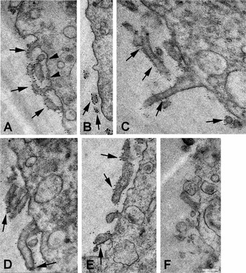
We wanted to know if the distribution of microvilli and the microvillar morphology in oolemma change after fertilization or artificial activation of the oocytes. We found that in ovulated oocytes the microvilli were thin and short and they were uniformly distributed on the surface of the oocyte (Fig. 1A), with the exception of the region above MII spindle, which was almost completely microvilli free (Fig. 1B). The microvilli-rich region of zygotes differed from that of ovulated oocytes. In zygotes the microvilli were irregular thick (Fig. 1C), and very long (Fig. 1C, D). Zygotic microvilli were distributed less evenly than microvilli present on the surface of the oocyte. In zygotes, clusters of microvilli were separated by large regions of smooth (microvilli-less) membrane (Fig. 1C, D). Such large regions of smooth membrane were very seldom seen on the surface of the ovulated oocytes. The surface of parthenogenotes looked more like the surface of ovulated oocytes than zygotes. In parthenogenotes tiny microvilli were distributed regularly, however their oolemma was smoother than the surface of ovulated oocytes (Fig. 1E).
Localization of CD9 protein on the surface of ovulated oocyte (A, B), zygote (C, D), and parthenogenote (E). (F) Control – ovulated oocyte incubated only in the presence of gold-conjugated secondary antibody. Arrows indicate CD9 localized to microvilli, arrowheads indicate CD9 present on the planar membrane between microvilli. Scale bar, 500 nm.

There is less CD9 protein in the membrane of zygotes than in the membrane of ovulated oocytes
To determine if fertilization or artificial activation affects the amount and the localization of CD9 in mouse oocytes, we labelled oocytes with a monoclonal antibody to CD9 and colloidal gold-conjugated secondary antibody. In ovulated oocytes, zygotes and parthenogenotes the majority of the label indicating the presence of CD9 molecules was localized on the microvilli of the plasma membrane, but some CD9 molecules were also present between the microvilli on the regions of smooth membrane. (Fig. 1A–E). In one of the ovulated oocytes we detected a single region, which almost completely lacked microvilli. In this region only very sparse label was detected (Fig. 1B). As this region of the oocyte probably corresponded to the microvilli-free area of the cell membrane located above the metaphase II spindle (Johnson et al., Reference Johnson, Eager, Muggleton-Harris and Grave1975; Kaji et al., Reference Kaji, Oda, Shikano, Ohnuki, Uematsu, Sakagami, Tada, Miyazaki and Kudo2000), we omitted this region in our calculation of the distribution of gold particles within the plasma membrane.
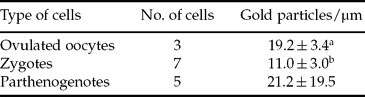
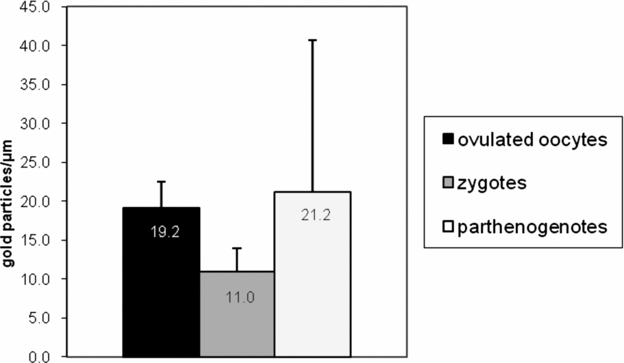
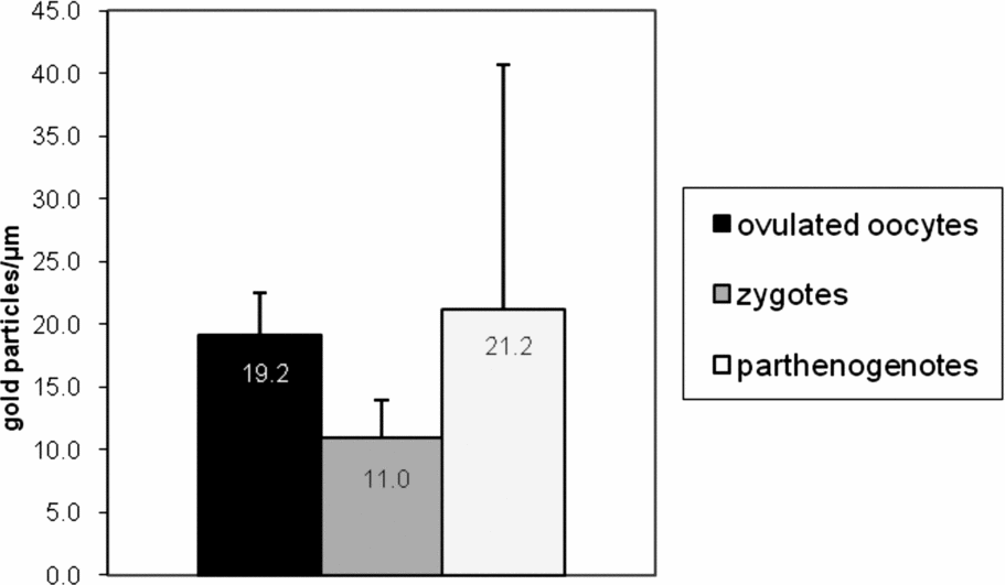
Table 1 and Fig. 2 show the mean number of gold particles (indicating the level of CD9) in membrane of ovulated oocytes, zygotes and parthenogenotes. We found that in zygotes the level of CD9 protein in oolemma was almost two times lower than in ovulated, unfertilized oocytes (11.0 vs. 19.2 gold particles/μm, respectively). In four examined parthenogenotes the mean number of gold particles per μm of sectioned membrane (21.2 particles/μm) was close to that of ovulated oocytes, however in one of these parthenogenotes there was 50.2 of gold particles/μm (mean value from all analysed images from this cell), and was almost five times greater than the mean value for other three parthenogenotes (11.5 particles/μm). Due to high standard deviation the mean number of gold particles per μm of membrane for parthenogenotes was not significantly different from the value of this parameter for ovulated oocytes and zygotes.
Number of gold particles per linear micron of sectioned membrane.
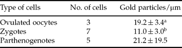
a, bValues with different superscript are significantly different (Student's t-test, p ≤ 0.05).
Mean number of gold particles per linear micron of sectioned membrane (±SD) of ovulated oocytes, zygotes and parthenogenotes.
In all experiments the negative controls (oocytes, zygotes and parthenogenotes incubated only with the secondary antibody) lacked gold particles in the membrane (Fig. 1F).
Discussion
Microvilli of the oolemma play an important role in gamete fusion during fertilization both in vertebrates and invertebrates (Longo, Reference Longo1997; Wilson & Snell, Reference Wilson and Snell1998). It is known that sperm incorporation in mammals occurs along the region of the oolemma containing microvilli (Yanagimachi & Noda, Reference Yanagimachi and Noda1970; Nicosia et al., Reference Nicosia, Wolf and Inoue1977; Shalgi & Phillips, Reference Shalgi and Phillips1980; Santella et al., Reference Santella, Alikani, Talansky, Cohen and Dale1992). In the mouse, the CD9 is the only oocyte protein currently known to be required for sperm–oocyte fusion (Chen et al., Reference Chen, Tung, Coonrod, Takahashi, Bigler, Chang, Yamashita, Kincade, Herr and White1999; Kaji et al., Reference Kaji, Oda, Shikano, Ohnuki, Uematsu, Sakagami, Tada, Miyazaki and Kudo2000; Miyado et al., Reference Miyado, Yamada, Yamada, Hasuwa, Nakamura, Ryu, Suzuki, Kosai, Inoue, Ogura, Okabe and Mekada2000; Le Naour et al., Reference Le Naour, Rubinstein, Jasmin, Prenant and Boucheix2000). It was suggested that microvilli can provide a scaffold for the presentation of adhesion/fusion proteins (Wilson & Snell, Reference Wilson and Snell1998) and CD9 has been recently found to be preferentially localized to the oocyte microvillar membrane (Runge et al., Reference Runge, Evans, He, Gupta, McDonald, Stahlberg, Primakoff and Myles2007). Microvilli of the oocyte are dynamic structures (Byrd & Belisle, Reference Byrd and Belisle1985), and it was shown that after fusion with sperm (Cline et al., Reference Cline, Schatten, Balczon and Schatten1983; Bronson, Reference Bronson1998) or resumption of cleavage (Dale et al., Reference Dale, Tosti and Iaccarino1995) they can change their structure and/or distribution. The purpose of this study was to investigate whether the loss of oolemma fusibility in zygotes is linked to its ultrastructural modifications, particularly to the changes in the amount and localization status of CD9.
We found that the majority of the surface of ovulated oocytes and, to lesser degree, the surface of parthenogenotes, was very rough i.e. covered with abundant, thin and rather short microvilli. When compared with the surface of ovulated oocytes, the oolemma of zygotes was much smoother and sparse in microvilli. Microvilli in zygotes were thicker and usually longer than microvilli of ovulated oocytes and parthenogenotes. These observations suggest that artificial activation of the oocyte results in limited modification of the morphology of the cell membrane, whereas the fusion with sperm and natural activation by the sperm factor (Swann et al., Reference Swann, Saunders, Rogers and Lai2006) induces pronounced reorganization of the oolemma structure. In addition, in the zygotes we observed clusters of microvilli, which were thicker than oocyte microvilli. It cannot be excluded that these thick microvilli resulted from the fusion of several adjacent thinner microvilli. Similar pattern of microvilli distribution was observed in the oolemma of the oocytes from mice with reduced fertility (Wabik-Sliz & Kujat, Reference Wabik-Sliz and Kujat1979). It is also known that the low diameter (thin) microvilli present in the oolemma facilitate its fusion with spermatozoa, as the cylindrical shape and narrow diameter provide the low radius of curvature required for the cell fusion (Wilson & Snell, Reference Wilson and Snell1998; Runge et al., Reference Runge, Evans, He, Gupta, McDonald, Stahlberg, Primakoff and Myles2007). Thus, we predict that the thick microvilli, which we observed on the surface of zygotes, are probably unable to promote fusion with spermatozoa.
The observation that the region where the gametes fuse forms actin filament rich fertilization cone (Tilney & Jaffe, Reference Tilney and Jaffe1980), and the fact that the fertilization cone does not form in artificially activated mouse oocytes, indicate that the fusion of the gametes induces reorganization of the microfilaments of the oocyte. It is known that the core of microvilli is formed by a dense bundle of cross-linked actin filaments (Longo, Reference Longo1987; Wilson & Snell, Reference Wilson and Snell1998). It is very likely that the fusion of the gametes induces modification of the microvilli by initiation of alterations of the actin microfilaments in a whole cortex, not only in the region of the sperm penetration. The observation that in aged oocytes, which were fertilized in vitro 22 h after hCG injection, in which the actin cytoskeleton differs from the cytoskeleton of freshly ovulated oocytes, the oolemma block is only partially effective indicates that oolemma block involves modification of actin filaments (Wortzman & Evans, Reference Wortzman and Evans2005). Additionally, it is well known that different inhibitors of the actin filaments affect the ability of the oocyte to fuse with spermatozoa in various ways – most of them block fusion, whereas cytochalasin D impairs development of the oolemma block to sperm penetration (McAvey et al., Reference McAvey, Wortzman, Williams and Evans2002). This indicates that the changes in the surface of the oocytes, which we observed after fertilization, are a consequence of the reorganization of the cortical actin, and that these changes are responsible for the loss of fusiblity of the zygote with sperm.
We detected tetraspanin CD9 on the surface of ovulated oocytes, zygotes and parthenogenotes. However, we found that the CD9 was more abundant in the oolemma of ovulated oocytes than in zygotes. We also observed some differences in distribution of CD9 between unfertilized oocytes and zygotes. In both types of cells this protein was predominantly localized on microvilli, but CD9 was also present on the smooth regions of the membrane, between the microvilli. However, there was more CD9 localized in the smooth regions of the oocyte membrane than in the smooth regions of the zygote membrane.
It is known that membrane CD9 protein interacts, via EWI proteins, with actin filaments (Sala-Valdes et al., Reference Sala-Valdes, Ursa, Charrin, Rubinstein, Hemler, Sanchez-Madrid and Yanez-Mo2006; Ziyyat et al., Reference Ziyyat, Rubinstein, Monier-Gavelle, Barraud, Kulski, Prenant, Boucheix, Bomsel and Wolf2006). Thus, modifications of actin cytoskeleton of the oocyte can be associated with the changes in the localization of CD9 in oolemma. Thus, we hypothesize that the development of the membrane block to sperm penetration may be related to the reorganization of microvilli and to relocation of their CD9 between microvilli and smooth region between microvilli.
The reduction in the amount of CD9 present on the surface of the oocyte after fertilization indicates that the sperm induces modification of oolemma. We found that the level of CD9 in zygotes was almost two times lower than in unfertilized oocytes. There is a possibility, that reduction of the amount of CD9 in the membrane of microvilli, together with its relocation may be responsible for the development of membrane block to sperm penetration after fertilization. However, we previously observed (using in situ immunofluorescence and western blot techniques) that in mouse zygotes the loss of oolemma fusibility with spermatozoa is not related to the decrease of CD9 protein in the oolemma (Komorowski et al., Reference Komorowski, Szczepanska and Maleszewski2003). Alternative explanation for this phenomenon is that fertilization causes an aggregation of CD9 molecules in the oolemma, thus lowering the number of the epitopes accessible to anti-CD9 antibody binding.
Modification of the structure of the oolemma, as well as the changes in localization of CD9, which we observed after fertilization, likewise the oolemma block to sperm penetration (Komar, Reference Komar1982; Borsuk & Tarkowski, Reference Borsuk and Tarkowski1989; Horvath et al., Reference Horvath, Kellom, Caulfield and Boldt1993; Maleszewski & Bielak, Reference Maleszewski and Bielak1993; Maleszewski et al., Reference Maleszewski, Kimura and Yanagimachi1996; Pyrzynska et al., Reference Pyrzynska, Maleszewski and Maluchnik1996; Wortzman-Show et al., Reference Wortzman-Show, Kurokawa, Fissore and Evans2007), must be only partially related to the oocyte activation, as these phenomena do not occur in parthenogenetically activated oocytes. The changes in the structure of oolemma and, in consequence, the membrane block to sperm penetration (Maleszewski et al., Reference Maleszewski, Kimura and Yanagimachi1996; Gardner & Evans, Reference Gardner and Evans2006), can be induced by the fusion of the membranes of the gametes. However, the mechanisms, by which the incorporation of the sperm membrane into oolemma induces oolemma block to sperm penetration still remains elusive.
Acknowledgements
Authors thank Prof. M. Kloc and Dr Katarzyna Szczepańska for their valuable comments on the manuscript.