The first human transcatheter pulmonary valve replacement was performed in 2000, and the procedure has been performed in the United States since 2007. Reference Bonhoeffer1,Reference Zahn2 Since that time, transcatheter pulmonary valve placement has become an accepted alternative to surgical pulmonary valve replacement in select patients. Reference Cheatham3–Reference Vezmar8 The Melody valve (Medtronic, Minneapolis, MN) was initially indicated for patients ≥30 kg with dysfunctional right ventricular to pulmonary artery conduits, and in conduits with a nominal diameter of 16 mm or greater at time of implantation. Off-label use of the Melody valve has been performed successfully in a variety of clinical scenarios. Reference Zahn2,Reference Boshoff9–Reference Martin15 Historically, it was suggested that right ventricle to pulmonary artery conduits not be expanded with balloons exceeding the nominal conduit diameter by more than 110%. Reference Powell16 However, the successful use of larger balloons in small conduits has been reported. Reference Zahn2,Reference Bensemlali10,Reference Shahanavaz14,Reference Powell16–Reference Carr18 We sought to determine the feasibility and safety of transcatheter pulmonary valve placement in right ventricle to pulmonary artery conduits with balloon dilation to >110% of the nominal conduit diameter (overdilation), and to compare patient characteristics, procedural success, complications, and short- and mid-term outcomes in those with conduit overdilation and those without.
Materials and methods
This retrospective cohort study included patients at two centres (University of Utah at Primary Children’s Hospital and Texas Children’s Hospital) who underwent transcatheter pulmonary valve placement using the Melody TPV (Medtronic, Minneapolis, MN) within a right ventricle to pulmonary artery conduit or stentless bioprosthetic valve between November 2010 and March 2017. The study was approved by the institutional review boards at the University of Utah and at Baylor College of Medicine. Study participants were identified from institutional databases, and all patients undergoing attempted transcatheter pulmonary valve placement during that time period were evaluated for inclusion in the study. We excluded those who went to the catheterization lab for transcatheter pulmonary valve placement, but in whom an implant was not attempted secondary to coronary artery or aortic root compression during balloon testing, and those without a previously placed right ventricle to pulmonary artery conduit or stentless bioprosthetic valve. Demographic data, previous surgical and catheter-based right ventricular outflow tract interventions, baseline echocardiographic data, procedural data at the time of transcatheter pulmonary valve placement, hospital length of stay, follow-up echocardiographic data, and complications were collected. The degree of conduit calcification was assessed angiographically at each individual institution and graded as follows: grade 1 had no calcification or mild calcification, grade 2 had moderate calcification, and grade 3 had severe calcification. The angiograms for all patients were reviewed at each respective center. Contained conduit tears were identified angiographically during the procedures and during angiographic review for the study. Of note, a subset of the patients in this study (<15%) are also represented in a recent multicenter study of Melody valve placement in small conduits. Reference Shahanavaz14 Data were managed using REDCap (Research Electronic Data Capture) electronic data capture tools hosted at the University of Utah. REDCap is a secure, web-based application designed to support data capture for research studies. Reference Harris19 Subjects were separated into two groups. The study group consisted of those who had a final transcatheter valve diameter >110% of the original conduit diameter (overdilation group), and the second group consisted of those who had a final valve diameter of ≤110% of the original conduit diameter (control group). A control group was used in this study to enable a comparison of the potential procedural risks and valve performance in the study group to that of transcatheter pulmonary valve placement without overdilation by the same operators. The control group comprised mostly patients with larger conduits that did not require over-expansion in order to reach adult-sized pulmonary valve diameters. Given the retrospective nature of the study, we did not attempt to evaluate which conduits could or could not be expanded.
Statistical methods
Statistical analyses were conducted using Stata 14, College Station, TX. Patient demographics and characteristics were summarized using descriptive statistics. Continuous variables were summarized as a mean (standard deviation) or median (interquartile range) according to their distribution. Frequency and percentage were reported for categorical variables. Unpaired t-tests were used for continuous data, and Fisher’s exact tests, Chi-squared tests, or Wilcoxon–Mann–Whitney tests were used for categorical data.
Results
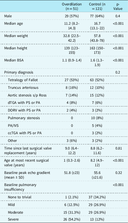
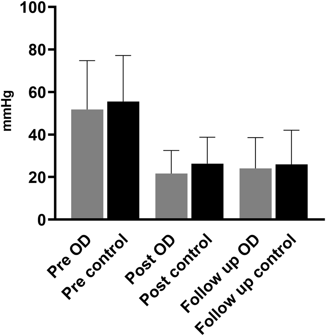
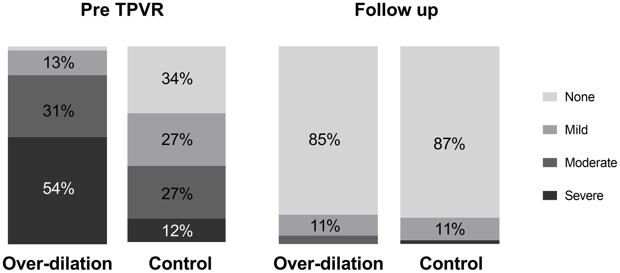
A total of 172 patients were included in this study (51 in the overdilation group and 121 in the control group). Patient demographics are summarized in Table 1. Briefly, the overdilation group was younger (11.2 years versus 16.7 years, p < 0.001) and smaller (32.8 kg versus 57.8 kg, p < 0.001) than the control group at the time of transcatheter pulmonary valve placement. The diagnoses were similar in both groups with approximately half of the patients in both groups having variants of tetralogy of Fallot. While the time interval since surgical valve placement was not different between the groups (9.0 years versus 8.8 years, p = 0.81), the overdilation group was significantly younger at the time of the most recent surgical valve implantation (1 year versus 8.2 years, p < 0.001). The primary indication for transcatheter pulmonary valve placement was different between groups: combined pulmonary stenosis/pulmonary insufficiency for the majority of the overdilation patients (57%), and pulmonary stenosis for the majority of the control patients (54%). There was no difference in the echocardiographic peak gradients in the overdilation versus the control groups at baseline (51.8 versus 55.6, p = 0.32). The overdilation group had more pulmonary insufficiency at baseline with 54% of the patients in that group having severe insufficiency.
Demographics.

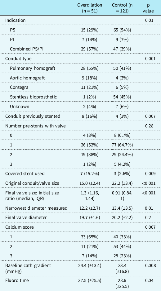
Conduit data are summarised in Table 2. The breakdown of the types of conduits differed between the two groups. In the overdilation group, the majority (73%) of the conduits were homografts, whereas in the control group, fewer than half (44%) were homografts, and half were stentless bioprosthetic valves. In the overdilation group, the non-homograft conduits included 11 Contegra valves and 1 Freestyle valve. In the patient with the Freestyle valve, the conduit was stented with two pre-stents, the Melody valve was placed, then the conduit was post-dilated with a high-pressure balloon after Melody valve implantation in order to over-expand the valve. In the control group, there were 6 Contegra valves, 25 Prima valves, 12 Freestyle valves, and 17 unknown. The unknown valves were documented as stentless bioprosthetic valves in the medical records, but the surgical record with the exact type of valve were not available. The nominal diameter of the conduits at time of surgical implantation was significantly smaller in the overdilation compared to the control group (15 mm versus 22.2 mm, p < 0.001). The range of conduit sizes at implant in the overdilation group was 9–20 mm, and the range of conduit sizes in the control group were 14–29 mm. The conduits were less calcified in the overdilation group versus the control group. There were eight patients (16%) in the overdilation group who had their conduit stented prior to the transcatheter pulmonary valve placement procedure, and there were four patients (3%) from the control group with previously stented conduits. Notably, the conduits in the control group had shrunk considerably more than those in the overdilation group (smallest angiographic diameter of 12.2 mm in overdilation versus 13.4 mm in the control groups); however, because of their larger size at implant, these valves did not require over-expansion to achieve adequate adult size.
Procedural data.

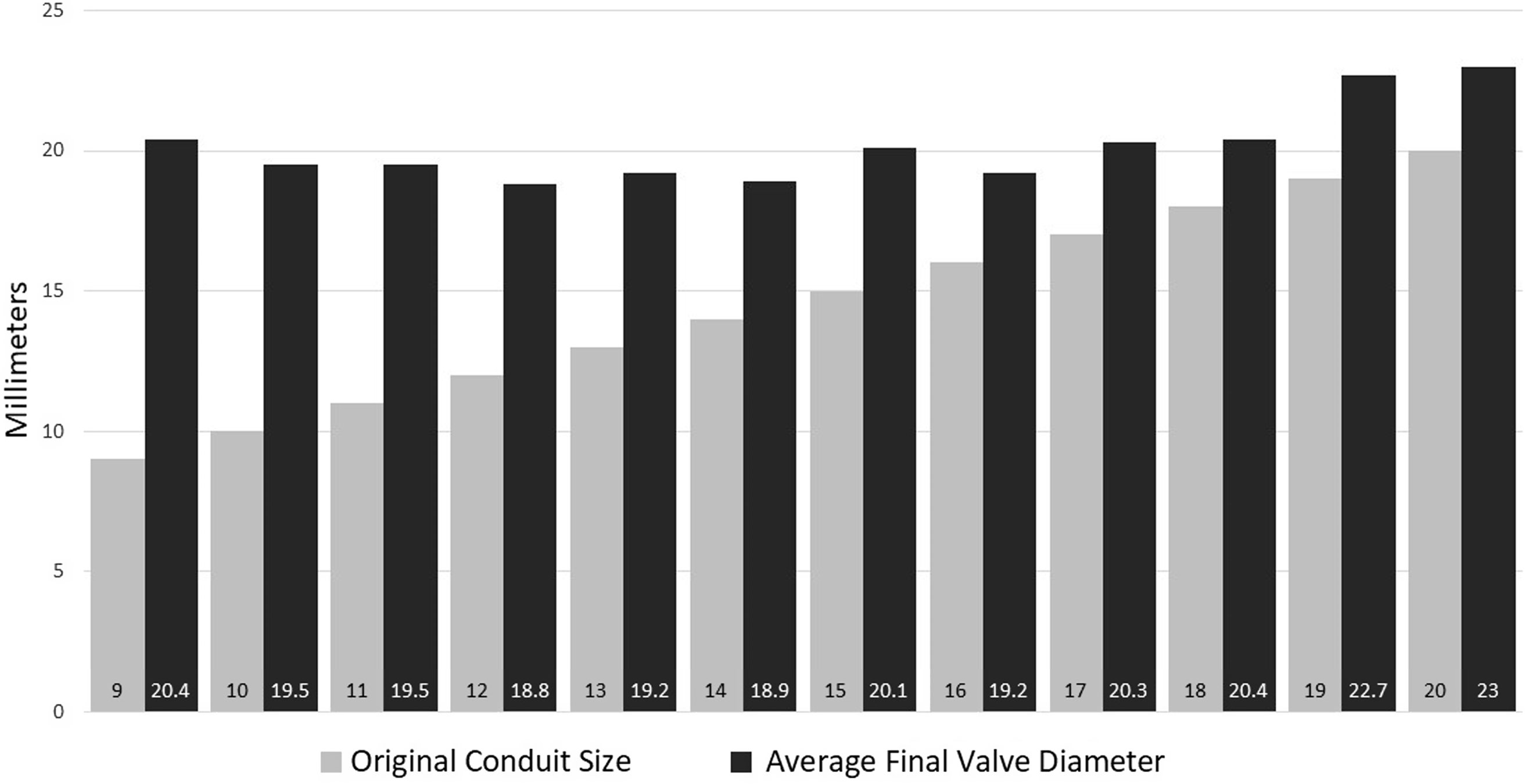
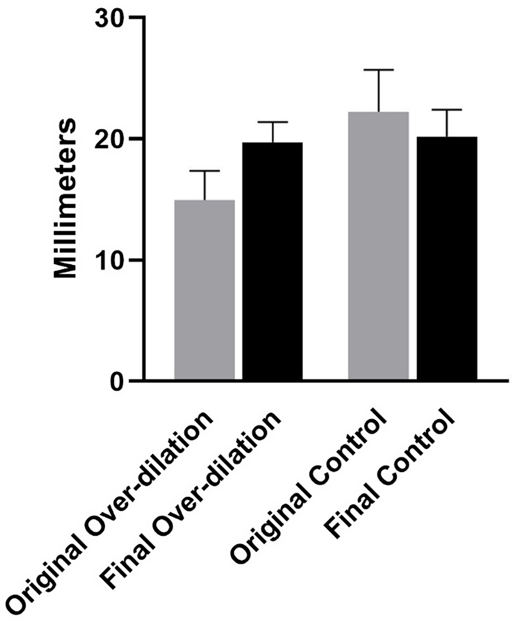
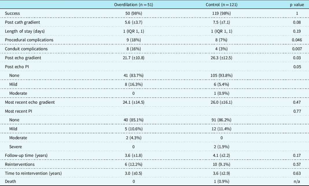
Transcatheter pulmonary valve placement was successful in 50/51 (98%) of the patients in the overdilation group and was successful in 119/121 (98%) of the patients in the control group. Procedural data are summarized in Table 2. As above, the narrowest angiographic diameter at the time of transcatheter pulmonary valve placement was slightly smaller in the overdilation group, but the final valve diameter was no different between the groups (19.7 mm versus 20.2 mm, p = 0.2). The valves were a median of 130% (IQR 116–144%) of the nominal conduit size in the overdilation group (Figs 1 and 2) and 91% (IQR 84–100%) of the nominal conduit size in the control group (Fig 3). The final valve diameter was measured based on the measurements of the Melody valve stent on the final angiogram. There was not a significant difference in the number of pre-stents used between the two groups, but a higher percentage of the overdilation group got two pre-stents (38% versus 24%). There was a significant difference in the percentage of patients who got a covered stent with a higher percentage in the overdilation group (15.2% versus 2.6%, p = 0.009). The overdilation group had longer fluoroscopy times than the control group (37.5 versus 28.6 minutes, p = 0.04). Hospital length of stay was not significantly different between groups with the median length of stay of 1 day in both groups. The overdilation group had two outliers, one who stayed 7 days after the emergent surgical pulmonary valve replacement for the uncontained tear, and the other who stayed 13 days for non-cardiac problems. The control group had a length of stay range from 1–4 days.
Lateral projections of angiography in an right ventricle to pulmonary artery conduit before and after transcatheter pulmonary valve placement. The left panel shows a 9-mm RV to PA conduit that is narrowest just above the valve in the conduit. The right panel shows the same conduit following transcatheter pulmonary valve placement. The final valve measured 20 mm in diameter.

Bar graph showing the size of the original conduit for each of the 12 sizes of conduits in the overdilation group compared to the average final valve diameter. There was one 9-mm conduit, one 10-mm conduit, two 11-mm conduits, five 12-mm conduits, four 13-mm conduits, five 14-mm conduits, eight 15-mm conduits, fourteen 16-mm conduits, three each of the 17-, 18-, and 19-mm conduits, and one 20-mm conduit.

Bar graph showing the mean original compared to final valve diameters of the overdilation and control groups with error bars indicating standard deviation.

Procedural complications were more frequent in the overdilation compared to the control group (Table 3, p = 0.046). In the overdilation group, nine patients (18%) had procedural complications. One patient had an uncontained conduit tear that was unable to be rescued with a covered stent and required ECMO cannulation and emergent surgical valve replacement. This was one of the eight patients with a previously stented conduit. Seven patients had contained tears that were either treated successfully with covered stents or with the Melody valve itself (Fig 4), and one patient had a posterior main pulmonary artery pseudoaneurysm that was treated with an 8-mm Amplatzer Vascular Plug II. The patient who had the conduit rupture had a calcium score of 1. Of those that had a contained tear the majority of them had a calcium score of 1, one had a calcium score of 2, and two had a calcium score of 3.
AP cranial projections of a conduit tear before and after covered stent placement. The panel on the left shows the tear on the left side of the 13-mm conduit following 16-mm balloon angioplasty. The panel on the right shows the conduit after the tear has been covered with a covered stent followed by a bare metal stent.

Outcomes.

In the control group, eight patients (7%) had procedural complications. One patient had a significant longitudinal conduit tear that was treated with an endovascular covered stent graft, within which a Melody valve was placed 8 months later. This patient had a calcium score of 2. Three patients had contained tears that were successfully covered with the Melody valve. One of these patients had a calcium score of 1, and two had a calcium score of 2. One patient developed a hemothorax that required chest tube placement. One patient had peripheral vessel damage to a pelvic vein that was treated with a 10-mm Amplatzer Vascular Plug II. One patient (on anticoagulation for a mechanical mitral valve) had a bleed from the femoral artery access site requiring a blood transfusion. One patient developed ventricular arrhythmias after transcatheter pulmonary valve placement requiring the initiation of beta-blocker therapy.
Follow-up time for the two groups was similar with mean follow-up time of 3.6 years in the overdilation group and 4.1 years in the control group (p = 0.17). Follow-up data are included in Table 3. There was no difference in the percentage of patients requiring reintervention (12.2% versus 9.1% in overdilation versus control, p = 0.57), or in the time to reintervention (3 versus 3.6 years in overdilation versus control, p = 0.63) (Fig 5). There was no difference in echocardiographic gradients between the overdilation and control groups at most recent follow-up prior to reintervention if applicable (peak gradients 24.1 versus 26 mmHg, p = 0.47, Fig 6). There was no difference in the degree of pulmonary insufficiency in the overdilation group compared to the control group at most recent follow-up (Fig 7). In the overdilation group, 85% of patients had no or trivial insufficiency and 10.6% had mild insufficiency (no patients with severe insufficiency). In the control group, 86% of patients had no or trivial insufficiency, 11.4% of patients had mild insufficiency, and 2% of patients had severe insufficiency.
Kaplan–Meier curve showing freedom from intervention over time between the overdilation and control groups with the dashed lines representing the 95% confidence intervals.

Bar graph showing the peak gradient by echocardiography before, immediately following, and at most recent follow-up prior to reintervention for the overdilation and control populations. The mean values are shown with error bars indicating standard deviation.

The degree of pulmonary insufficiency in the overdilation and control groups prior to transcatheter pulmonary valve placement and at most recent follow-up.

In the overdilation group, there were six reinterventions (12% of patients) that were all surgical pulmonary valve implantations for endocarditis. These were Melody valves implanted in four Contegra valves, one pulmonary homograft, and one aortic homograft. In the control group, there were four patients who underwent surgical pulmonary valve replacement for endocarditis (two pulmonary homografts and two surgical bioprosthetic valves), one patient who underwent surgical pulmonary valve replacement secondary to presumed nickel allergy from pre-stents, three patients who underwent a second transcatheter pulmonary valve placement for pulmonary stenosis (one who had developed a stent fracture), and two patients who underwent balloon angioplasty for recurrent stenosis (total reintervention rate of 9%). The patients who had reintervention for recurrent stenosis had variable conduit types, including aortic homograft, pulmonary homograft, Freestyle valve, and Contegra. There was one patient in the control group who died 8 years following transcatheter pulmonary valve placement at an outside institution. The details of the death were largely unknown other than he died from apparent complications of open-heart surgery at an outside facility.
Discussion
Despite the general attitude that right ventricle to pulmonary artery conduits should not be dilated to >110% of their nominal diameter, this study demonstrates that it is feasible to over-expand conduits in some patients, and that outcomes are similar to those not requiring conduit over-expansion during transcatheter pulmonary valve placement. Although contained conduit tears occur more often during the procedure, serious complications were not different in those who had over-expansion versus those who did not. To our knowledge, this study is the largest to date focused on the degree of over-expansion of right ventricle to pulmonary artery conduits during transcatheter pulmonary valve placement and adds important information to the growing body of knowledge of transcatheter pulmonary valve placement. A recent French study looked at Melody valve placement in 11 patients with conduits <16 mm in diameter. They showed 100% procedural success in their small population of slightly older, slightly larger patients with a similar initial conduit size (15 mm in our study compared to 16 mm in their study). Reference Bensemlali10 Another recent study of nine centers in the United States examined Melody valve placement within small (<16 mm) diameter conduits. In that study, 78% of the total group of 140 patients were able to undergo transcatheter pulmonary valve placement with dilation of their right ventricle to pulmonary artery conduits from a median diameter of 15 mm to a median diameter of 19 mm (as mentioned above, our centers contributed a small number of patients to this study). The freedom from reintervention was 89% at 4 years. Reference Shahanavaz14 The overdilation group in our study had an 88% freedom from reintervention at 3.6 years. The median age and weight of the patients in that study were similar to the overdilation group in our current study. Our study, like the two others, highlighted above, adds to the body of knowledge, and shows promising data to expand the use of transcatheter pulmonary valve placement in patients with smaller right ventricle to pulmonary artery conduits. Our study contrasts the other two studies in that it evaluates overdilation in a wider range of conduit sizes (not just limited to <16 mm), which may be particularly useful now that the larger diameter Edwards SAPIEN valves (Edwards Lifesciences LLC, Irvine California) are being used as well. Also, placing a larger valve in a mid-sized conduit has the potential to pave the way for more future valve-in-valve procedures, delaying or in some cases avoiding repeat surgical pulmonary valve replacement.
One of the most significant concerns with overdilation of right ventricle to pulmonary artery conduits during transcatheter pulmonary valve placement is causing a conduit tear during conduit preparation, prior to valve implantation. This is likely the reason that the complication rate was higher in the overdilation compared to the control group, as nearly all of the complications seen in the overdilation group were contained conduit tears. Because the control groups larger diameter conduits did not necessitate over-expansion to reach adult size final valve diameters, it is not surprising that there was a lower percentage of conduit tears seen in this group. The percentage of conduit tears in the control group (at 3%) was similar to the reported literature. Reference Bishnoi20 It is also worth mentioning that there are operators who would not consider a contained conduit tear a complication; however, for this study, we considered contained tears complications because we felt that it is difficult to predict how a conduit would tear, and progression of a contained tear could lead to a more serious complication. In this study, there was one patient in each group with a conduit tear significant enough to require surgical intervention. Interestingly, most of those that had either contained or uncontained tears had calcification scores of 1–2 with only two patients in the overdilation group (that had contained tears) having a calcium score of 3.
We recognize that a small contained tear may not present a danger to the patient; however, in our practice if a contained tear is recognized, we feel it is prudent to cover that contained tear in order to prevent a more serious complication. The body of literature with regard to conduit tears does support that the majority of these can be treated in the catheterization laboratory at the time of the transcatheter pulmonary valve placement. Reference Bishnoi20,Reference Hainstock21 Though there was not a difference in the number of stents placed prior to the valve, there was a higher percentage of patients in the overdilation group that got a covered stent during the procedure. It is not the standard practice of either institution to prophylactically place a covered stent, so the patients who had covered stents placed in this study represent those that had a contained tear identified during the procedure.
Prophylactic stenting of right ventricle to pulmonary artery conduits with covered stents that may be at a higher risk for conduit injury has been suggested by some investigators. Reference Eicken5 However, one of our concerns with stenting the conduit prior to dilation is causing coronary compression with an already stented conduit. If one were to stent the conduit at a small diameter and then dilate it, coronary compression could be a possibility. There was a previous study that explored stenting the conduit (not with covered stents) prior to conduit dilation using 3-D rotation angiography for coronary evaluation as a way to potentially more safely dilate the conduit. In this study with relatively a relatively small number of 12 patients, there were no procedural complications. Reference Pockett, Moore and El-Said22 These were all done as part of the same procedure and not as staged procedures as our previously stented patients were. Although covered stents are not as strong as the bare metal stents used in the study referenced above, one would have to keep coronary compression in mind if using this technique.
Our series of overdilation patients seems to have a fairly high rate of conduit tear as well as endocarditis. Our procedural complication rate of 18% and conduit complication rate of 16% in the overdilation group are similar to complication rates found in previous studies of smaller conduits and smaller children (9–36%) and higher than what has been reported in other recent studies in older patients (6–14%). Reference Zahn2,Reference McElhinney4,Reference Butera6,Reference Bensemlali10,Reference Berman11,Reference Shahanavaz14,Reference Martin15 One of the unexpected findings was the relatively higher incidence of Melody valve explants for endocarditis in the overdilation group as compared to the control group, with an incidence of endocarditis in the overdilation group of 16% and in the control group of 5%. The rate of infective endocarditis following transcatheter pulmonary valve placement is an area of current investigation worldwide, and the incidence varies significantly between studies (3–25%). Reference McElhinney23–Reference Abdelghani27 Our rates of endocarditis fall into that reported in the literature, and it is unclear if other technical modifications such as that used in the study mentioned above Reference Pockett, Moore and El-Said22 would result in a lower incidence of endocarditis in patients where an over-expansion strategy is used. The clinical implications and morbidity associated with infective endocarditis are significant, and this remains an area of concern. Although prior studies have shown increased rates of endocarditis with higher gradients following transcatheter pulmonary valve placement, we did not see higher gradients in the overdilation group during the study period. However, one theory is that it is not the higher gradient itself that has led to endocarditis in other studies, but rather another characteristic of the patients that had a higher gradient. One possible common characteristic between patients with a high gradient in other studies and the overdilation patients in our cohort is that in both cases the patient had a smaller conduit to start. The reason for this finding in this study remains unclear and warrants further investigation in small conduits.
It is notable that all of the reinterventions in the overdilation group were surgical valve replacements for endocarditis, and 50% of the reinterventions in the control group were surgical valve replacements (four for endocarditis and one for a presumed nickel allergy). The reintervention rate in the overdilation group was 12%, and the reintervention rate in the control group was 9%. In the small series by Bensemlali et al of transcatheter pulmonary valve placement in small conduits, 3/11 patients (27%) required surgical replacement of the pulmonary valve within 3 years following transcatheter pulmonary valve placement. Reference Bensemlali10 A larger more recent study by Shahanavaz et al. noted a transcatheter-based reintervention rate after Melody valve placement of 14% with median follow-up of 5 years. Reference Shahanavaz28 In the same cohort of patients, Cabalka et al. reported 25% of patients with a stentless conduit underwent reintervention (either catheter-based or surgical) within 5 years. Reference Cabalka29 Another large recent study by McElhinney et al. looking at both Melody and Sapien valves revealed a cumulative reintervention rate of 25.1% at 8 years, and surgical reintervention rate of 14.4% at 8 years, Reference McElhinney30 though this study has a longer follow-up than our data.
One of the most encouraging findings in our study is that we were able to achieve a final valve diameter of nearly 20 mm in the overdilation group, with no significant difference in the final valve diameter compared to the control group, no difference in valve function by echocardiography at similar follow-up intervals, and no difference in reintervention rates between groups. We recognize that the patient populations and conduits are different between the two groups but believe that the comparison of the follow-up data shows the ability to achieve the desired result when over-expanding a conduit during transcatheter pulmonary valve placement. Whether or not a conduit was over-expanded was largely based on conduit size and the ability to achieve a final valve diameter of roughly 20 mm. Our goal in every patient is to implant a valve that will be suitable in size for an adult, as most patients will be expected to reach adult size before requiring reintervention on their pulmonary valve. Therefore, we aimed to over-expand conduits whose diameters were smaller at implant than would be appropriate for an adult. Due to the retrospective nature of this study, the intent to over-expand could not be determined. There were not conduits that could not be expanded per se, other than the patient in the over-expansion group who had a conduit rupture requiring urgent surgical intervention. Although it was somewhat surprising that the final valve diameter of the control group was often smaller than the original conduit size, many of these conduits exceeded the 22–24 mm diameter that the Melody valve is able to achieve. It should be noted that this group of conduits was more heavily calcified, and that the minimum angiographic diameter of the conduits was 13.4 mm in this group and was only about a millimeter larger than in the overdilation group. Additionally, we are reporting the final valve size, not the size of the balloon used to implant the valve, and likely the former is slightly smaller than the latter due to recoil of the valve in the conduit or incomplete expansion of the valve over the implantation balloon. Though there is a notable difference in the degree to which the conduit luminal diameter decreased between the two groups, many of the patients in the overdilation group had likely outgrown their conduits due to the smaller sizes at implant.
The final valve size to original conduit size ratio in the overdilation group was similar in our study to that found in other studies examining valve placement in small conduits (125–130%). Reference Bensemlali10,Reference Shahanavaz14 Our strategy of overdilating these conduits to final diameters similar to that of the control group potentially allows for the avoidance of an additional sternotomy and surgical valve replacement in these young patients while setting them up for additional transcatheter pulmonary valve placement attempts in the future. The durability of these promising initial results will need to continue to be examined, as previous studies have shown that good immediate term outcomes do not necessarily guarantee good longer-term haemodynamic outcomes, especially in those with smaller conduits. Reference Bensemlali10,Reference Boudjemline12,Reference Shahanavaz14
While these initial results are promising, the authors recognise that there is inherent risk in overdilation, and that all conduits and bioprosthetic valves will eventually tear if incrementally dilated. While a contained tear may have no acute clinical consequences for the patient, it is difficult to predict whether a tear will be contained. It should be noted that a small subset of patients in the overdilation group had significant over-expansion of their conduits, with a small number increasing the conduit to as much as twice the size of the initial conduit. In the opinion of the authors, careful, stepwise dilation with frequent angiographic evaluation of the conduits is important when undertaking overdilation, especially for those in whom the conduit will be significantly over-expanded. Further investigation is needed to understand which conduits and bioprosthetic valves may be better suited to safe overdilation.
Limitations
Our study is limited by a small sample size and its retrospective design. Because of the retrospective design, there was no way for the authors to determine the intent of the operators with regard to planning over-expansion or not. It is also limited by a lack of longer-term follow-up data. There may also be referral bias in our centers, leading to an incomplete sample of patients with small conduit diameters requiring valve replacement. We have included all patients in whom an attempt at transcatheter pulmonary valve placement was made. However, it is likely that some patients who underwent right ventricle to pulmonary artery conduit stent placement could have undergone an “attempt” at transcatheter pulmonary valve placement but did not due to operator preference. This may decrease the true success rate of transcatheter pulmonary valve placement in this patient population.
Conclusions
Transcatheter pulmonary valve placement can be performed in dysfunctional right ventricle to pulmonary artery conduits dilated to >110% of the original conduit diameter during valve placement. Procedural complications were more frequent in the overdilation than control group; however, there were no procedural deaths, and nearly all of the procedural complications were immediately and successfully managed in the catheterization laboratory in both groups. Over-expanding small right ventricle to pulmonary artery conduits to similar diameters to those of larger conduits allows for transcatheter pulmonary valve placement to be performed in these patients rather than a repeat surgical intervention. The longer-term outcomes of transcatheter pulmonary valve placement in conduits requiring over-expansion requires further study; however, our initial follow-up data show no difference in outcomes.
Acknowledgements
None.
Financial support
This research received no specific grant from any funding agency, commercial, or not-for-profit sectors.
Conflicts of interest
None.
Ethical standards
The authors assert that all procedures contributing to this work comply with the ethical standards of the relevant national guidelines on human experimentation (please name) and with the Helsinki Declaration of 1975, as revised in 2008, and have been approved by the institutional committees at the University of Utah and Baylor College of Medicine.