Case
A 39-year-old woman with history of D-transposition of the great arteries presented to the clinic in 2010 to establish care after previously being treated at a different institution. Her past medical history was remarkable for D-transposition of the great arteries, which initially required a Blalock–Hanlon septectomy in 1971 followed by a Mustard procedure in 1972. Owing to complex pulmonary stenosis and mitral valve chordal attachments into the ventricular septum, she underwent placement of a left ventricular apex-to-pulmonary artery Hancock valved conduit in 1996. In 2005, a routine 24-hour Holter monitor demonstrated long sinus pauses with sinus node dysfunction. She had a dual-chamber pacemaker placed in 2005 at another institution.
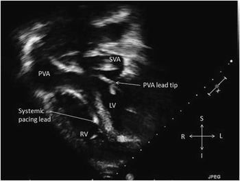
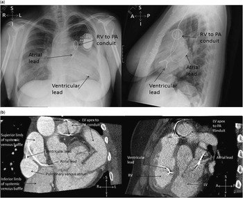
At this visit in 2010, she had no medical concerns. Her electrocardiogram was remarkable for sinus rhythm with first-degree atrioventricular block and non-specific intraventricular conduction delay. Pacemaker interrogation at this visit demonstrated that she was not pacemaker dependent with a predominately sinus rhythm 87% of the time. Her echocardiogram demonstrated mild left ventricular outflow tract obstruction with a peak systolic gradient of ~15 mmHg, mild tricuspid and mitral regurgitation, mild obstruction of the superior limb of the systemic venous baffle, small pulmonary venous baffle leak, and mildly depressed systemic right ventricular function. In addition, the echocardiogram demonstrated that the atrial lead was located in the systemic venous atrium superior to the mitral valve. The ventricular lead was demonstrated to course across the atrial baffle, from the systemic venous atrium entering the pulmonary venous portion of the atria, and then coursing through the tricuspid valve with its tip in the systemic right ventricular apex (Fig 1). The misplaced systemic ventricular lead was not seen on previous echocardiograms at the outside institution. A chest X-ray and cardiac CT scan performed after the echocardiogram confirmed lead placement in the more anterior systemic right ventricle (Fig 2).
Apical four chamber demonstrating the course of the pacing leads. Both leads course inferiorly through the superior vena cava. The atrial lead courses through the superior limb of the systemic venous atrial baffle. The ventricular lead courses across the systemic venous baffle to enter the pulmonary venous portion of the atria, then courses through the tricuspid valve with its tip in the apex of the right ventricle. SVA = systemic venous atrium; PVA = pulmonary venous atrium; LV = left ventricle; RV = right ventricle; S = superior; I = inferior; R = right; L = left.

Image (a) is a 2-view chest x-ray demonstrating the course of the pacing leads. Both leads course inferiorly through the superior vena cava. The atrial lead courses through the superior limb of the systemic venous atrial baffle with its tip terminating in the posterior and left portion of the systemic venous atrium. The ventricular lead courses across the systemic venous baffle to enter the pulmonary venous portion of the atria, then courses through the tricuspid valve with its tip in the apex of the anterior systemic right ventricle. Image (b) is a coronal and sagittal image from the patient’s CT angiogram included to further illustrate the path of pacing leads through the venous and systemic venous baffles. S = superior; I = inferior; R = right; L = left; A = anterior; P = posterior; LV = left ventricle; RV = right ventricle; PA = pulmonary artery.

The abnormal placement of her ventricular pacing lead in the systemic right ventricle raised concern for thrombus formation and future thromboembolic events. Once we determined that the lead was inadvertently placed in the systemic ventricle by echocardiography, chest X-ray, and CT scan, it was felt that lead removal would be safest with surgery but carried significant operative risk compared with the current risk of thromboembolism. We elected to continue long-term anticoagulation following closely for thromboembolic events. Antithrombotic options included warfarin and aspirin with aspirin being selected secondary to patient preference. Before selecting aspirin, this patient did not have any cerebral imaging to evaluate for clinically silent thromboembolic phenomena, which may have been useful for guiding anticoagulation therapy. She has been followed-up yearly in the clinic over the past 5 years during which she has not had any significant thromboembolic events.
Discussion
Before the development of the arterial switch procedure, a significant number of patients with transposition of the great vessels were managed with the atrial switch procedure. Late complications of the procedure include atrial arrhythmias and systemic right ventricular dysfunction.Reference Dos, Teruel and Ferreira 1 In addition, around 20% of patients require pacemaker placement secondary to sinus node dysfunction.Reference Puley, Siu and Connelly 2 During generator placement, improper placement of ventricular leads is a rare complication that is often under-reported and under-represented in the literature.Reference Sharifi, Sorkin, Sharifi and Lakier 3 In patients with normal cardiac anatomy or D-transposition after arterial switch operation, proper lead placement is into the morphologically right ventricle. As this patient had D-transposition palliated with the atrial switch procedure, the morphologically right ventricle is the systemic ventricle. The fact that this misplaced lead was discovered 5 years after placement shows how this can be a challenging diagnosis in patients with this complex anatomy.
There are limited data on the management and intervention strategies for incorrectly placed transvenous pacing leads in the systemic ventricle of patients with transposition of the great arteries after atrial switch operation. There is only one report of improper lead placement in the setting of an atrial switch; this was an adult patient with a Senning repair for D-transposition of the great arteries who had a misplaced ventricular lead diagnosed 3 years after implantation. Although this patient was asymptomatic from thromboembolic complications, he did have his misplaced lead extracted secondary to concerns for being at higher risk for future thromboembolic events.Reference Kuppahally, Green, Michaels, Ishihara, Freedman and Litwin 4 His case, like ours, was associated with a delay in diagnosing a misplaced lead, emphasising the need for thorough, compulsive imaging. There are multiple case studies available on patients with lead placement in the left ventricle through an atrial connection such as a patent foramen ovale or atrial septal defect without having other significant heart disease.Reference McManus, Mattei, Rose, Raskin and Rosenthal 5 In this population, patients with ventricular leads placed in the systemic ventricle are often asymptomatic. The incorrectly placed leads should be detected by chest X-ray when the ventricular lead is placed in the more anterior morphologically right ventricle as opposed to the more posterior morphologically left ventricle.Reference Konings, Dekkers, Groenink, Bouma and Mulder 6 In the event this is missed on the chest X-ray, such as in our patient, these patients can be diagnosed by having a paced rhythm with right bundle branch block or direct visualisation of the misplaced lead on echocardiogram.Reference Anastacio, Castillo-Sang and Lawton 7 There is significantly increased risk of thromboembolic events in patients with interatrial communications after having misplaced ventricular leads.Reference Rodriguez, Baltodano and Tower 8 , Reference Khairy, Landzberg, Gatzoulis, Mercier and Fernandes 9 To mitigate the risk of thromboembolic events in asymptomatic patients, some recommend lead removal in patients without haemodynamically significant heart disease by lead extraction versus surgical removal, whereas others recommending lifetime anticoagulation with warfarin.Reference Wynn, Weston, Cooper and Somauroo 10 , Reference Van Gelder, Bracke and Oto 11 In this population, there have been reports of some patients with chronically implanted leads being treated with aspirin therapy having no thromboembolic events for over 10 years.Reference Van Gelder, Bracke and Oto 11
This case of ventricular lead misplacement in the systemic right ventricle of a patient with D-transposition of the great arteries and complex pulmonary stenosis after Mustard procedure helps highlight the importance of ensuring the ventricular lead is in proper position in patients with complex cardiac anatomy. We recommend that lead placement in patients with this anatomy should be performed by only experienced implanters familiar with CHD. Diagnosis of misplaced ventricular leads should be by either fluoroscopy during lead implantation or by post-case chest X-ray. This patient’s misplaced leads were diagnosed by echocardiography, which should not be the initial test of choice to demonstrate misplaced leads; however, the astute interpretation by the echocardiographer allowed for this unexpected problem to be diagnosed. Although all patients with misplaced leads in the systemic ventricle warrant anticoagulation therapy, consideration should also be given to obtain cerebral imaging to evaluate for subclinical thromboembolic events to help guide decisions about lead removal. In addition, MRI-compatible pacemakers should be used in patients with this complex cardiac anatomy as they often later need cardiac MRI for additional information that can be difficult to acquire with echocardiography alone. This report hopes to increase consciousness about proper lead positioning in patients with transposition of the great arteries who have been treated with an atrial switch operation and to demonstrate the diagnostic challenges it can pose.
Acknowledgements
None.
Financial Support
This research received no specific grant from any funding agency or from commercial or not-for-profit sectors.
Conflict of Interest
None.