Introduction
Chorioamnionitis or inflammation of the amniotic fluid and fetal membranes, caused by multiple bacterial species, is the leading cause for preterm delivery and neonatal complications.Reference Romero, Espinoza, Chaiworapongsa and Kalache 1 , Reference Goldenberg, Hauth and Andrews 2 Even in the absence of a proven infection, fetuses and neonates may have systemic inflammation referred to as the fetal inflammatory response syndrome (FIRS).Reference Gotsch, Romero and Kusanovic 3 Antenatal exposure to inflammation can result in adverse pulmonary, neurological and other organ development.Reference Gantert, Been and Gavilanes 4 Most studies in the field of chorioamnionitis and FIRS have focused on the lungs and brain.Reference Romero, Espinoza and Goncalves 5 , Reference Been and Zimmermann 6 Information regarding the effects of chorioamnionitis on the liver is limited. Nevertheless, effects on the liver may play an important role in the development of metabolic diseases.Reference Wouters, van Gorp and Bieghs 7
Previously, we demonstrated that chorioamnionitis-induced FIRS sensitizes the fetus to develop hepatic inflammation, leading to hepatocellular damage in preterm fetal sheep.Reference Bieghs, Vlassaks and Custers 8 The livers also had disturbed glucose and lipid metabolism.Reference Bieghs, Vlassaks and Custers 8 Numerous studies have shown that there is an association between fetal nutritional status and the risk for developing disease during later life.Reference Roseboom, van der Meulen and Ravelli 9 Hence, antenatal hepatic inflammation and disturbed metabolism might trigger long-term hepatic abnormalities. We therefore hypothesized that chorioamnionitis would cause postnatal inflammation-related liver injury and disturbed lipid metabolism.
To test our hypothesis, we examined whether LPS-induced chorioamnionitisReference Kramer, Moss and Willet 10 – Reference Newnham, Kallapur and Kramer 16 causes postnatal hepatic inflammation and lipid changes at term and at 7 weeks of age. We used a model of chronic chorioamnionitis with intra-amniotic LPS (4 mg) injections at 90, 100 and 110 days of gestation to mimic the physiological conditions of babies born at term after chronic chorioamnionitis exposure. We evaluated hepatic cell infiltrations, hepatocellular damage and lipid levels in the liver and plasma. Further, we measured lipid storage by the measurement of mRNA expression of several genes involved in lipid metabolism, levels of sphingolipids in the liver and plasma and hepatic cytochrome c oxidase subunit 4 (COX-4) as a marker for mitochondrial function.
Methods
Animals, intra-amniotic injections and tissue processing at delivery
The study protocol was approved by and was performed according to the Animal Ethics Committees of the University of Western Australia (Perth, Australia) and the Cincinnati Children's Hospital (Ohio, USA). The pulmonary outcome of these animals was reported previously.Reference Lee, Lambermont and Pillow 17 Time-mated Merino ewes randomly received 4 mg of lipopolysaccharide (LPS – Escherichia coli 055:B5; Sigma Chemical) or the equivalent volume of saline by ultrasound-guided intra-amniotic injections at 90, 100 and 110 days of gestation.Reference Kramer, Moss and Willet 10 All animals were kept on pasture land with the same access to food and the same environment. At 147 days of gestation (term gestation is 150 days), lambs were delivered surgically and humanely killed (seven LPS-exposed and three saline controls). A second group of lambs were allowed to deliver spontaneously at term and were humanely killed (pentobarbital 100 mg/kg) at 7 weeks postnatally (five LPS-exposed and five saline controls; Supplementary Figure S1). Blood plasma and liver were sampled and snap-frozen in liquid nitrogen for further analysis.
Immunohistochemistry and histological analysis
Inflammatory cell infiltration was identified in 7-μm sections by immunohistochemical staining for T-lymphocytes (CD3) and for myeloperoxidase (MPO), predominantly synthesized by neutrophils. Endogenous peroxidase activity was blocked with 0.3% hydrogen peroxide and nonspecific binding sites were blocked with serum. After antigen retrieval (DAKO S2031, dilution 1:10), sections were incubated overnight with primary antibody (polyclonal rabbit anti-human CD3, DAKO A0452, dilution 1:200 and polyclonal rabbit anti-human MPO, DAKO A0398, dilution 1:500). Biotinylated polyclonal swine anti-rabbit immunoglobulin G (DAKO E0353, dilution 1:200/1:500, respectively) was the secondary antibody.
Hepatic apoptosis was determined by evaluating terminal transferase dUTP nick end-labeled (TUNEL) cells using a commercially available kit (In Situ Cell Death Detection Kit, Roche Applied Science).
Sections were photographed at 200× magnification using a Nikon digital camera DMX1200 and ACT-1 v2.63 software (Nikon Corporation). For the CD3 and MPO immunohistochemical staining, cell numbers were counted as cells per square millimeter in six randomly selected fields. TUNEL-stained sections were scored in a blinded manner by an animal pathologist (M.J.G.). The sections from each animal were scored semiquantitatively as follows: 0 if there were no apoptotic hepatocytes present, 1 for a few apoptotic hepatocytes (<3 cell clusters), 2 for moderate cell apoptosis (3–7 cell clusters), 3 for a large number of apoptotic hepatocytes (7–9 cell clusters) and 4 if apoptosis was widespread (>9 cell clusters).
RNA extraction and RT-PCR
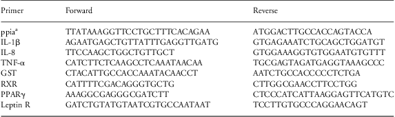
Total RNA was isolated from frozen liver samples with Tri-reagent of Sigma, as described previously.Reference Shiri-Sverdlov, Wouters and van Gorp 18 Reverse transcription was performed using the iScript cDNA synthesis kit (Bio-Rad) according to the manufacturer's instructions. Real-time PCR was performed on a Bio-Rad MyIQ using the IQ SYBR Green Supermix with fluorescein (Bio-Rad) and 10 ng of cDNA. Primers for target genes interleukin (IL)-1β, IL-8, tumor necrosis factor alpha (TNFα), glutathione S-transferase (GST), leptin receptor, retinoid X receptor (RXR) and proliferator-activated receptor-γ (PPARγ) were developed using Primer Express version 2.0 (Applied Biosystems) using default settings. To standardize for the amount of cDNA, cyclophylin A (peptidylprolyl isomerase A) was used as the housekeeping gene. Primer sequences are given in Table 1. The liver real-time PCR results were obtained using the relative standard curve method.
Ovine primer sequences used in RT-PCR

RT-PCR, reverse transcription-PCR; ppia, peptidylprolyl isomerase A; IL, interleukin; TNF, tumor necrosis factor alpha; GST, glutathione S-transferase; RXR, retinoid X receptor; PPARγ, proliferator-activated receptor-γ.
aHousekeeping gene.
Analysis of hepatic lipids
Approximately 50 mg of frozen liver tissue was homogenized in SET buffer (sucrose 250 mM, EDTA 2 mM and Tris 10 mM). Protein content was measured using the bicinchoninic acid method (Pierce). Triglycerides and total cholesterol levels were measured according to the manufacturer's instructions on a Benchmark 550 Micro-plate reader (Bio-Rad).
Plasma measurements
Triglycerides and total cholesterol were measured with a Beckman Coulter Synchron LX20 PRO Clinical Chemistry analyzer (Beckman Coulter). The measurements were validated in adult and fetal sheep. Alanine aminotransferase (ALT) and aspartate aminotransferase (AST) activities were determined by an enzymatic rate method on the Beckman Coulter Synchron LX20 PRO Clinical Chemistry analyzer (Beckman Coulter).
Western blots
Liver sections were homogenized in ice-cold lysis buffer containing a cocktail of proteinase inhibitors and phosphatase inhibitors. 12% SDS-PAGE gels were loaded with 25 μg of total protein and transferred to a 0.45-μm nitrocellulose membrane. Membranes were incubated overnight at 4°C with monoclonal antibody to COX-4 (Cell Signaling, dilution 1:1000). For loading control, the membranes were probed with β-actin antibody (Sigma Chemicals, dilution 1:1000).
Sphingolipid analysis in liver and plasma
Liver tissue samples (20 mg) were homogenized in Aqua bidest using the Ultra-Turrax (ICA Labortechnik). Sphingolipids were extracted from liver homogenates or 20 μl plasma (diluted 1:10 in Aqua bidest), respectively, and prepared for liquid chromatography-electrospray ionization/multi-stage mass spectrometry (LC ESI-MS/MS) as described previously,Reference Shaner, Allegood and Park 19 except that a larger volume (0.4 ml) of LC mobile phase solvent was used to redissolve the final dried residue. Complex sphingolipids (ceramide, monohexosylceramide, sphingomyelin and dihydroceramide) were separated by normal phase LC using a Supelco 2.1 (i.d.) × 50 mm LC-NH2 and a binary solvent system. Sphingoid bases and sphingoid base 1-phosphates were separated by reverse phase LC using a Supelco 2.1 (i.d.) × 50 mm Discovery C18 column (Sigma Chemical) and a binary solvent system at a flow of 1.0 ml/min. The sphingolipids were analyzed by LC ESI-MS/MS in positive ionization mode using an ABI 3000 triple quadrupole (QQQ) tandem mass spectrometer for complex sphingolipids (ceramide, monohexosylceramide, sphingomyelin, dihydroceramide), and the sphingoid bases and sphingoid base 1-phosphates were analyzed using an ABI 4000 QTrap as previously described.Reference Shaner, Allegood and Park 19 Quantitation was performed by multiple reaction monitoring and comparison with the spiked internal standard (Sphingolipid Mix II, Avanti Polar Lipids), as well as correction for differences in ion yield because of chain length. Sphingolipid quantities are expressed as pmol/mg protein.
Data analysis
Data were statistically analyzed using Statistical Package for Social Sciences (SPSS 17.0 Software). Comparisons between controls and LPS-injected groups were performed using two-way ANOVA controlling for age. Significant effects were analyzed using post-hoc Bonferroni corrections. Scoring of apoptotic cells was calculated by Pearson's χ 2-test. Results are presented as mean ± s.e.m. and values were considered statistically significant if P < 0.05.
Results
Hepatic homeostasis and lipid metabolism after LPS-induced chorioamnionitis were evaluated at term gestation and at 7 weeks of age (Supplementary Figure S1). The body weights of LPS-exposed lambs were not different from controls at term (5.0 kg ± 1.0 v. 5.5 kg ± 1.0) and at 7 weeks of age (16.2 kg ± 1.3 v. 15.5 kg ± 1.5).
No liver inflammation in 7-week-old animals
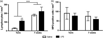
After LPS exposure, CD3-positive T-lymphocytes increased in the livers of term animals relative to controls. The number of CD3-positive cells was increased at 7 weeks of age relative to term animals but there was no difference between the 7-week-old groups (Fig. 1a). The numbers of MPO-positive neutrophils were not changed by LPS exposure at either age (Fig. 1b). In addition, there were no differences in the mRNA levels of IL-1β, IL-8 and TNF between groups (Table 2).
Hepatic inflammation based on immunohistochemistry. T-lymphocyte (CD3) staining (a) and neutrophil (myeloperoxidase, MPO) staining (b) of frozen liver sections. Results are expressed as cells/mm2. **P < 0.01 significantly different from the saline-injected group. LPS, lipopolysaccharide.

Representation of IL-1β, IL-8 and TNF mRNA expression relative to cyclophylin A in the four different groups

IL, interleukin; TNF, tumor necrosis factor alpha; LPS, lipopolysaccharide.
Values are normalized to the levels of control animals and expressed as mean ± s.e.m.
No hepatocellular damage in 7-week-old animals
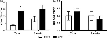
At term birth, TUNEL-positive hepatocytes were elevated after LPS-induced chorioamnionitis (Fig. 2a). However, no changes were observed in the plasma markers for liver damage ALT and AST (data not shown). In addition, no indications for oxidative stress could be observed based on GST mRNA expression (Fig. 2b). In the 7-week-old animals, no indication for liver damage was found (Fig. 2a and 2b).
Quantification of hepatocellular damage. Apoptotic score of terminal transferase dUTP nick end-labeled-positive cells from saline and lipopolysaccharide (LPS)-exposed animals (a). Relative mRNA expression of glutathione S-transferase (GST) normalized to cyclophylin A (b). *P < 0.05 significantly different from the saline-injected group.

Increased hepatic lipid levels in 7-week-old animals
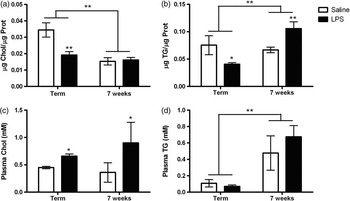
After LPS exposure, term lambs had decreased hepatic cholesterol and triglyceride levels compared with controls (Fig. 3a and 3b). Seven weeks after birth, hepatic cholesterol levels were comparable between control and LPS-exposed animals (Fig. 3a). Hepatic triglyceride levels, on the other hand, were significantly increased compared with controls in the 7-week-old sheep (Fig. 3b). These changes in hepatic lipid levels were age-related (Fig. 3a and 3b).
Liver and plasma lipid levels of saline and lipopolysaccharide (LPS)-exposed animals. Hepatic cholesterol (Chol) levels (a) and hepatic triglyceride (TG) levels (b) corrected for their protein (Prot) content. Plasma total cholesterol (c) and plasma TG levels (d). *P < 0.05 and **P < 0.01 significantly different from the saline-injected group.

In addition, both in term and in 7-week-old animals, plasma cholesterol levels were higher after LPS-induced chorioamnionitis (Fig. 3c). Plasma triglyceride levels were not different between groups (Fig. 3d). Plasma triglycerides increased in 7-week-old animals compared with term animals (Fig. 3d).
Increased leptin receptor mRNA levels, COX-4 levels and plasma ceramides in 7-week-old animals
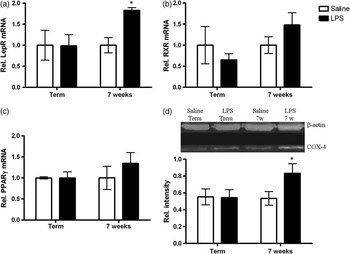
Despite the decreased triglyceride levels, intra-amniotic LPS did not change the mRNA levels of leptin receptor, RXR and PPARγ in term animals (Fig. 4a–4c). However, 7 weeks after birth, the relative expression of leptin receptor mRNA, but not RXR and PPARγ mRNA, was higher in the LPS-exposed animals compared with controls (Fig. 4a–4c).
Relative mRNA expression level of leptin receptor (a), retinoid X receptor (RXR) (b) and proliferator-activated receptor-γ (PPARγ) (c) normalized to cyclophylin A of saline- and LPS-exposed animals. Representative Western blots showing cytochrome c oxidase subunit 4 (COX-4) in saline- and lipopolysaccharide (LPS)-exposed animals (top) and bars representing relative band intensity of COX-4 with β-actin as loading control (bottom) (d). *P < 0.05 significantly different from the saline-injected group.

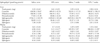
As increasing evidence demonstrates that elevated ceramide biosynthesis contributes to augmented lipid storage,Reference Yang, Badeanlou and Bielawski 20 , Reference Alkhouri, Dixon and Feldstein 21 we measured sphingolipids in liver and plasma. Nevertheless, there were no significant changes in hepatic ceramide metabolites for the term animals or for the 7-week-old animals (Table 3). In plasma, however, levels of ceramide and dihydroceramide decreased after LPS exposure at birth but increased at 7 weeks of age (Table 3). Plasma sphingosine levels increased in LPS-exposed sheep both at term as at 7 weeks of age (Table 3).
Liver and plasma sphingolipid levels (pmol/mg protein) in saline and LPS-exposed animals at 147 days of gestation and postnatal age of 7 weeks

LPS, lipopolysaccharide.
*P < 0.05 significantly different from saline injected group.
To investigate whether the changes in lipid levels could be due to changes in hepatic energy metabolism,Reference Alkhouri, Dixon and Feldstein 21 we measured the expression of COX-4, a mitochondrial membrane enzyme fundamentally involved in the respiratory electron transport system and lipid oxidation.Reference Pejznochova, Tesarova and Hansikova 22 , Reference Wei, Clark and Thyfault 23 Although we did not observe any significant changes in the term animals, Western blot analysis showed that 7-week-old LPS-exposed animals had higher COX-4 protein content in liver compared with control animals (Fig. 4d).
Discussion
In the current study, we asked whether the antenatal inflammation-related liver injury caused by LPS-induced chorioamnionitis could provoke hepatic disturbances at term and at 7 weeks of age. Interestingly, the acute antenatal disturbances in lipid metabolism that were found in previous work,Reference Bieghs, Vlassaks and Custers 8 persisted for up to 7 weeks of age. These are remarkable findings, as the last antenatal LPS exposure was ∼37 days before birth and the effect was still observed 7 weeks after birth. Moreover, we found that these postnatal changes in lipid levels might be correlated with hepatic increases in lipid biosynthesis, mitochondrial biogenesis (COX-4) and circulating ceramide levels.
Fetuses exposed to chronic chorioamnionitis are at higher risk for postnatal morbidity and mortality, with the most important target organs being the brain and lungs.Reference Jobe and Ikegami 24 Various animal models using intra-amniotic LPS or Ureaplasma suggest that antenatal inflammation primes the fetal lung leading to increased postnatal lung inflammation.Reference Normann, Lacaze-Masmonteil and Eaton 25 , Reference Yoder, Coalson and Winter 26 In addition, it has been suggested that exposure to intra-uterine inflammation might be associated with cognitive impairments.Reference Normann, Lacaze-Masmonteil and Eaton 25 However, to the best of our knowledge, the effects of chorioamnionitis on postnatal liver function have not been studied. In our previous work, we demonstrated that chorioamnionitis-induced FIRS can cause hepatic inflammation, consequently leading to disturbances in glucose and lipid metabolism.Reference Bieghs, Vlassaks and Custers 8 Therefore, fetuses exposed to chorioamnionitis may be more susceptible to develop metabolic diseases in adulthood. This might be a new twist to the Developmental Origins of Health and Disease (DOHaD) paradigm where intra-uterine metabolic conditions are correlated with outcomes in later life.Reference Godfrey, Gluckman and Hanson 27
Although the fetal liver was inflamed shortly after fetal exposure to LPS-induced chorioamnionitis,Reference Bieghs, Vlassaks and Custers 8 the only inflammatory effect at term was increased hepatic lymphocyte infiltration. As these animals received three LPS injections that were completed 37 days before delivery, this might be an indication of chronic inflammation. However, other markers for inflammation such as the mRNA expression of pro-inflammatory cytokines were not increased at term. This is in line with the observation that IL-1β and TNF mRNA levels were not increased 2 weeks after LPS-induced chorioamnionitis in fetal sheep.Reference Bieghs, Vlassaks and Custers 8 A plausible explanation could be that these cytokines are not important in chronic inflammation, as they mediate particularly the early inflammatory response.Reference Caille, Bossi, Grimaldi and Vieillard-Baro 28
Further, no effect on inflammation could be observed with respect to the MPO staining. It has been suggested that MPO-expressing cells are involved in acute, as well as in chronic inflammation.Reference Loria, Dato, Graziani and Biasucci 29 This chronic role of MPO was seen in the gut, where a massive influx of MPO-positive cells was observed 30 days after intra-amniotic LPS injection.Reference Wolfs, Buurman and Zoer 30 In addition, antenatal exposure to LPS also caused increased postnatal lung MPO levels.Reference Kramer, Moss and Willet 10 , Reference Normann, Lacaze-Masmonteil and Eaton 25 As MPO plays a postnatal role in other tissues after LPS-induced chorioamnionitis, we speculate that the postnatal liver can mitigate the inflammatory response.
The mechanisms involved in this postnatal inflammatory process may include alterations of the innate immune system.Reference Normann, Lacaze-Masmonteil and Eaton 25 The inflammatory response fully develops during the first few weeks of postnatal life.Reference Schwarzenberg, Potter and Berry 31 , Reference Patricolo, Zangari and Paolocci 32 Therefore, we speculate that in the postnatal liver, the immune system is maturing so that the liver can clear the inflammation.
Our current study reveals a novel finding: hepatic cholesterol and triglyceride levels in chorioamnionitis-exposed animals decreased at term but hepatic triglyceride and plasma cholesterol levels increased at the age of 7 weeks. To our knowledge, nothing is known about the effects of fetal chorioamnionitis on hepatic lipid metabolism after birth. Emerging evidence indicates that inflammation and oxidative stress may be fundamental to lipid homeostasis.Reference Mundt, Wirth and Zender 33 , Reference Chikano, Sawada and Shimoyama 34 Therefore, it is likely that the observed changes in lipid levels are triggered by the chorioamnionitis-induced FIRS. Remarkably, lipid levels did not increase but decreased after LPS exposure in term animals. This effect is also seen in growth-restricted fetal sheep, because of placental insufficiency.Reference Duffield, Vuocolo and Tellam 35 However, when these animals are then exposed to a nutrient-rich environment after birth, lipid levels increase considerably.Reference Duffield, Vuocolo and Tellam 35 We did not manipulate or control the feeding during pregnancy. Yet, the fetal adaptation to inflammation in utero may have comparable effects on the metabolism and the subsequent adaptation postnatally.
To gain more insights into the mechanism of increased lipid levels, we assessed whether chorioamnionitis changes the expression of genes that regulate adipogenesis (PPARγ and RXR) and adipokine signaling (leptin receptor). Although RXR and PPARγ were not significantly increased, they might have played a role as well, because the heterodimerization of these receptors induces the transcription of the leptin receptor.Reference Duffield, Vuocolo and Tellam 35 Changes in leptin receptor were expected as this gene regulates accumulation of lipids,Reference Duffield, Vuocolo and Tellam 35 and indeed the mRNA levels of leptin receptor were increased in 7-week-old animals exposed to prenatal chorioamnionitis.
Hepatic lipid metabolism can also be affected by impaired mitochondrial fatty acid oxidation.Reference Wei, Clark and Thyfault 23 Hence, we measured the expression of COX-4 as a mitochondrial loading control.Reference Pejznochova, Tesarova and Hansikova 22 Surprisingly, COX-4 levels did not decrease after LPS exposure but instead increased in the 7-week-old animals. As mitochondrial lipid oxidation can reduce gene transcription and peptide synthesis,Reference Demeilliers, Maisonneuve and Grodet 36 we speculate that the increased COX-4 levels might be a mechanism to compensate for the increased lipid accumulation due to increased leptin receptor mRNA levels.
Finally, we looked at sphingolipid levels as more and more evidence implicates ceramides to be responsible for the cytokine-induced metabolic changes.Reference Yang, Badeanlou and Bielawski 20 , Reference Alkhouri, Dixon and Feldstein 21 , Reference Memon, Holleran and Moser 37 Although it is known that LPS can induce hepatic sphingolipid synthesis,Reference Zimmermann, Ginis and Furuya 38 we did not observe significant changes in hepatic ceramide metabolites in term or 7-week-old animals. Nevertheless, ceramides synthesized in the liver could be released into the plasma because plasma sphingolipid levels were higher after LPS exposure. Interestingly, the changes in ceramide and dihydroceramide levels correlated with the hepatic triglyceride levels, supporting the idea that LPS can both induce triglyceride and cholesterol synthesis and cause changes in lipoproteins.Reference Memon, Holleran and Moser 37
In summary, antenatal chorioamnionitis has consequences for postnatal hepatic metabolism. Although hepatic lipid peroxidation increased in response to the initial prenatal inflammatory provocation, hepatic triglycerides and plasma cholesterol were increased at 7 weeks after birth. As these lipid changes were correlated with changes in sphingolipid levels, these animals might be more susceptible to develop further hepatic inflammation and even insulin resistance or metabolic disorders in later life.Reference Ruvolo 39 , Reference Browning and Horton 40 However, the long-term consequences in adult life of the antenatal changes seen in hepatic inflammation and metabolism remain to be established explicitly.
Acknowledgements
This study was supported by AI-069716 from the National Institute of Health; a Viertel Senior Medical Research Fellowship (J.J.P.), a NHFA/NHMRC Fellowship (G.R.P.) and the Women and Infants Research Foundation, Western Australia; VENI BWK 016.096.141 from the Dutch Scientific Research Organization and the Research School for Oncology (GROW); a grant of the Maag Lever Darm Stichting (MLDS): WO 08-16 (R.S.). The authors would like to thank Andrea J.X. Lee (School of Women's and Infant's Health, University of Western Australia) for conducting the animal experiments, Claudia Clorefice (Clinical Chemistry, University Hospital Maastricht) for helping us with performing the plasma measurements and Maria Nikiforou (Neuropsychology, Maastricht University) for performing the Western blots.
Supplementary materials
For supplementary material referred to in this article, please visit http://dx.doi.org/doi:10.1017/S2040174412000049.