Introduction
The Pterobranchia are a group of colonial or pseudocolonial hemichordates with a long evolutionary history ranging from the Cambrian to the present day. Extant pterobranchs are benthic and classified into two major orders: the Cephalodiscida and the Rhabdopleurida. A close relationship between rhabdopleurid pterobranchs and graptolites was suggested by some previous studies (Urbanek, Reference Urbanek1986, Reference Urbanek1994; Urbanek and Dilly, Reference Urbanek and Dilly2000; Mierzejewski and Kulicki, Reference Mierzejewski and Kulicki2003) and confirmed by a cladistic investigation (Mitchell et al., Reference Mitchell, Melchin, Cameron and Maletz2013), which indicated that the extinct Graptolithina should be included in the Pterobranchia. On the basis of scanning electron microscope back-scatter electron (SEM-BSE) analyses (Maletz et al., Reference Maletz, Steiner and Fatka2005), some fossils previously presumed to represent Cambrian algae have been reinterpreted as pterobranchs (Maletz et al., Reference Maletz, Steiner and Fatka2005; Maletz, Reference Maletz2014; LoDuca et al., Reference LoDuca, Caron, Schiffbauer, Xiao and Kramer2015a, Reference LoDuca, Wu, Zhao, Xiao, Schiffbauer, Caron and Babcock2015b; Maletz and Steiner, Reference Maletz and Steiner2015), greatly improving our knowledge of the early fossil record of the Pterobranchia. However, the age of the oldest pterobranchs and even of the Hemichordata is still not settled. The earliest potential candidate for hemichordate fossils so far known is acid-isolated organic fragments of Sokoloviina costata Kirjanov, Reference Kirjanov1968 representing tubaria with zigzag-shaped fusellar collars from the basal Cambrian Rovno Formation of Ukraine (Sokolov, Reference Sokolov1997, pl. 8.4). Other remains have been reported from Cambrian Stage 5 (Series 3), including the potential colonial pterobranch Yuknessia simplex Walcott, Reference Walcott1919 (LoDuca et al., Reference LoDuca, Caron, Schiffbauer, Xiao and Kramer2015a) and the tubicolous enteropneust hemichordates Spartobranchus tenuis Caron, Conway Morris, and Cameron, Reference Caron, Conway Morris and Cameron2013 and Oesia disjuncta Walcott, Reference Walcott1911 (Caron et al., Reference Caron, Conway Morris and Cameron2013; Nanglu et al., Reference Nanglu, Caron, Conway Morris and Cameron2016) from the Burgess Shale, and undescribed fragments from the Kaili Formation in Southwest China (Harvey et al., Reference Harvey, Ortega-Hernández, Lin, Zhao and Butterfield2012). Frond-like fossil remains with a pair of tentacles from the Chengjiang Lagerstätte were described as Galeaplumosus abilus Hou et al., Reference Hou, Aldridge, Siveter, Derek, Williams, Zalasiewicz and Ma2011 and interpreted as a possible hemichordate zooid (Hou et al., Reference Hou, Aldridge, Siveter, Derek, Williams, Zalasiewicz and Ma2011). The assignment of the sole known specimen to the hemichordates, however, was questioned by recent studies (LoDuca et al., Reference LoDuca, Caron, Schiffbauer, Xiao and Kramer2015a; Maletz and Steiner, Reference Maletz and Steiner2015; Ou et al., Reference Ou, Han, Zhang, Shu, Sun and Mayer2017). Another potential rhabdopleurid pterobranch from the Niutitang Formation of China (Cambrian Stage 2; Zhao et al., Reference Zhao, Steiner, Yang, Erdtmann, Guo, Zhou and Wallis1999) requires further study, and its pterobranch affinity remains uncertain (Maletz, Reference Maletz2014; LoDuca et al., Reference LoDuca, Caron, Schiffbauer, Xiao and Kramer2015a; Maletz and Steiner, Reference Maletz and Steiner2015). The earliest widely accepted occurrences of pterobranchs are from the basal part of Stage 5 of the Cambrian Period (Maletz, Reference Maletz2014; LoDuca et al., Reference LoDuca, Caron, Schiffbauer, Xiao and Kramer2015a; Maletz and Steiner, Reference Maletz and Steiner2015). In this study, we reinterpret Malongitubus kuangshanensis Hu, Reference Hu2005 from the early Cambrian Chengjiang Konservat-Lagerstätte (Series 2, Stage 3) as a possible pterobranch, judging mostly from the identification of a resistant internal stolon system.
Geological setting, materials, and methods
All material is from a single bedding plane in a small quarry on the northwest slope of a hillside near Kuangshan village, ~10 km southwest of the county town Malong, 100 km east of Kunming in Yunnan Province, China (25°20'15''N, 103°30'20''E). Stratigraphically, this interval belongs to the upper part of the Eoredlichia–Wutingaspis Zone within the Yu’anshan Formation and is equivalent to the strata containing the renowned Chengjiang Biota in Chengjiang County and the Haikou district of Kunming (Luo et al., Reference Luo, Jiang and Tang1994; Zhang et al., Reference Zhang, Babcock, Xiang, Sun, Luo and Jiang2001). Associated fossils from the same interval include arthropods (trilobites, bradoriids, and other arthropods), sponges, cnidarians, anomalocaridids, lobopodians, priapulids, hyolithids, brachiopods, chancelloriids, vetulicolians, and algae (a detailed list of fossils is available in the appendices of Hu, Reference Hu2005 and Zhao et al., Reference Zhao, Hu, Caron, Zhu, Yin and Lu2012), representing a local assemblage of the Chengjiang biota. The lithology of the section and the occurrence of the pterobranch-bearing layer were illustrated previously (Hu, Reference Hu2005, text-fig. 9; Wang et al., Reference Wang, Zhang, Holmer, Hu, Wang and Li2012, fig. 1B). The preservation of the probable pterobranchs and other associated fossils was interpreted to be the result of rapid burial by a distal storm event (Hu, Reference Hu2005; Zhao et al., Reference Zhao, Hu, Caron, Zhu, Yin and Lu2012).
The specimens of Malongitubus kuangshanensis are compressed, often fragmentary, and commonly partly superimposed on one another on the bedding surface. In most cases, the tubes are whitish or pale in color due to intense weathering, with occasional dark remains of original organic material. The materials were prepared using a sharp blade under a binocular microscope. Overall images of the specimens were obtained with a Canon Mark II Camera with an EF 100 mm f/2.8L IS USM close-up lens under direct light. Enlargements of details at the millimeter scale were photographed with a Zeiss Smartzoom 5 microscope system under fiber-optic lights. BSE pictures were obtained using a HITACHI SU3500 in the Nanjing Institute of Geology and Palaeontology, Chinese Academy of Sciences, China, with an accelerating voltage of 20–30 kV. SEM microphotographs were obtained and energy-dispersive spectroscopic analysis was conducted at the State Key Laboratory of Continental Dynamics, Northwest University, Xi’an, China, with a ZEISS-SUPRA 40VP.
Repositories and institutional abbreviations
Nanjing Institute of Geology and Palaeontology, Chinese Academy of Sciences, China (NIGP); Early Life Institute of Northwestern University (ELI). A specimen of Dalyia racemata from the Burgess Shale was also examined in this study (specimen number USNM 194121, Smithsonian Institution, Washington DC, USA). The nomenclature applied in this study follows that of recent works on pterobranchs (Maletz et al., Reference Maletz, Steiner and Fatka2005; LoDuca and Kramer, Reference LoDuca and Kramer2014; Maletz, Reference Maletz2014; LoDuca et al., Reference LoDuca, Caron, Schiffbauer, Xiao and Kramer2015a; Maletz and Steiner, Reference Maletz and Steiner2015).
Systematic paleontology
? Phylum Hemichordata Bateson, Reference Bateson1885
? Class Pterobranchia Lankester, Reference Lankester1877
? Subclass Graptolithina Bronn, Reference Bronn1849, emend. Mitchell et al., Reference Mitchell, Melchin, Cameron and Maletz2013
Genus Malongitubus Hu, Reference Hu2005
Type species
Malongitubus kuangshanensis Hu, Reference Hu2005.
Diagnosis (emended)
Colonial organism with long, slender, and branching thecal tubes. Thecal tubes parallel-sided, each with an internal continuous thread. There are usually four, or less commonly five, terminal thecal tubes, which are free and in similar widths and lengths. Fine annulations present on the surface of thecal tubes. Terminal thecal tubes narrow distally, commonly with thickened apertures.
Remarks
Malongitubus closely resembles Dalyia Walcott, Reference Walcott1919 from the Burgess Shale (Cambrian Series 3, Stage 5) in overall colony shape and in having nearly parallel-sided thecal tubes and inner threads of relatively constant dimensions, herein interpreted as a stolon system. The close similarity between the two taxa led to the suggestion of possible synonymy (Maletz and Steiner, Reference Maletz and Steiner2015). However, Malongitubus differs in having a greatly variable branching distance, from millimeters to centimeters. In addition, the presence of round structures at the branching points of Dalyia (Maletz and Steiner, Reference Maletz and Steiner2015) allows differentiation of the two taxa. On the basis of this difference, Malongitubus is retained as a separate taxon. Holdfast structures and initial (larval) development of both taxa are unknown. The development of fuselli cannot be proven, but is likely. No zooid remains have been preserved. Comparing to Yuknessia from the Cambrian of British Columbia and Utah, the thecal tubes of Malongitubus are completely free and parallel-sided or slightly expanding, whereas those of Yuknessia conspicuously widen distally. In addition, the presence of repent and erect thecae in Yuknessia also distinguish it from Malongitubus. The absence of more differentiated thecal types, such as autothecae, bithecae, and stolothecae, in M. kuangshanensis indicates that it differs from dendroid graptolites, which have been reported from the traditional “middle Cambrian” and younger strata (Rickards et al., Reference Rickards, Baillie and Jago1990; Johnston et al., Reference Johnston, Johnston and Powell2009; LoDuca and Kramer, Reference LoDuca and Kramer2014).
Malongitubus kuangshanensis Hu, Reference Hu2005
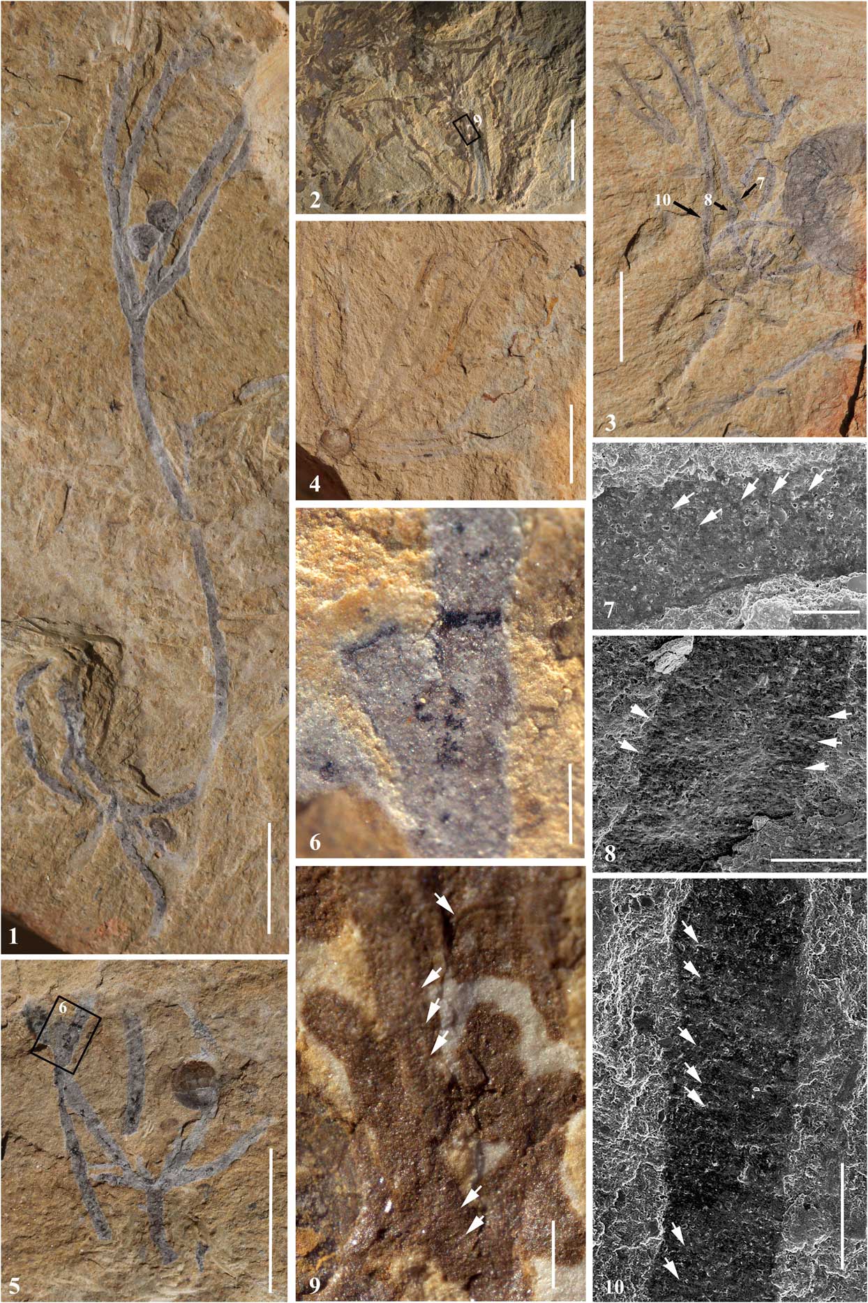
Malongitubus kuangshanensis from the upper part of the Yu’anshan Formation. (1) NIGP-165029: a colony with a bent lower portion and a branched upper portion. (2) NIGP-105030: dense tubaria overlapping each other. The framed area is enlarged in (9). (3) NIGP-105031: a branched colony with a priapulid worm, Maotianshania cylindrica, to the right. The areas indicated by the black arrows are enlarged in (7, 8, 10). (4) ELI-B CLP K007A: a radiating colony. Note the brachiopod to the bottom left. (5) ELI- B CLP K010, part: a colony with a branching tubarium and a stolon system. (6) Thecal apertures. Close-up of the area framed in (5). (7, 8) SEM photograph showing the weak imprints of annulations. White arrows indicate the annulations interpreted as traces of possible fusellar construction. (9) Close-up of the area framed in (2), showing annulations of the tubarium. Some of the annulations are indicated by white arrows. (10) SEM photograph showing the weak imprints of annulations. (1–5) Scale bars=5mm; (6, 9, 10) scale bars=0.5 mm; (7) scale bar=200 μm; (8) scale bar=300 μm.

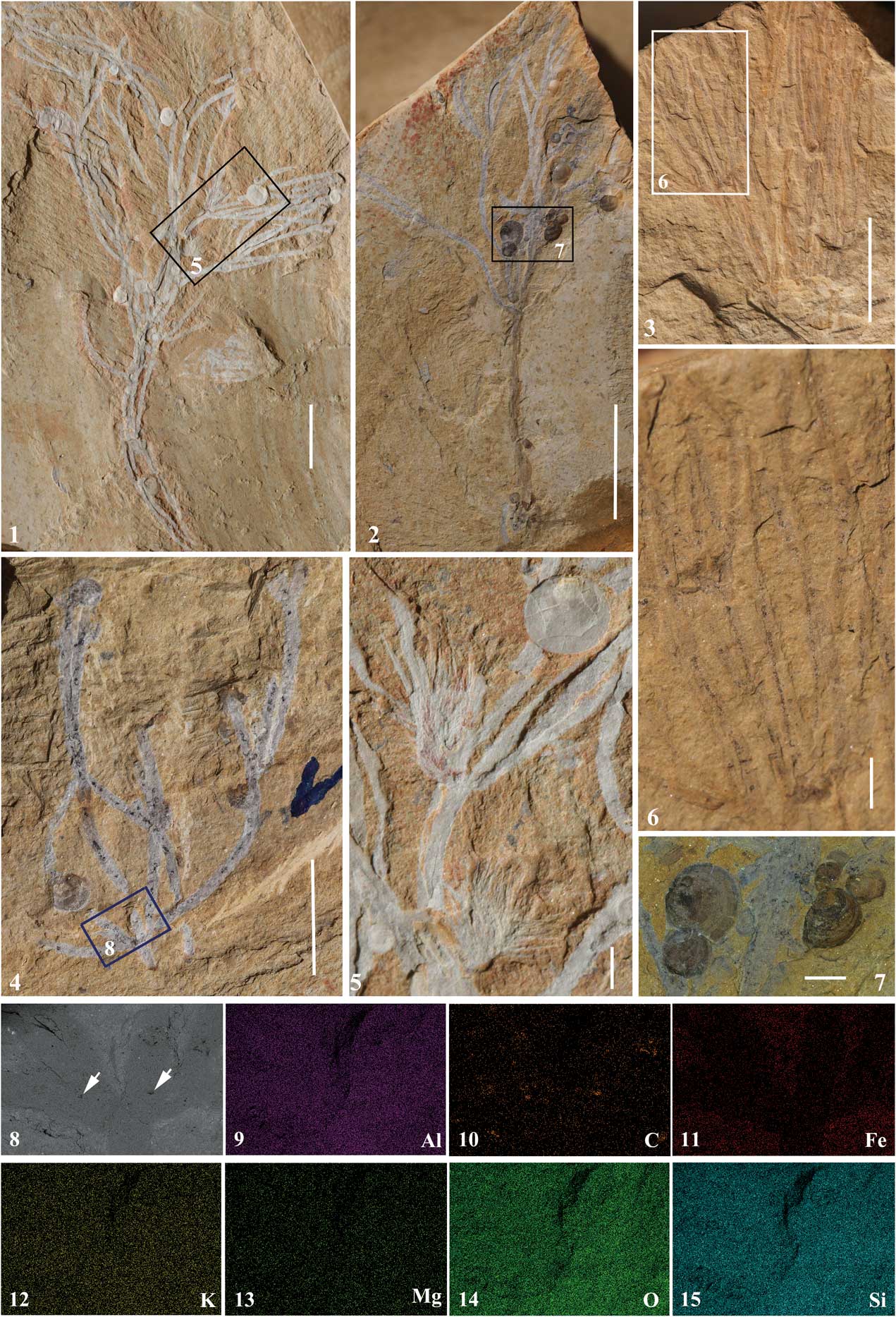
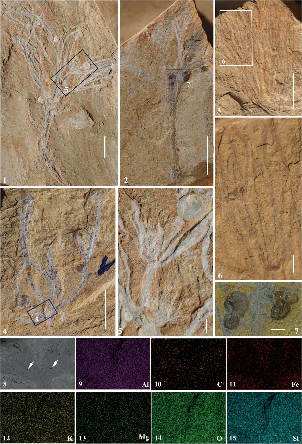
Malongitubus kuangshanensis and attached brachiopods and sponges as secondary tierers. (1) Holotype, NIGP-165028, showing the associated brachiopods and sponges. (2) NIGP-165032, showing the attachment of numerous brachiopods onto the tubes. (3) NIGP-165033. Densely arranged tubes. Close-up of the area framed in (6). (4) ELI-B CLP K010B, counterpart: a colony with both adult and juvenile brachiopods. (5) Close-up of the area framed in (1), showing two sponges and one brachiopod on the tubes. (6) Close-up of the area framed in (3), showing the dark, continuous stolon. (7) Close-up of the two juvenile brachiopods indicated by the framed area in (2). (8) BSE image of the area marked by the frame in (4). White arrows point to the organic remains. (9–15) Energy-dispersive spectroscopic analysis of the tubarium in (8). (1–3) Scale bars=10 mm; (4) scale bar=5 mm; (5, 6) scale bars=1 mm; (7) scale bar=0.5 mm.
2005 Malongitubus kuangshanensis Reference HuHu, p. 187, pl. 18, figs. 1–7.
2012 Malongitubus kuangshanensis; Reference Wang, Zhang, Holmer, Hu, Wang and LiWang et al., p. 62, fig. 2A–G.
Holotype
NIGP-165028 (labeled as Kuangshan-01 in Hu, Reference Hu2005), a large, nearly complete (base missing) multibranched tubarium with four terminal thecal tubes (Fig. 2.1).
Occurrence
Kuangshan village, Malong County, Yunnan Province, China. Upper part of the Eoredlichia–Wutingaspis Zone, Yu’anshan Formation, Cambrian Stage 3, Series 2, early Cambrian.
Description
Maximum height of the tubarium is 6 cm. Tubarium branches are 0.4 to 0.5 mm wide. Distances between bifurcations vary from 1 mm to several centimeters. Remains of soft-bodied zooids are not evident. Thecal tubes mostly straight or curved, sometimes bent (Fig. 1.1). The angle of the thecal tubes to the stipe varies from 15 to 70 degrees. The width of the terminal thecal tubes varies from 250 μm at the aperture to 400 μm at the location just above the branching point. The number of terminal thecal tubes is usually four, or less commonly five (Hu, Reference Hu2005, pl. 18, figs. 4, 5). The terminal thecal tubes are free with similar widths and lengths. The apertures of the thecal tubes appear to narrow slightly and are thickened (Fig. 1.6; Hu, Reference Hu2005, pl. 18, fig. 5). Energy-dispersive spectroscopic analysis reveals that the dark remains on the tubes are organic carbon (Fig. 2.9–2.15).
A number of remains of tubaria show a darker internal thread of relatively constant width, herein interpreted as remains of an original stolon system (Figs. 1.5, 2.3, 2.6). As a result of weathering and early decay, the stolon system is preserved as fragments (Figs. 1.5, 2.4), with only a few cases in which the stolon is visible as a continuous thread for a distance of more than a few millimeters (Fig. 2.6). The stolon is divided where the tubarium branches. The width of the stolon is about 10 μm. Transverse annulations of the tubaria are sometimes faintly preserved (Fig. 1.7–1.10) and may have resulted from previously existing fusellar structures. The heights of the annulations are about 20 μm. However, no unequivocal fusellar structures are presently documented.
Due to fragmentary preservation of all available specimens, no basal parts of tubaria are observed; thus, the means of colony attachment to the substrate cannot be determined.
Other material
About 20 specimens with incomplete parts of the tubarium and a large number of incomplete thecal tubes or branches.
Remarks
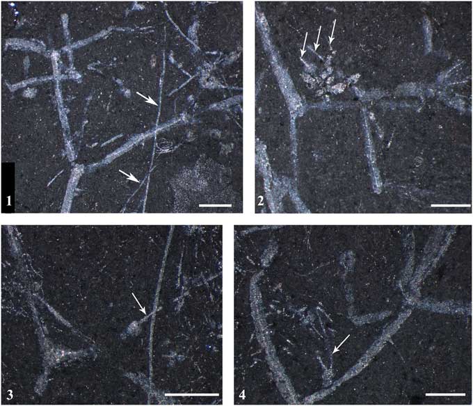
When the taxon Malongitubus kuangshanensis was erected, the phylogenetic affinity of the species was kept open, and the similarity with Dalyia racemata from the Burgess Shale was discussed briefly. At that time, the latter was considered to be an alga, but is now recognized as a possible pterobranch hemichordate (Maletz and Steiner, Reference Maletz and Steiner2015). The possible pterobranch affinity of M. kuangshanensis was also mentioned by Maletz and Steiner (Reference Maletz and Steiner2015) but not discussed in detail. The identification of a resistant stolon system indicates that it has a close affinity to the Pterobranchia and more generally that it is a colonial organism. A “stolon” has sometimes been reported from extant hydrozoan colonies, which are morphologically and functionally differentiated into hydrorhiza, hydrocaulus, and especially swollen hydrotheca (Brusca and Brusca, Reference Brusca and Brusca2003; Ruppert et al., Reference Ruppert, Fox and Barnes2004). However, in colonial hydrozoans, the “stolon” represents a thin extension of the hydropolyp, which is not sclerotized as in pterobranchs and is thus differentiated as coenosarc. While the chitinous perisarc and hydrothecae of colonial hydrozoans may have some fossilization potential, it is rather unlikely that the coenosarc would be preserved in the fossil record. By contrast, decay experiments on modern pterobranchs have demonstrated that the sclerotized stolon system is one of the most durable structures of pterobranch colonies and may prevail until most of the zooids and tubarium have decayed (Beli et al., Reference Beli, Piraino and Cameron2017). In Malongitubus, and notably also in the closely related Dalyia racemata from the Cambrian Stage 5 Burgess Shale, the putative stolon system is the most resistant structure of the fossil remains. This is also shown by the fact that sometimes only stolons remain after decay or parts of stolons extend out of decayed tubaria (Fig. 3). Dalyia was originally interpreted as a red alga (Walcott, Reference Walcott1919), but was recently reinterpreted as a colonial graptolite by Maletz and Steiner (Reference Maletz and Steiner2015) from the identification of a possible stolon system. Therefore, M. kuangshanensis is also interpreted as a probable pterobranch due to the existence of a sclerotized stolon system. Its placement within the Cephalodiscida can be excluded, since the latter are noncolonial and lack a stolon system. Further assignment within the Graptolithina is not attempted herein.
Dalyia racemata from the Cambrian Stage 5 Burgess Shale, No. USNM 194121, Smithsonian Institution. (1) Repeated dichotomous branching of tubarium with distinct threads within the partly decayed tubarium, interpreted as a sclerotized stolon system; thinner filaments besides the branched tubarium represent separated stolons from decayed colonies (arrows). (2) Branched tubarium with internal stolons; separated, decayed branching shows that tube material was decayed and only thickened round basal part of tube and fragments of stolons are left (arrows). (3) Partly decayed tubarium with retained left tube with inner stolon and decayed right tube with exposed stolon (arrow). (4) Branched tubarium with stolon in the left tube; remains of stolons of decayed colonies (arrow). Scale bars=1.0 mm.

Attempts at identifying fusellar structures by SEM-BSE were unsuccessful in the current study. This is probably due to the loss of most of the original organic material of the tube wall as a result of early decay and weathering, leaving only weak imprints of annulations, which are likely to be difficult to detect. The loss of organic material in the main part of the tubarium is also confirmed by the energy-dispersive spectroscopic analysis (Fig. 2.9–2.15). This interpretation also can be applied to Dalyia racemata from the Burgess Shale. As indicated by Maletz and Steiner (Reference Maletz and Steiner2015), all available specimens of D. racemata are diagenetically altered and pale in color, and no original organic material is present.
Taphonomic experiments on modern pterobranchs (Briggs et al., Reference Briggs, Kear, Baas, de Leeuw and Rigby1995; Beli et al., Reference Beli, Piraino and Cameron2017) show different rates of decay between zooids, tubaria, and the stolon system. The zooids decay rapidly and become unrecognizable after a few days, whereas the tubes and the interior stolon system can last for several months. This decay process of modern pterobranchs can be applied to M. kuangshanensis, which shows well-preserved tube outlines and stolon system and the absence of any zooids. Preservation of zooids is extremely rare in the fossil record of pterobranchs. The most substantial record is the putative reports by Durman and Sennikov (Reference Durman and Sennikov1993) and Sennikov (Reference Sennikov2016) of soft tissue in a middle Cambrian (Drumian) rhabdopleurid from Siberia. Remarkably, a few specimens of arthropods (e.g., Naraoia) from the same bedding plane as M. kuangshanensis show well-preserved diverticula (Hu, Reference Hu2005, pl. 13, fig. 7; Zhao et al., Reference Zhao, Hu, Caron, Zhu, Yin and Lu2012, fig. 7N), indicating favorable conditions for soft-tissue preservation. It seems likely that individuals of M. kuangshanensis were exposed on the seafloor and subjected to microbial decay before final burial; consequently, the zooids disappeared due to protracted decay.
Paleoecology
Tiny brachiopods are observed on some of the specimens (Fig. 2.1, 2.2, 2.4, 2.5, 2.7). The brachiopods range from 0.8 to 2.2 mm in diameter. The shells of the brachiopods are complete and articulated, with the two valves fitted together. These tiny brachiopods are called Kuangshanotreta malungensis Zhang, Holmer, and Hu in Wang et al., Reference Wang, Zhang, Holmer, Hu, Wang and Li2012 and are assigned to the acrotretoids (Wang et al., Reference Wang, Zhang, Holmer, Hu, Wang and Li2012). All individuals of K. malungensis are directly in contact with the tubes and are oriented with the posterior margin toward the tubes (Fig. 2.5, 2.7). Judging from the overall colony arrangement and the attachment of brachiopods, it seems likely that M. kuangshanensis was a benthic, erect pterobranch that was attached to a firm substrate while alive. The attached small brachiopods are interpreted as secondary tierers, which are benthic suspension feeders that use primary tierers to reach higher water levels above the seafloor to obtain a feeding advantage. This interpretation is also supported by the fact that these brachiopods are very rare in the surrounding matrix. Some other brachiopod species have also been interpreted as secondary tierers (Zhang et al., Reference Zhang, Han, Wang, Emig and Shu2010), but they were only able to occupy lower levels (less than 5 cm above the seafloor; Wang et al., Reference Wang, Zhang, Holmer, Hu, Wang and Li2012), whereas those on M. kuangshanensis could reach slightly higher elevations. In addition to the miniature brachiopods, five juvenile individuals of the sponge Choia sp. are attached to the tubarium of the holotype of M. kuangshanensis (Fig. 2.1, 2.5). The sponges are 4–5 mm tall (Fig. 2.5), implying probable secondary tiering. Benthic pterobranchs may have occurred in some early Cambrian shallow marine communities in dense accumulations and provided firm substrates and shelter for other benthic metazoans as secondary tierers. Pterobranchs played an important role in early Cambrian marine benthic communities. The interpretation of M. kuangshanensis as a pterobranch also indicates that a brachiopod–pterobranch association had likely developed by the Cambrian Series 2, representing an early form of metazoan commensalism.
Acknowledgments
This work was supported by the National Basic Research Program (2013CB835006); the Strategic Priority Research Program (B) of the Chinese Academy of Sciences (XDB18030304); and the National Natural Science Foundation of China (Grant Nos. 41425008, 41472012). We are grateful to M. Zhu, Nanjing Institute of Geology and Palaeontology, Chinese Academy of Sciences, and Z. Zhang, Northwest University, for constructive discussion and suggestions. The help of J. Thompson, University of Southern California, USA, and L. Muir, Amgueddfa Cymru – National Museum of Wales, UK, in improving the English of the manuscript is gratefully acknowledged. We also thank J. Jin and two reviewers for their most valuable comments and criticisms.