Introduction
Liposarcoma that accounts for approximately 20 per cent of soft tissue sarcomas, is one of the most common soft-tissue sarcomas in adults.Reference Toro, Travis, Wu, Zhu, Fletcher and Devesa1 They typically occur in the middle aged to older adult, and there is a male preponderance.Reference Toro, Travis, Wu, Zhu, Fletcher and Devesa1, Reference Engström, Bergh, Gustafson, Hultborn, Johansson and Löfvenberg2 They usually occur in the lower extremities and retroperitoneum, and rarely occur in the head and neck region.Reference Engström, Bergh, Gustafson, Hultborn, Johansson and Löfvenberg2 The parapharyngeal space is a very rare site for liposarcomas, and only a few case reports exist. We present a case of a 19-year-old female with a well-differentiated liposarcoma arising in the parapharyngeal space. Through our case and previously reported studies, we review the features of head and neck liposarcomas.
Report of a case
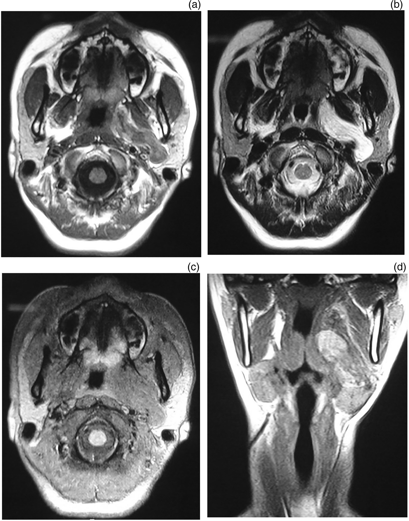
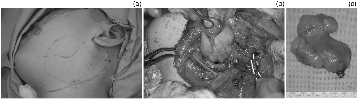
A 19-year-old female presented with a two months' history of an enlarging, slightly painful mass in the left submandibular region. Physical examination revealed a soft and smooth mass which was 3 cm in size under the left mandible with intact skin. The patient had no lower cranial nerve palsy or other systemic symptoms. A computed tomography (CT) scan with contrast revealed a dumbbell-shaped tumour adjacent to the left submandibular gland expanding to the parapharyngeal space, reaching to the anterior wall of the external auditory canal. Most parts of the tumour showed low density in the CT scan, but partially showed heterogeneous density. On magnetic resonance imaging (MRI), the tumour showed low signal intensity partially with high signal intensity areas on T1-weighted images. On T2-weighted images, most of the tumour showed high signal intensity. The tumour showed low signal intensity generally on fat suppressing T1-weighted MR images (Figure 1). From the anatomical site and radiological findings of the tumour, we considered lymphangioma, salivary gland tumour, neurilemmoma and dermoid cyst as principal differential diagnosis. The patient underwent an en bloc resection. The tumour was excised utilising a cervical–parotid approach. The submandibular gland was removed and the posterior belly of digastric muscle and stylohyoid muscle were resected to improve access to the parapharyngeal space. Findings during intra-operative inspection revealed a lobulated lipomatous mass surrounded with a smooth intact capsule. The size of the excised tumour was 7 × 7 cm2 (Figure 2). Pathological examination of the tumour revealed a well-differentiated liposarcoma. The patient has had no recurrence after five years and nine months of follow up.
On magnetic resonance imaging, the tumour showed low signal intensity (SI) partially with high SI areas on axial T1-weighted imaging (T1WI) (a), and most of the tumour showed high SI on T2-weighted imaging (T2WI) (b). The tumour showed low SI on fat suppressing T1WI (c). Coronal T2WI demonstrated a dumbbell-shaped tumour in the parapharyngeal space (d).

A sigmoid-curved skin incision line from the pre-auricular area to the submandibular area was designed for cervical–parotid approach (a). The submandibular gland was removed and the posterior belly of digastric muscle and stylohyoid muscle were resected to improve access to the parapharyngeal space. The tumour was removed without injury to adjacent neurovascular components (b). The excised specimen showed a lobulated lipomatous mass surrounded by a smooth intact capsule. The size of the excised tumour was 7 × 7 cm2 (c).
Pathological findings
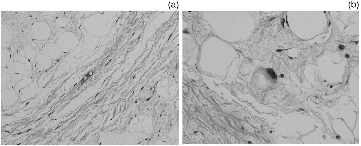
The histological section showed a proliferation of lobules of various-sized adipocytes with irregular nuclei, some atypical multi-vacuolated lipoblasts and stromal spindle to polygonal-shaped cells embedded in a fibromyxoid background (Figure 3).
Histological sections of the well-differentiated sclerosing liposarcoma. The section showed a proliferation of lobules of various-sized adipocytes with irregular nuclei, some atypical multi-vacuolated lipoblasts and stromal spindle to polygonal-shaped cells embedded in a fibromyxoid background. (haematoxylin & eosin; ×200 (a), ×400 (b)).
The histological diagnosis was well-differentiated sclerosing liposarcoma.
Discussion
In the presented case, the patient's age and the tumour site made it difficult for us to make a qualitative diagnosis before the operation. In the CT findings, the tumour showed a well-circumscribed, low-density mass. On MRI, the tumour showed heterogeneous intensity on T1-weighted imaging, and predominantly high signal on T2-weighted imaging. On fat suppressed imaging, the mass showed a uniform low signal. In previous studies, the characteristic appearances of well-differentiated liposarcomas on MRI are predominantly lipomatous tumours with thick septa (>2 mm), globular and/or nodular areas and/or associated masses.Reference van Vliet, Kliffen, Krestin and van Dijke3 In our case, the thick septa in the tumour were unapparent. But retrospectively, the CT and MRI findings indicated an adipose prominent solid mass with nodular or patchy non-adipose components, and these are consistent with liposarcoma.
To confirm the diagnosis of liposarcoma, an incisional biopsy or surgical removal of the tumour is usually necessary. We used a cervical–parotid approach to remove this parapharyngeal tumour. The tumour was a capsule-covered lesion, and could be easily separated from the surrounding tissues. The histological diagnosis was well-differentiated sclerosing liposarcoma.
The World Health Organization classification recognises three biological types encompassing five subtypes of liposarcoma: well-differentiated/dedifferentiated, myxoid/round cell and pleomorphicReference Fletcher, Unni and Mertens4. Well-differentiated liposarcoma accounts for 40–45 per cent of all liposarcomas. Dedifferentiation occurs in up to 10 per cent of well-differentiated liposarcomas, 90 per cent of dedifferentiated liposarcomas arise de novo while 10 per cent occur in recurrence. Myxoid/round cell liposarcoma is the second most common subtype, accounting more than 30–40 per cent of liposarcomas. Pleomorphic liposarcoma represents the rarest subtype, accounting for 5–10 per cent of all liposarcomas.Reference Engström, Bergh, Gustafson, Hultborn, Johansson and Löfvenberg2, Reference Fletcher, Unni and Mertens4
The clinical behaviour of liposarcoma reflects its histological appearance, so the histological subtype remains the most important prognostic factor. Well-differentiated and myxoid tumours are considered low-grade lesions, and on the other hand, dedifferentiated, round cell and pleomorphic types are high-grade lesions. It is reported that the five-year survival rates are 83–100 per cent for well-differentiated,Reference Kooby, Antonescu, Brennan and Singer5–Reference Chang, Hajdu, Collin and Brennan7 20 per cent for dedifferentiated,Reference Singer, Antonescu, Riedel and Brennan6 88–100 per cent for myxoid,Reference Chang, Hajdu, Collin and Brennan7, Reference Antonescu, Tschernyavsky and Decuseara8 83 per cent for round cellReference Antonescu, Tschernyavsky and Decuseara8 and 56 per cent for pleomorphic tumours.Reference Chang, Hajdu, Collin and Brennan7 Low-grade tumours rarely develop distant metastasis. Well-differentiated liposarcomas have minimal metastatic potential.Reference Singer, Antonescu, Riedel and Brennan6, Reference Chang, Hajdu, Collin and Brennan7, Reference Weiss and Rao9
In contrast to the low incidence of distant metastasis, well-differentiated liposarcoma often recur locally, approximately in 40–60 per cent of casesReference Singer, Antonescu, Riedel and Brennan6, Reference Weiss and Rao9, Reference Lucas, Nascimento, Sanjay and Rock10 (Table I). The recurrence rates are different according to the location. Tumours located in the subcutaneous layer and in the extremities have lower local recurrence rates than those in the retroperitoneum.Reference Weiss and Rao9 It is considered that the difference in the recurrence rates by location is caused by the difficulties in obtaining adequate surgical margins. Dalal et al. reported 12-year disease-specific survival rates by margin status in overall subtypes of liposarcoma. Patients with microscopically negative and positive margins had 12-year disease-specific survival rates of 74 and 68 per cent, respectively, whereas those with grossly positive margins had a significantly decreased 12-year disease-specific survival rate of 25 per cent.Reference Dalal, Kattan, Antonescu, Brennan and Singer11 As surgical excision remains the dominant modality of curative therapy for liposarcoma, wide marginal excision with a 1–2 cm margin of normal tissue is strongly recommended. But in many cases, especially when the tumours are located in deep soft-tissue sites, neurovascular components and critical organs adjacent to the tumour prevent wide excisions.
Summary of published reports on well-differentiated liposarcoma
DDLS = dedifferentiated liposarcoma; DSS = disease-specific survival rate, – not stated
In the head and neck region, liposarcoma is a rare tumour; approximately 1 per cent of head and neck sarcomas. Golledge et al. reviewed 76 cases, including their own cases.Reference Dalal, Kattan, Antonescu, Brennan and Singer11 They reported that the five-year survival by life table analysis was 67 per cent, and concluded that the overall prognosis for head and neck liposarcoma is better than for liposarcoma arising elsewhere, particularly in the retroperitoneum.Reference Golledge, Fisher and Rhys-Evans12 The difference of the prognosis between a retroperitoneal and head and neck lesion might be caused by the tumour size when they are detected. In the head and neck lesion, many tumours are found at about 5 cm in size as a painless mass,Reference Golledge, Fisher and Rhys-Evans12 while most retroperitoneal liposarcoma are large (>10 cm) at the time of diagnosis.Reference Singer, Antonescu, Riedel and Brennan6
The histological diagnosis of our case was well-differentiated sclerosing liposarcoma. Well-differentiated liposarcoma can be classified as lipoma-like, sclerosing, inflammatory and spindle cell type.Reference Fletcher, Unni and Mertens4 Kooby et al. reviewed a total of 91 well-differentiated liposarcoma patients, including 28 sclerosing types and reported that the sclerosing subtype and the margin status are associated with the risk of local recurrence.Reference Kooby, Antonescu, Brennan and Singer5 They found that patients with sclerosing tumours had a 10-year local recurrence-free survival of 53 per cent compared with 100 per cent for patients with non-sclerosing tumours. Patients with sclerosing type and microscopic positive margins had a high local recurrence rate, the 10-year local recurrence-free survival was 17 per cent. They recommended re-excision or adjuvant radiotherapy (RT) be considered in this high recurrent risk group.
The margin status of our case was microscopically positive. The tumour was covered by a capsule and some tumour cells were seen in the connective tissue outside the capsule. In our case, utilising skull base surgery can be considered. However, it may decrease the quality of life of the patient due to lower cranial nerve palsy, etc. We also considered adjuvant RT, which was not performed since the benefit of adjuvant RT to well-differentiated liposarcoma is still not clear. Although it is known that the subgroup of myxoid liposarcoma is highly radiosensitive,Reference de Vreeze, de Jong, Haas, Stewart and van Coevorden13 little is reported about well-differentiated liposarcoma. In the same way, very few data are available with regard to the use of chemotherapy in liposarcoma. Further investigations are required about the benefit of the adjuvant therapy.
Since our case is considered to be in the high recurrent risk group, careful follow up will be necessary. Lucas et al. reviewed 58 patients of well-differentiated liposarcoma and reported that 53 patients required at least one additional operation for local recurrence. The average time between initial operation and the second operation was 5.2 years and delayed recurrences were common.Reference Lucas, Nascimento, Sanjay and Rock10 Long-term follow up will be necessary for well-differentiated liposarcoma.
• This paper reported a rare case of a 19-year-old female with a well-differentiated liposarcoma arising in the parapharyngeal space
• Cervical–parotid approach was used to remove this parapharyngeal tumour. Although wide local excision is recommended for treatment of liposarcoma, taking a wide surgical margin is often difficult in the head and neck region
• In contrast to the low incidence of distant metastasis, well-differentiated liposarcoma often recur locally, in approximately 40–60 per cent of cases. Long-term follow up will be necessary because delayed local recurrences are not rare in well-differentiated liposarcoma
In conclusion, we present a rare case of well-differentiated liposarcoma arising in the parapharyngeal space. Although wide local excision remains the mainstay for treatment for liposarcoma, taking a wide surgical margin is often difficult in the head and neck region because of adjacent neurovascular components. The benefits of adjuvant therapy are still not proven. Long-term follow up will be necessary because delayed local recurrences are not rare.