INTRODUCTION
Cryptosporidium parvum is an obligate intracellular apicomplexa protozoan parasite that primarily infects the epithelial cells of the small intestine and is responsible for the diarrhoeal illness in a number of mammals including humans (Priest et al. Reference Priest, Xie, Arrowood and Lammie2001; Tzipori and Ward, Reference Tzipori and Ward2002). Most cases of human infection are usually asymptomatic or characterized by self-limiting diarrhoea in immunocompetent or healthy individuals, but the infection can become a chronic and life-threatening disease in immunocompromised individuals, most notably patients with acquired immunodeficiency syndrome (AIDS) (Peterson, Reference Peterson1992; Fayer et al. Reference Fayer, Morgan and Upton2000; Kosek et al. Reference Kosek, Alcantara, Lima and Guerrant2001). Although intensive studies into the biology and biochemistry of the parasite have been carried out in recent years, effective therapeutic or preventive interventions for C. parvum infection are still not available. New drugs or vaccines against the parasite are urgently needed, but poor experimental tractability of the parasite is one of the major obstacles to developing effective vaccines and/or treatment approaches. Identification and characterization of target molecules that play pivotal roles in the parasite's life cycle can be one of the approaches to vaccine and chemotherapeutic agent development.
Leucine aminopeptidases (LAP; EC 3.4.11.1) are one of the major neutral exopeptidases that catalyse the removal of leucine residues from the amino termini of proteins or polypeptides, and they have been identified in numerous microorganisms, plants, invertebrates, and vertebrates (Taylor, Reference Taylor1993; Matsui et al. Reference Matsui, Fowler and Walling2006). LAPs are involved in diverse essential biological and physiological roles, including processing, maturation, activation or degradation of substrates, or interactions with peptide-dependent signalling (Matsui et al. Reference Matsui, Fowler and Walling2006; Rawlings et al. Reference Rawlings, Morton and Barrett2006). Accumulated evidence from apicomplexa parasitic protozoa suggests crucial roles for LAPs in parasite biology. Plasmodium falciparum LAP (PfLAP) participates in the terminal stages of haemoglobin hydrolysis and generates free amino acids required for protein synthesis and maintenance of osmotic balance (Gavigan et al. Reference Gavigan, Dalton and Bell2001; Gardiner et al. Reference Gardiner, Trenholme, Skinner-Adams, Stack and Dalton2006; Dalal and Klemba, Reference Dalal and Klemba2007; Stack et al. Reference Stack, Lowther, Cunningham, Donnelly, Gardiner, Trenholme, Skinner-Adams, Teuscher, Grembecka, Mucha, Kafarski, Lua, Bell and Dalton2007). LAPs of P. vivax (PvLAP), Toxoplasma gondii (TgLAP), and Babesia gibsoni (BgLAP) have been recently identified, and their essential roles in the life cycles of the parasites have been proposed (Jia et al. Reference Jia, Terkawi, Aboge, Goo, Luo, Li, Yamagishi, Nishikawa, Igarashi, Sugimoto, Fujisaki and Xuan2009, Reference Jia, Nishikawa, Luo, Yamagishi, Sugimoto and Xuan2010; Lee et al. Reference Lee, Song, Seok, Jha, Han, Song, Yu, Hong, Kong and Chung2010). Due to their essential role in parasite biology, LAPs of apicomlexan parasitic protozoa have been recognized as drug targets.
In this study, we identified a gene encoding an M17 leucine aminopeptidase of C. parvum (CpLAP) and characterized the biochemical properties of the recombinant protein. CpLAP was found to share similar structural and biochemical properties with LAPs of other apicomplexa parasitic protozoa. This is the first report on aminopeptidase of C. parvum and provides insights into the protein metabolism of the parasite.
MATERIALS AND METHODS
Identification and cloning of the gene encoding for CpLAP
A gene putatively encoding an M17 leucine aminopeptidase of C. parvum (Gene ID: cgd5_2600) was identified by data mining the Cryptosporidium genomic resource (CryptoDB, http://cryptodb.org). The open reading frame (ORF) of CpLAP was amplified by polymerase chain reaction (PCR) using forward (5′-ATGGTAAATCCAATACATCCAGAATGCATT-3′) and reverse (5′-TCAACATTTTGAGGAAAGCTCTTTAATAAT-3′) primers and C. parvum Iowa II genomic DNA. The amplification reaction was performed using the following thermal cycling profile: 94°C for 5 min, 30 cycles at 94°C for 1 min, 52°C for 1 min, and 72 °C for 2 min, followed by a 72°C extension for 10 min. The PCR product was analysed by 1·2% agarose gel electrophoresis, purified from the gel using a gel extraction kit (Qiagen, Valencia, CA, USA), ligated into a T&A cloning vector (Real Biotech Cooperation, Banqiaa City, Taiwan), and then transformed into Escherichia coli DH5α. Sequencing reactions were conducted using a BigDye Terminator Cycle Sequencing Ready Reaction Kit with an ABI 377 automatic DNA sequencer (Applied Biosystems, Foster City, CA, USA). Analysis of the primary structure of the deduced amino acid sequence was conducted with DNASTAR (DNASTAR, Madison, WI, USA) and Signal P (http://www.cbs.dtu.dk/services/SignalP). TargetP1.1 (http://www.cbs.dtu.dk/services/TargetP) and PSORT (http://www.psort.org) were used to predict protein localization. Phylogenetic analysis was conducted using the neighbor-joining method with MEGA 4 (http://www.megasoftware.net). To determine the strength of the groupings, bootstrap values for nodes were calculated by analysing 1000 bootstrap replicate data sets.
Expression and purification of recombinant CpLAP
To express recombinant CpLAP, the entire sequence of CpLAP was amplified using the primers 5′-GGATCCATGGTAAATCCAATACATCCA-3′, which contained a 5′ Bam HI site, and 5′-CTGCAGTCAACATTTTGAGGAAAGCTC-3′, which harboured a 5′ Pst I site. The PCR product was purified, ligated into a T&A cloning vector (Real Biotechnology Cooperation), and transformed into E. coli DH5α. The resulting plasmid DNA was digested with the appropriate restriction enzymes and ligated into the pQE-30 expression vector (Qiagen), which was then transformed into E. coli M15 [pREP4] cells (Qiagen). Selected clones were grown and induced with 1 mm isopropyl-1-thio-β-D-galactopyranoside (IPTG). The cells were collected, suspended in native lysis buffer (50 mm NaH2PO4, 300 mm NaCl, 10 mm imidazole, pH 8·0), sonicated on ice, and then centrifuged at 4°C for 20 min at 12 000 g. The recombinant protein was purified by nickel-nitrilotriacetic acid (Ni-NTA) chromatography (Qiagen) by following the manufacturer's instructions. The purification and purity of the recombinant protein were monitored by sodium dodecyl sulfate-polyacrylamide gel electrophoresis (SDS-PAGE).
Production of specific antibody for CpLAP (anti-CpLAP) and Western blot analysis
Anti-CpLAP was produced by immunization of BALB/c mice with the purified recombinant CpLAP (50 μg) 3 times at 2-week intervals. Two weeks after the final boosting, the mice were sacrificed and the sera were collected. The immunoglobulin G (IgG) fraction was isolated from the sera with a Protein G-Sepharose column (Amersham Biosciences, Piscataway, NJ, USA). For Western blot analysis, the extract of C. parvum sporozoites was prepared as previously described (Na et al. Reference Na, Kang, Cheun, Cho, Moon, Kim and Sohn2009). In brief, 109 sporozoites were suspended in phosphate-buffered saline (PBS; pH 7·4) which contained a complete protease inhibitor cocktail™ (Roche, Mannheim, Germany) and sonicated on ice using 5-sec pulses at 50% output power for up to 3 min in a W-380/385-series SONICATOR Ultrasonic Liquid Processor (Misonix Inc., Farmingdale, NY, USA). The sonicated C. parvum sporozoite extract (20 μg) was subjected to SDS-PAGE, and the proteins were transferred to a nitrocellulose membrane (Bio-Rad, Hercules, CA, USA). The membrane was blocked with PBS supplemented with 0·05% Tween 20 (PBST; pH 7·4) and 3% skim milk for 1 h. The membrane was then incubated with anti-CpLAP diluted 1:1000 in PBST at room temperature for 2 h. After several washes with PBST, the membrane was incubated with 1:1000 diluted horseradish peroxidase-conjugated goat anti-mouse IgG (Sigma, St Louis, MO, USA). The blot was visualized with 4-chloro-1-naphthol (Sigma) and the reaction was stopped by washing the membrane with distilled water.
Enzyme assay
The aminopeptidase activity of CpLAP was assayed fluorometrically based on hydrolysis of l-Leucine 4-methyl-coumaryl-7-amide (Leu-MCA; Peptide International, Louisville, KY, USA). Reactions were carried out in 96-well microtitre plate format. In brief, 10 μl of enzyme solution was added to 190 μl of Tris-HCl buffer (pH 8·5) containing 5 μ m Leu-MCA and 1 mm MnCl2, and the release of fluorescence was assessed at an excitation wavelength of 355 nm and an emission wavelength of 460 nm for 30 min at 37°C with a Fluoroskan Ascent FL fluorometer (Thermo, Vantaa, Finland).
Optimal pH and stability of CpLAP
The optimal pH for the maximum activity of CpLAP was assessed in sodium phosphate (pH 6·0–6·5), Tris-HCl (pH 7·0–8·5), and Glycine-NaOH (pH 9·0–9·5). Recombinant CpLAP (50 nm) was added to each pH buffer containing 5 μ m Leu-MCA and 1 mm MgCl2, and the enzyme activity measured as described above. For each pH, an appropriate blank was separately measured as a control. The pH stabilities of recombinant CpLAP at pH 5·0, 6·0, 7·0, 8·0 or 9·0 were also examined by incubating the enzyme at 37°C in appropriate buffers.
Effects of metal ions and inhibitors
The requirement of metal ions for optimal CpLAP activity was analysed by assaying the enzyme activity after pre-incubation at room temperature for 20 min in 50 mm Tris-HCl (pH 8·5) containing one of the following metal chlorides: CaCl2, CoCl2, CuCl2, FeCl2, MgCl2, MnCl2, and ZnCl2. The effects of different protease inhibitors or metal chelators on CpLAP activity were assayed by pre-incubating the enzyme with different concentrations of each inhibitor or metal chelator for 20 min at room temperature, followed by measuring residual enzyme activity using Leu-MCA as a substrate. Results are expressed as percentage relative activity with respect to the control as 100%, which did not contain any inhibitor or chelator. The inhibitors and metal chelators used in this study were as follows: phenylmethylsulphonyl fluoride (PMSF), pepstatin A, trans-epoxy-succinyl-l-leucylamido(4-guanidino)butane (E-64), 1,10-phenanthroline, ethylenediaminetetraacetic acid (EDTA), bestatin, and amastatin. The inhibitors and metal chelators used in this study were purchased from Sigma or Peptide International. To further analyse the binding characteristics of metal ions to CpLAP, the inhibition profile of CpLAP by EDTA was investigated in 96-well microplate format as previously described (Maric et al. Reference Maric, Donnelly, Robinson, Skinner-Adams, Trenholme, Gardiner, Dalton, Stack and Lowther2009). CpLAP (30 nm) was incubated with different concentrations (0–100 mm) of EDTA in 50 mm Tris-HCl (pH 8·5) supplemented with 100 μ m ZnCl2 at room temperature for 15 min, after which Leu-MCA (5 μ m) was added. Rates of hydrolysis were monitored based on increases in fluorescence. Inhibition was observed by plotting percentage relative activity against log of EDTA concentration, where 100% relative activity is the rate in the absence of EDTA (Maric et al. Reference Maric, Donnelly, Robinson, Skinner-Adams, Trenholme, Gardiner, Dalton, Stack and Lowther2009). Reactivation of EDTA-treated CpLAP was also analysed by addition of either MnCl2 or ZnCl2 to EDTA-treated enzyme, which was prepared by dialysing against 50 mm Tris-HCl (pH 8·5) to remove residual EDTA, for 15 min before addition of Leu-MCA (5 μ m). Enzyme activity was measured and compared to the control without addition of any metal ion. All experiments were performed in triplicate, and the mean and standard deviation were calculated.
Substrate specificity
Substrate specificity of CpLAP was determined by using l-Alanine-MCA (Ala-MCA), l-Methionine-MCA (Met-MCA), Leu-MCA, l-Lysine-MCA (Lys-MCA), and l-Phenylalanine-MCA (Phe-MCA) as substrates. All substrates were purchased from Peptide International. All assays were carried out in triplicate, and the mean and standard deviation were calculated. The kinetic constants, Michaelis–Menten constant (Km) and maximum velocity (Vmax), of CpLAP for each substrate were determined using GraphPad software (GraphPad Software Inc., La Jolla, CA, USA).
RESULTS
Cloning and molecular characterization of CpLAP
The CpLAP gene consisted of a 1575 bp sequence that encoded for 524 amino acids. Analysis of the deduced amino acid sequence of CpLAP using Signal P (http://www.cbs.dtu.dk/services/SignalP) revealed that CpLAP did not possess a signal peptide sequence. Predictions regarding CpLAP localization by TargetP1.1 (http://www.cbs.dtu.dk/services/TargetP) and PSORT (http://www.psort.org) found that CpLAP was localized to the cytosol with 0·939% and 56·5% probabilities, respectively. Multiple sequence alignment of the amino acid sequence of CpLAP with other presently known LAPs from other apicomplexan protozoan parasites revealed that the N-terminal portion was less conserved between the sequences, but the catalytic C-terminal domain was more conserved (Fig. 1). CpLAP showed highest sequence similarity (98·7%) with LAP of C. hominis (only 7 amino acids were different). The overall sequence similarity between CpLAP and LAPs of other amicomplexan protozoan parasites was 29·6–98·7%, whereas that within the C-terminal domain was 42·1–99·1%. The octapeptide M17 aminopeptidase family signature sequence (NTDAEGRL) was found in CpLAP (residues 376–383). The amino acid residues essential for metal-binding and substrate-binding were also highly conserved in CpLAP (Fig. 1). Phylogenetic analysis revealed that CpLAP was closely related to LAPs of other apicomplexa, including C. hominis, Plasmodium spp., T. gondii, and Babesia spp. (Fig. 2). The apicomplexan group, in turn, formed a larger clade with the M17 LAPs of plants, cyanobacteria, and algae, which indicates their close association and common ancestry.
Multiple sequence alignment of CpLAP. The deduced amino acid sequence of CpLAP was aligned with those from other apicomplexa parasites. The dashes represent gaps introduced to maximize alignment. The substrate binding sites are marked as asterisks and essential metal coordinating residues are indicated as boxes. The octapeptide M17 aminopeptidase family signature sequence is represented as a bold line. Percentage sequence similarity is represented as shading: black (100%), dark grey (57–86%), light grey (28–43%) and no shading (<15%). Cp, C. parvum LAP (XM_626197); Ch, C. hominis LAP (XP_667960); Pf, Plasmodium falciparum LAP (XP_001348613); Pv, P. vivax LAP (XP_001615930); Pk, P. knowlsei LAP (XP_002260412); Tg, Toxoplasma gondii LAP (XM_002368420); Bg, Babesia gibsoni LAP (AB490782).

Phylogenetic analysis. The phylogeny tree was constructed with the neighbor-joining method using the MEGA4 program. Numbers on the branches indicate bootstrap proportions (1000 replicates).

Expression and purification of recombinant CpLAP
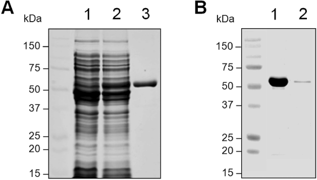
The full-length gene of CpLAP was amplified, cloned into the pQE-30 expression vector, and transformed into E. coli. Recombinant CpLAP was expressed in E. coli as a soluble protein with an apparent molecular weight of 58 kDa (Fig. 3A), which was consistent with the estimated molecular mass of the deduced amino acid sequence of CpLAP. The recombinant protein was purified by Ni-NTA affinity chromatography and then used in this study. To determine the expression of CpLAP in C. parvum sporozoites, Western blot analysis was performed. The anti-CpLAP was reacted with a protein with an approximate molecular weight of 58 kDa, which well coincided with the predicted molecular size of CpLAP, in the extract of C. parvum sporozoites (Fig. 3B).
Expression of recombinant CpLAP and Western blot analysis. (A) Expression and purification of recombinant CpLAP. Proteins were analysed by SDS-PAGE and stained with Coomassie blue. Lane 1, E. coli lysate control; lane 2, IPTG-induced E. coli lysate; lane 3, purified CpLAP by Ni-NTA affinity chromatography. (B) Western blot analysis. The extract of C. parvum sporozoites was separated on SDS-PAGE and the proteins were transferred to nitrocellulose membrane. The blot was probed with anti-CpLAP. Lane 1, recombinant CpLAP (5 μg); lane 2, extract of C. parvum sporozoites (20 μg).

Biochemical properties of recombinant CpLAP
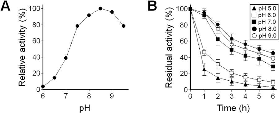
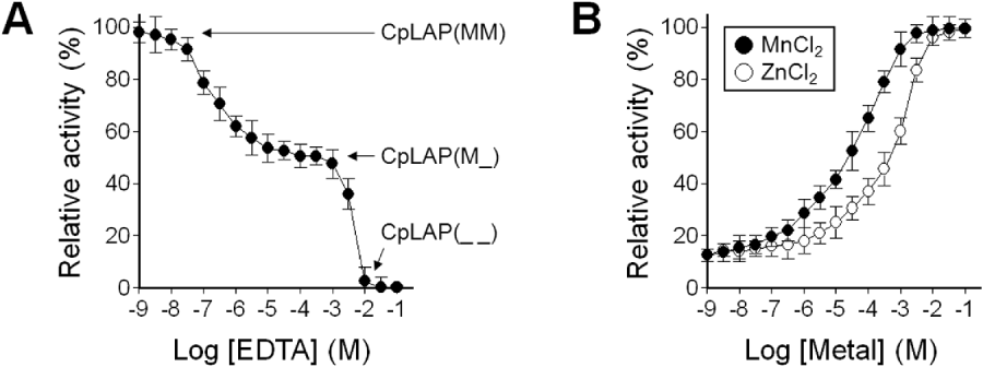
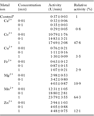
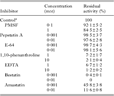
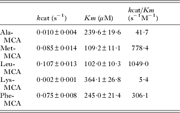
CpLAP showed a broad pH optimal range (pH 7·5 to 9·5), with a maximum at pH 8·5 (Fig. 4A). The enzyme was highly unstable in acidic pHs, but relatively stable at neutral and alkaline conditions (Fig. 4B). The effects of divalent metal ions on the activity of CpLAP were determined. CpLAP activity was highly increased in the presence of Mn2+ and Co2+ with progressive increases in accordance with the concentrations of these metal ions (Table 1). Mg2+ and Zn2+ also increased CpLAP activity up to 10-fold, whereas the other metal ions showed no or little effects on CpLAP activity compared to the control. Effects of protease inhibitors and metal chelators on CpLAP activity were also investigated. CpLAP activity was strongly inhibited by bestatin and effectively inhibited by both metal chelators, EDTA and 1,10-phenanthroline (Table 2). Amasatin, an inhibitor of aminopeptidases A/PS, also partially inhibited CpLAP activity. Meanwhile, inhibitors of other classes of proteases, including PMSF, pepstatin A, and E-64, did not show any inhibitory effects. Titrations of CpLAP with different concentrations of EDTA found that CpLAP activity was progressively inhibited by EDTA in a dose-dependent manner (Fig. 5A). This indicates that CpLAP possessed 2 metal ion binding sites, the readily exchangeable site (site 1) and the tightly bound site (site 2) (Kim and Lipscomb, Reference Kim and Lipscomb1993; Maric et al. Reference Maric, Donnelly, Robinson, Skinner-Adams, Trenholme, Gardiner, Dalton, Stack and Lowther2009). CpLAP activity was completely inhibited by 10 mm EDTA, which indicates an apoenzyme, in which metal ions were removed from both sites, designated as CpLAP(_ _). About 50% inhibition of CpLAP was observed at EDTA concentrations between 100 and 500 μ m, which indicates removal of metal ions from site 1, termed CpLAP(M_). Addition of metal ions to CpLAP(M_) resulted in successful restoration of activity (Fig. 5B), whereas CpLAP(_ _) was not reactivated by addition of metal ions (data not shown). The substrate specificity of CpLAP was characterized using several fluorogenic peptide substrates (Table 3). CpLAP readily hydrolysed Leu-MCA and showed highest activity against this peptide substrate. Substantial amidolytic activities against Met-MCA and Phe-MCA were also identified, but CpLAP showed no or little activity against Ala-MCA and Lys-MCA.
Characterization of biochemical properties of recombinant CpLAP. (A) Optimal pH. Enzyme activity was assayed in various pH buffers ranging from pH 6·0 to 9·5. Maximal activity was shown as 100%. (B) Enzyme stability. Recombinant enzyme was incubated in various pH buffers at 37°C for the indicated time and residual enzyme activity was assayed. The activity was assayed in 100 mm Tris-HCl (pH 8·5) in the presence of 1 mm MnCl2 with a fluorogenic substrate Leu-MCA.

Metal-binding characteristics of CpLAP. (A) Inhibition of CpLAP by EDTA. CpLAP (30 nm) was incubated with different concentrations (0–100 mm) of EDTA in 50 mm Tris-HCl (pH 8·5) supplemented with 100 μ m ZnCl2 for 10 min. After then, Leu-MCA (5 μ m) was added and the enzyme activity measured. (B) Reactivation of EDTA-treated CpLAP by addition of metal ions. Different concentrations (0–100 mm) of MnCl2 or ZnCl2 were incubated with CpLAP(M_) in 50 mm Tris-HCl (pH 8·5), respectively, and the enzyme activity of each sample was measured with Leu-MCA as a substrate.

Effect of divalent metal ions on the activity of CpLAP

a Control, without any metal ion.
Effect of protease inhibitors and metal chelators on the activity of CpLAP

a Control, without any inhibitor or chelator.
Kinetic parameters for CpLAP

DISCUSSION
Proteases of apicomplexa parasitic protozoa play essential roles in parasite physiology, including protein catabolism, host cell invasion, and host cell egress (Que et al. Reference Que, Ngo, Lawton, Gray, Liu, Engel, Brinen, Ghosh, Joiner and Reed2002; Rosenthal, Reference Rosenthal2002; Shaw et al. Reference Shaw, Roos and Tilney2002; Kim, Reference Kim2004; Goldberg, Reference Goldberg2005; Stack et al. Reference Stack, Lowther, Cunningham, Donnelly, Gardiner, Trenholme, Skinner-Adams, Teuscher, Grembecka, Mucha, Kafarski, Lua, Bell and Dalton2007; Blackman, Reference Blackman2008; Roiko and Carruthers, Reference Roiko and Carruthers2009; Skinner-Adams et al. Reference Skinner-Adams, Stack, Trenholme, Brown, Grembecka, Lowther, Mucha, Drag, Kafarski, McGowan, Whisstock, Gardiner and Dalton2009; Wegscheid-Gerlach et al. Reference Wegscheid-Gerlach, Gerber and Diederich2010). Consequently, extensive studies have been carried out to characterize the biochemical and functional properties of numerous proteases of ampicomplexa parasites, especially Plasmodium spp. and T. gondii, and to investigate them as promising drug targets, but only a few studies have been made in C. parvum. Serine and cysteine protease activities have been identified in several developmental stages of C. parvum, and they are proposed to be associated with excystation and host cell invasion by the parasite (Nesterenko et al. Reference Nesterenko, Tilley and Upton1995; Forney et al. Reference Forney, Yang, Du and Healey1996). A subtilisin-like serine protease (CpSUB1) and a papain-family cysteine protease (cryptopain-1) have recently been characterized, and their possible roles in host cell invasion suggest their potential as drug targets (Feng et al. Reference Feng, Akiyoshi, Widmer and Tzipori2007; Na et al. Reference Na, Kang, Cheun, Cho, Moon, Kim and Sohn2009; Wanyiri et al. Reference Wanyiri, Techasintana, O'Connor, Blackman, Kim and Ward2009). The recently released Cryptosporidium genomic resource (CryptoDB) (Heiges et al. Reference Heiges, Wang, Robinson, Aurrecoechea, Gao, Kaluskar, Rhodes, Wang, He, Su, Miller, Kraemer and Kissinger2006) indicates that Cryptosporidium spp. also have a number of different classes of proteases, but almost all of them remain to be characterized.
In this study, we identified an M17 LAP of C. parvum and characterized the biochemical properties of the recombinant protein. CpLAP is a typical enzyme that belongs to the M17 family LAP, which share similar structural and biochemical properties. CpLAP showed optimum activity at neutral or slight alkaline pH, and its activity was significantly enhanced by Mn2+ and Co2+ at millimolar concentrations, which are similar to LAPs of other apicomplexa parasites (Stack et al. Reference Stack, Lowther, Cunningham, Donnelly, Gardiner, Trenholme, Skinner-Adams, Teuscher, Grembecka, Mucha, Kafarski, Lua, Bell and Dalton2007; Jia et al. Reference Jia, Terkawi, Aboge, Goo, Luo, Li, Yamagishi, Nishikawa, Igarashi, Sugimoto, Fujisaki and Xuan2009, Reference Jia, Nishikawa, Luo, Yamagishi, Sugimoto and Xuan2010; Lee et al. Reference Lee, Song, Seok, Jha, Han, Song, Yu, Hong, Kong and Chung2010). The M17 family of LAPs contain 2 metal-binding sites, a readily exchangeable site (site 1) and a tight binding site (site 2), with different affinities for metal ions (Kim and Lipscomb, Reference Kim and Lipscomb1993; Sträter and Lipscomb, Reference Sträter and Lipscomb1995; Maric et al. Reference Maric, Donnelly, Robinson, Skinner-Adams, Trenholme, Gardiner, Dalton, Stack and Lowther2009; McGowan et al. Reference McGowan, Oellig, Birru, Caradoc-Davies, Stack, Lowther, Skinner-Adams, Mucha, Kafarski, Grembecka, Trenholme, Buckle, Gardiner, Dalton and Whisstock2010). PfLAP retains residual activity when the metal ion is removed from site 1, whereas removal of metal ions from both sites results in an inactive apoenzyme that cannot be reactivated by the addition of divalent metal ions (Maric et al. Reference Maric, Donnelly, Robinson, Skinner-Adams, Trenholme, Gardiner, Dalton, Stack and Lowther2009). The metal ion at site 1 can be readily exchanged for other divalent metal ions, but the type of metal ion at site 1 influences the catalytic efficiency of PfLAP for peptide substrates as well as the binding of the inhibitors (Maric et al. Reference Maric, Donnelly, Robinson, Skinner-Adams, Trenholme, Gardiner, Dalton, Stack and Lowther2009). A similar metal-binding pattern was also observed in CpLAP. When EDTA was added to CpLAP at different concentrations, the enzyme was inactivated in a dose-dependent manner. When the activity of CpLAP was inhibited by 50% by EDTA, which involved removal of metal ion from site 1 (Maric et al. Reference Maric, Donnelly, Robinson, Skinner-Adams, Trenholme, Gardiner, Dalton, Stack and Lowther2009), the enzyme activity was fully restored by sequential addition of metal ions. Meanwhile, the activity of the apoenzyme, in which metal ions were removed from both sites, was not restored by the addition of metal ions. For PfLAP, it is postulated that the concentration of intracellular Zn2+ may be an important regulator for activity, and Mg2+ is influential for PfLAP efficiency by occupying metal-binding sites in vivo (Maric et al. Reference Maric, Donnelly, Robinson, Skinner-Adams, Trenholme, Gardiner, Dalton, Stack and Lowther2009). The binding mode of metal ions and their significance to the binding of substrates and inhibitors to CpLAP is not fully understood, and more comprehensive studies are currently underway.
The activity of CpLAP was effectively inhibited by the aminopeptidase inhibitor, bestatin. Bestatin and its derivatives effectively inhibit PfLAP (Gardiner et al. Reference Gardiner, Trenholme, Skinner-Adams, Stack and Dalton2006; Stack et al. Reference Stack, Lowther, Cunningham, Donnelly, Gardiner, Trenholme, Skinner-Adams, Teuscher, Grembecka, Mucha, Kafarski, Lua, Bell and Dalton2007) and block the growth of both P. falciparum and P. chabaudi chabaudi in culture (Nankya-Kitaka et al. Reference Nankya-Kitaka, Curley, Gavial, Bell and Dalton1998; Stack et al. Reference Stack, Lowther, Cunningham, Donnelly, Gardiner, Trenholme, Skinner-Adams, Teuscher, Grembecka, Mucha, Kafarski, Lua, Bell and Dalton2007). Bestatin also showed broad-spectrum inhibitory effects on the M17 LAPs from B. gibsoni, T. gondii and P. vivax (Jia et al. Reference Jia, Terkawi, Aboge, Goo, Luo, Li, Yamagishi, Nishikawa, Igarashi, Sugimoto, Fujisaki and Xuan2009, Reference Jia, Nishikawa, Luo, Yamagishi, Sugimoto and Xuan2010; Lee et al. Reference Lee, Song, Seok, Jha, Han, Song, Yu, Hong, Kong and Chung2010). Although it was reported that bestatin induces immunomodulatory effects on certain immune effector cells, it has very low toxicity in experimental animals and humans (Umezawa, Reference Umezawa1980; Schorlemmer et al. Reference Schorlemmer, Bosslet and Sedlacek1983; Iwaki et al. Reference Iwaki, Nakamura and Koyama1986; Abe et al. Reference Abe, Alvord, Koyama, Matsuda and Talmadge1989), and consequently has been used as a safe therapy for certain cancers such as squamous cell carcinoma in humans (Scornik and Botbol, Reference Scornik and Botbol1997, Reference Scornik and Botbol2001). Although the in vivo therapeutic efficacy and safety of bestatin should be elucidated further, the fact that bestatin showed a broad-spectrum inhibition of LAPs from human-infecting apicomplexa parasites suggests that bestatin or its derivatives could be an effective therapeutic agent. Consistent with M17 leucyl aminopeptidases of apicomplexa protozoan parasites, CpLAP showed substrate preference for N-terminally exposed hydrophobic amino acids, most markedly Leu, although a slight difference was also identified. CpLAP more efficiently cleaved the hydrophobic amino acid Phe as compared to LAPs of other apicomplexa parasites. This indicates that CpLAP has a broader substrate spectrum than those of other apicomplexa parasites. This different substrate preference of CpLAP is probably due to the different molecular structure of its active site and/or different amino acid requirements of C. parvum in different environments. However, a detailed study should be conducted.
Phylogenetic analysis revealed that LAPs of apicomplexa protozoa parasites clustered with M17 LAP members of plants, cynobacteria, and algae. This is not surprising given their close association and common ancestry, as suggested previously (Wilson et al. Reference Wilson, Williamson and Preiser1994; Stack et al. Reference Stack, Lowther, Cunningham, Donnelly, Gardiner, Trenholme, Skinner-Adams, Teuscher, Grembecka, Mucha, Kafarski, Lua, Bell and Dalton2007). Due to the technical shortcoming of the in vitro cultivation of C. parvum, we could not clearly determine the expression pattern and cellular localization of CpLAP in the life cycle of the parasite. We tried immunofluorescence histology using sporozoites of C. parvum, but we could not obtain solid data on the localization of the protein in the parasite. However, we confirmed its expression in C. parvum sporozoites by Western blot. Considering the general roles of LAPs in cells, it is reasonable to assume that the primary function of CpLAP is associated with the final stages of the general intracellular protein catabolism cascade, but this should be clarified further. Many questions including the biological significance of the enzyme and its potential as a drug target still remain to be answered. However, this is the first report on the characterization of CpLAP and therefore provides important insights into the proteolytic cascades of the parasite. Moreover, our study constitutes a solid foundation for subsequent studies on biology of CpLAP and its potential as a drug target.
ACKNOWLEDGEMENTS
This study was supported by a grant of the Korea Healthcare Technology R&D Project, Ministry for Health, Welfare & Family Affairs, Republic of Korea (A100119).