Introduction
Bone-anchored hearing aid (BAHA) systems are well-established, effective surgical treatment options that have been used for hearing rehabilitation for many years. Although some surgical modifications have been made over the years, the main components (including soft tissue reduction) have remained because they are considered effective in reducing infections around the abutment.
Despite the inclusion of extensive soft tissue reduction and changes in hardware, the most common complications associated with BAHA systems are still related to adverse skin reactions around the abutment. Additionally, soft tissue reduction adds complexity to a surgical procedure that otherwise consists of a routine skin incision.Reference de Wolf, Hol, Huygen, Mylanus and Cremers1, Reference Dun, Faber, de Wolf, Mylanus, Cremers and Hol2
Over the years, other modifications to the macro- and microstructure of the implants have been made to enhance their stability.Reference Gottlow, Sennerby, Rosengren and Flynn3, Reference Sennerby, Gottlow, Rosengren and Flynn4 Although such changes have strengthened osseointegration between implant and bone tissue, until recently, the skin and titanium surfaces have remained somewhat incompatible. This problem has now been addressed by using hydroxyapatite particles to coat the current abutments.Reference Larsson, Wigren, Andersson, Ekeroth, Flynn and Nannmark5
Recent studies have shown that implantation of BAHA systems that do not require soft tissue reduction has numerous benefits compared with routine surgical techniques (Flynn et al., unpublished data).Reference Hultcrantz6 For instance, a less-invasive technique that does not require skin thickness reduction provides a simpler, shorter surgical procedure. Furthermore, a technique that does not require permanent hair removal immediately around the abutment is likely to be more aesthetically appealing to patients. If soft tissue thickness is retained, then faster healing and less numbness (sensory loss or paraesthesia) at the implant site may also be expected. This multicentre, retrospective study evaluated the surgical and audiological outcomes of a new connective interface for the CochlearTM Baha® BA400 that can be implanted without a need for skin thinning.
The primary objectives of this report were to evaluate soft tissue complications after BAHA surgery, surgical time, implant stability, loading time and hearing performance. The secondary objectives were to assess the medication and procedures used to treat any soft tissue complications, measure the time taken for wound healing, and register serious events such as the loss of implant.
Materials and methods
This study involves a multicentre, non-blinded, retrospective case series. The research protocol was approved by the Kocaeli University Ethics Committee and conducted in accordance with the Declaration of Helsinki and in adherence to Turkish law and regulations. Patients implanted with the new abutments at three tertiary care institutions from December 2012 until March 2013 were examined. In total, 16 patients who had undergone BAHA surgery over this period were assessed for hearing results, implant stability and surgical complications. All patients met the following inclusion criteria: more than five years of age at implant; candidates for bone conduction implant; no history of uncontrolled diabetes mellitus; and no history of conditions that could jeopardise osseointegration and/or wound healing such as radiotherapy or Paget's disease. All patients were informed about alternative treatments and both the risks and benefits of this surgery; all provided informed consent. Information was collected on gender, current concomitant diseases, clinical issues, surgical details and events and post-operative outcomes.
Evaluations included pre-operative pure tone audiometric thresholds, and pre- and post-operative free-field warble tone thresholds and speech recognition thresholds under aided and unaided listening conditions. Participants were pre-operatively assessed under aided conditions using a sound processor fitted onto a Softband. Audiological tests were performed by an audiologist using an AC 40 clinical audiometer (Interacoustics, Assens, Denmark). The audiometer was calibrated according to International Organization for Standardization standards. Air-conduction hearing thresholds for 0.25–8 kHz and bone-conduction hearing thresholds at 0.5, 1, 2, 3 and 4 kHz were measured using TDH-39 earphones (Telephonics company, New York, USA) and B-71 bone vibrator (Radioear Corporation, New Eagle, USA), respectively.
The investigated devices conform to European Union specifications (Conformité Européenne (‘CE’) marking) for their intended use, as reported in this clinical investigation. The Baha BA400 abutment was designed with concave shaping at its lower aspect. The abutment is made from commercially pure titanium coated with a hydroxyapatite layer of up to 3 mm below the top surface (i.e. 2 mm below the top surface on 6 mm abutments) on the area that contacts soft tissue. The abutments are classified as MDD Class IIb medical devices, delivered sterile for single use and pre-mounted onto 3 and 4 mm Cochlear Baha BI300 implants.
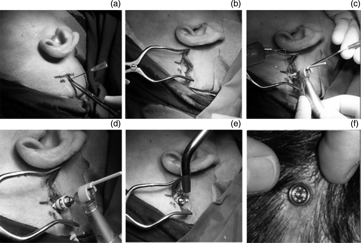
For adults, BAHA surgery was performed under local or general anaesthesia, according to the patient's preference. General anaesthesia was used for children. The implant was positioned 50–55 mm posterosuperiorly to the ear canal at the level of a line drawn posteriorly from the suprameatal crest. Skin thickness was measured using a needle prior to making a longitudinal incision of approximately 30 mm. The incision was opened using an automatic retractor, and approximately 10 mm of periosteum surrounding the planned implant site was removed. Finally, the temporal bone was drilled and the implant placed. Unlike in routine techniques, soft tissue reduction was not performed. The incision was closed primarily. The skin above the abutment was punctured using a dermal punch and a healing cap was placed on the abutment (Figure 1).
Series of photographs showing the one-stage surgical technique with no skin thickness reduction. (a) Skin thickness is first measured using a needle. (b) An incision is made using an automatic retractor, and approximately 10 mm of periosteum around the planned implant site is removed. (c), (d) The temporal bone is drilled and expanded, and the implant positioned. (e) The implant stability quotient is measured during surgery. (f) Implant at post-operative week four.

All patients were seen weekly for the first month after surgery. Patients started to use their sound processors in the fourth week after surgery and were reviewed at 3- and 6-month follow-up visits. Patients were examined for a possible soft tissue reaction around the implant at each visit; if present, tissue reactions were classified using Holgers scale (0–4). Additionally, two perpendicular measurements of the implant stability quotient were made at each site during every visit.
Results
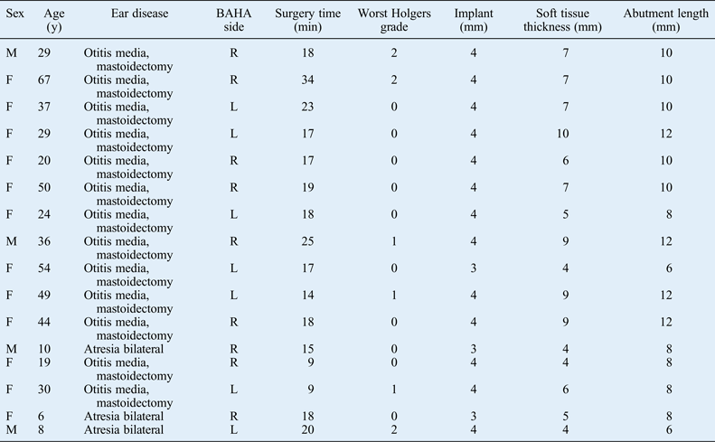
In total, 16 patients (3 males, 11 females and 2 children) were enrolled in this study. The mean age was 32 years and the range was 6–67 years. One patient had concomitant hypertension; no other patients had concomitant disease. The main surgical indication was chronic otitis media; however, three patients were treated for bilateral aural atresia. Nine patients were implanted on the right side and seven on the left. In 13 cases, 4 mm implants were used; in the other 3 cases, 3 mm implants were used. Abutments were 6 mm in length in two patients (12.5 per cent), 8 mm in five patients (31.25 per cent), 10 mm in five patients (31.25 per cent), and 12 mm in four patients (25.0 per cent): the choice of length was determined by the patient's skin thickness. The mean skin thickness and mean abutment length were 6.4 mm and 9.3 mm, respectively. The mean surgical time was 19.4 minutes (range 14–34 minutes; Table I).
Patient demographics
BAHA = bone-anchored hearing aid; y = years; min = minutes; M = male; R = right; F = female; L = left
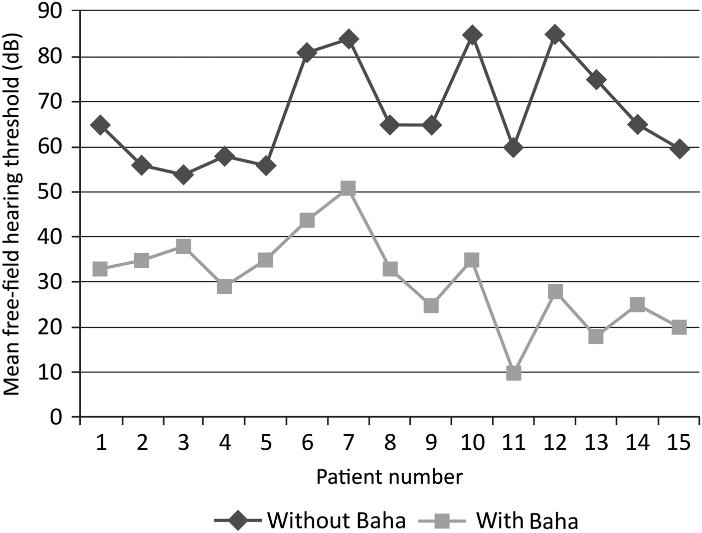
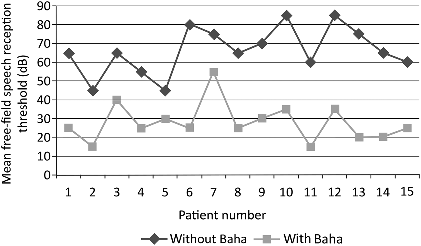
Five patients had mixed hearing loss, and the remaining 11 had primarily conductive hearing loss. Of the latter patients, three had congenital ear canal atresia with almost normal cochlear function. One patient did not use her BAHA consistently for aesthetic reasons. Therefore, as a result of missing data, she was excluded from the analysis of audiological evaluation data. The mean free-field hearing threshold (i.e. four-frequency average for 0.5, 1, 2 and 4 kHz) for the remaining 15 patients was 67.60 ± 11.3 dB under unaided conditions and 30.60 ± 10.3 dB under aided conditions with the BAHA (Figure 2). The mean free-field speech reception thresholds were 66.33 ± 12.5 dB without the BAHA and 27.87 ± 10.2 dB with the BAHA (Figure 3). The Wilcoxon signed rank test showed speech data to be significantly different with and without the BAHA (p < 0.001): there was an average gain of 38 dB in the speech reception threshold with the BAHA compared with unaided speech.
Graph showing the mean free-field hearing threshold with and without the bone-anchored hearing aid for each patient.
Graph showing the free-field speech reception threshold with and without the bone-anchored hearing aid bone-anchored hearing aid for each patient.
All patients received the BAHA monaurally. Two patients stopped using their device. Of these, one did not want to use her device for psychological reasons: the device was removed leaving the sleeping fixture in place. The other patient, a musician, uses a conventional hearing aid during concerts because of feedback problems with the BAHA; however, he uses his device at all other times. A third patient, an eight-year-old child, lost his implant because of trauma. The remaining 13 patients are still using their device and benefit from speech recognition in quiet conditions, sound comfort and a reduced occurrence of ear infections.
For all patients, wounds had healed by the seventh day after surgery, and only one patient complained of pain around the abutment. There were no reports of numbness. A total of 118 observations of the skin around the abutment were recorded during the follow-up period. Holgers grade 2 reactions were noted in six observations (5.08 per cent) for three implanted patients (18.75 per cent). All reactions resolved with local treatment within one or two weeks. No skin overgrowth was noted for any patient.
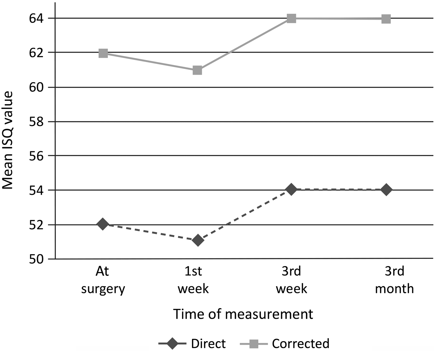
Mean implant stability quotient values were measured as 52, 51, 54 and 57 in the operating theatre, and after one week, three weeks and three months, respectively (Figure 4). Five patients had lower implant stability quotient values in the first post-operative week compared with the initial measurement. Only two patients had an implant stability quotient value that was even lower than the initial measurement in the third week. However, the values were relatively stable, and so we loaded the sound processors in the fourth week for all patients.
Corrected and uncorrected mean implant stability quotient (ISQ) values measured in the operating theatre and after the first week, third week and third month.
Discussion
The surgical implantation technique has gradually been simplified over the years. The initial surgical technique for bone conduction hearing implants was developed in Gothenburg, Sweden, and described by Tjellström et al. in 1981.Reference Tjellström, Lindström, Hallén, Albrektsson and Brånemark7 The original two-stage surgical procedure was simplified to a single-stage procedure in 1989, and this has since remained the surgical standard in adults.Reference de Wolf, Hol, Huygen, Mylanus and Cremers1, Reference Tjellström and Granström8 In 2001, a dermatome technique was developed to provide the correct cutaneous thickness for BAHA surgery.Reference Stalfors and Tjellström9 Finally, a simplified technique without a skin flap has been described: it comprises only a longitudinal incision in contrast to the previous (semi-)circular incision or flap techniques.Reference de Wolf, Hol, Huygen, Mylanus and Cremers1, Reference Bovo10 The common feature of all of these surgical techniques is the reduction of subcutaneous tissue around the abutment to minimise infections.Reference Hultcrantz6
A very limited number of published studies have described the use of BAHA surgical techniques without skin thinning and combined with the use of longer abutments. In 2011, Hultcrantz reported the only example of this surgical method.Reference Hultcrantz6 His study included 14 patients, divided equally into control and test groups and then compared for aesthetic outcome, infection rate, surgical time and several other measures. There was no significant difference in infection rate between the two groups. Furthermore, use of the new technique led to improved surgical times and aesthetic outcomes; however, it did not include a comparison of hearing performance between groups.Reference Hultcrantz6 Our study made no formal analysis of the aesthetic outcome. However, the apparent preservation of the hair follicles in the skin immediately surrounding the abutment has the clear potential to provide improved aesthetic outcomes.
One problem with the commonly described BAHA surgical technique is pocket formation and subsequent epidermal down-growth due to incomplete integration between the titanium abutment and adjacent skin. These histological formations are thought to represent complications of infection around the abutment. This problem was addressed in a previous study comparing four abutments (two coated with hydroxyapatite and two of pure titanium) in an experimental sheep model.Reference Larsson, Wigren, Andersson, Ekeroth, Flynn and Nannmark5 It was shown that hydroxyapatite-coated abutments led to significantly reduced pocket depth and epidermal down-growth compared with pure titanium abutments. Moreover, soft tissue stability can be achieved without soft tissue removal. Thus, the currently recommended procedure for titanium BAHA abutments avoids soft tissue removal.Reference Larsson, Wigren, Andersson, Ekeroth, Flynn and Nannmark5
• Adverse skin reactions are a major problem for patients after bone-anchored hearing aid system implantation
• Soft tissue reduction is helpful but adds surgical complexity
• A simpler and shorter surgical procedure avoids skin thickness reduction
• A new abutment combined with less-invasive surgery improves aesthetics and reduces numbness around the abutment
• This technique has shorter surgical times with no increase in infection rate
• Healthy soft tissue around the abutment promotes shorter healing times following less-invasive surgery
Prior to more recent reports, subcutaneous tissue reduction was recommended by many authors to prevent soft tissue movement and thus reduce the development of scar tissue and infection.Reference Granström11, Reference Wazen, Young, Farrugia, Chandrasekhar, Ghossaini and Borik12 However, as reported by Hultcrantz, the infection complication rates for BAHA surgery without skin thinning is better than for the control group.Reference Hultcrantz6 In our study, infection at the abutment site (Holgers grade greater than 2) was noted for 3 out of 16 patients (18.75 per cent), or for 5.08 per cent of the total number of observations. These results are consistent with published infection rates.Reference de Wolf, Hol, Huygen, Mylanus and Cremers1 All cases healed following treatment with locally applied antibiotic ointment (2% mupirocin) within about a week. Although no clear information on the healing times of peri-implant infections has been reported, our clinical observations show that healing times are shorter for patients without soft tissue reduction than for those with soft tissue reduction. We hypothesise that the presence of healthy soft tissue immediately surrounding and in contact with the abutment contributes to the relatively shortened healing times observed.
Resonance frequency was measured for all BAHA patients at regular intervals using an Osstell AB, Gothenburg, Sweden implant stability quotient device and converted into an implant stability quotient value ranging from 1 to 100. In our case series, a reduction in the mean implant stability quotient values was observed during the initial healing period. Consistent with published reports, the values reached a stable level after three weeks.Reference D'Eredità, Caroncini and Saetti13 Therefore, we confidently recommend that the loading time for a BAHA sound processor should be as short as three weeks after implantation. In our case series, implant stability quotient values were lower compared with studies using 5.5 mm abutments.Reference D'Eredità, Caroncini and Saetti13 Therefore, a correction factor of ±3 units/mm must be incorporated into the implant stability quotient value to account for the influence of abutment length when comparing implant stability quotient reference values between studies (Figure 4).
In this case series, only one patient complained of pain around the abutment; however, no objective cause of the pain was found. In addition, the patient was not pleased with the aesthetics of her BAHA. Following subsequent removal of the implant, the area healed within a few days. Although no large case series has reported the incidence of numbness around the implant site, this is a temporary complication for patients undergoing soft tissue reduction. However, the published case series suggest that numbness is a rare complication for these patients.Reference Hultcrantz6
In this case series, the group mean free-field hearing thresholds and mean free-field speech reception thresholds significantly improved following BAHA implantation compared with unaided conditions. An average gain of 38 dB in the speech reception threshold was observed after BAHA implantation; this is similar to the value reported by Liepert and DiToppa.Reference Liepert and DiToppa14 According to their findings, patients implanted with the BAHA could understand speech at normal conversation levels, and this improvement is related to the size of the air-bone gap.Reference de Wolf, Hendrix, Cremers and Snik15
Conclusion
This case series demonstrates that the new abutment with a hydroxyapatite coating can be implanted percutaneously without soft tissue reduction. During the 12- to 16-month post-implantation follow-up period, we observed that the use of this surgical technique for Baha BA400 implantation resulted in a similar infection rate, improved aesthetics, a faster healing time, fewer reports of numbness and shorter surgery time compared with conventional surgery using bone conduction hearing implants.