sTAPVD is the commonest type of TAPVD in which an ascending vein drains all four pulmonary veins to the systemic circulation. The ascending vein is at risk of obstruction along its pathway before entering the systemic circulation. An obstructed drainage leads to pulmonary venous congestion and pulmonary venous hypertension with subsequent haemodynamic collapse imminent if the obstruction is not relieved. We present a case of a newborn with sTAPVD associated with an obstructed right-sided ascending vein. The baby had haemodynamic instability initially, which subsequently improved preoperatively.
Case report
A 4-hour-of life-term newborn with a birth weight of 2.6 kg was noted to be mildly cyanosed (SpO2 92%) upon postnatal screening in our centre. Otherwise, he appeared to be well with no respiratory distress. Clinically, there was a systolic cardiac murmur at the left sternal edge with no signs of heart failure. Initial clinical diagnosis was suggestive of a septal defect or valvular issues of the heart. Congenital pneumonia or sepsis was also considered. He was subsequently noted to have worsening cyanosis requiring mechanical ventilatory support within the next few hours associated with haemodynamic instability. Initial laboratory investigations did not reveal any conclusive evidence of sepsis or pneumonia although clinically, the infection cannot be ruled out. Chest X-ray showed mild cardiomegaly with mild pulmonary venous congestion and haziness over the right perihilar region predominantly. The child was started on a course of C-penicillin and gentamicin for 1 week.
Transthoracic echocardiography study revealed the likelihood of a supracardiac anomalous pulmonary venous drainage with a 5 mm size atrial septal defect. Pulmonary veins were not seen draining into the left atrium directly, but drained into an ascending vein arising posterior to the heart. The ascending vein then drained into the right SVC. Furthermore, there was an exaggerated blood flowing into the right atrium from SVC and the shunt across ASD was from RA to LA. These findings supported the diagnosis of sTAPVD. However, echocardiography was unable to ascertain conclusively whether the patient had a total or partial type of anomalous pulmonary venous drainage. The possibility of an obstructed ascending vein was also considered as an obstructive pattern was observed at SVC-ascending vein junction on Doppler echocardiography with a pressure gradient of 4–6 mmHg.
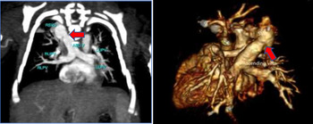
He required ventilatory support for 4 days with inotropic support (dopamine and adrenaline) as well. However, his condition stabilised after a few days with gradual weaning to room air and haemodynamic stability was achieved when inotropes were stopped. His oxygen saturation remained between 88 and 95% in room air. Computed tomographic scan done at day 20 of life confirmed the presence of supracardiac total anomalous pulmonary venous drainage (Fig 1). All four pulmonary veins joined a pulmonary venous confluence, which continued immediately as a right-sided ascending vein along the paravertebral gutter. The ascending vein then joined the SVC posteriorly. There was also mild stenosis of the ascending vein at the point of entry into SVC measuring 3.9 mm. A 6 mm in diameter ASD was also present.
Showing the drainage of all four pulmonary veins joining a venous confluence with subsequent ascending vein draining into right superior caval vein. Note the narrowing at the ascending vein SVC junction (red arrow). ASC V=ascending vein; LLPV=left lower pulmonary vein; LUPV=left upper pulmonary vein; RLPV=right lower pulmonary vein; RSVC=right superior caval vein; RUPV=right upper pulmonary vein.

Subsequent serial echocardiography and clinical assessments suggested that the obstruction of the SVC-ascending vein junction was mild. Baby was discharged back home after 3 weeks of life. At home, he was relatively stable, but has intermittent mild cyanosis during crying. He also has mild heart failure symptoms noted during feeding at home. Anti-failure medications (furosemide and spironolactone) were commenced at 1 month of life and surgical repair was then planned. The child underwent surgical repair at 2 months of life. Preoperatively assessment with CT scan suggested that the patient may have some difficulties during surgery due to the anatomy. PVC instead of being horizontal and posterior to the LA like in the majority of cases of sTAPVD was now oblique and posterior to LA. Part of the right pulmonary artery also lie in between the PVC and posterior wall of the LA. This anatomy may pose a problem in creating a communication between the PVC and LA surgically. There is a risk of post-operative obstruction at the PVC-LA communication due to these factors.
He underwent complete repair with the creation of this communication as well as partial ligation of the ascending vein and pericardial patch closure of atrial septal defect. Surgery involved incising the PVC horizontally from the anterior aspect with another horizontal incision made across the right atrium and interatrial septum into left atrium, within the same line. The opened PVC was then anastomosed to the opening of the pericardial reflection. A pericardial patch was then used to partition between the left atrium and right atrium. No significant obstruction was seen at the PVC-LA communication on post-operative echocardiography. He managed to overcome the initial post-operative issues of pulmonary hypertension and low cardiac output syndrome. He required inhaled nitric oxide for 3 days before commencing on sildenafil for 6 weeks duration. Inotropes were also given for the first 3 days post-operatively including milrinone, dopamine, and adrenaline. There were no other complications like SVC obstruction or atrial arrhythmias. He was subsequently discharged home 2 weeks after the surgery and was doing well on follow-up.
Discussion
Total anomalous pulmonary venous drainage is due to the failure of all pulmonary veins to drain directly into the left atrium. It is uncommon; Seale et al Reference Seale, Uemura and Webber1 reported only 7.1 cases per 100,000 live births. Supracardiac TAPVD is the commonest type with Burroughs et al Reference Burroughs and Edwards2 reported a frequency of 40% in his series. Embryologically, this is due to failure to establish a normal connection between the common pulmonary vein and left atrium, with the persistence of either a left or right common cardinal vein. The CCV represent a communication between the pulmonary venous system with systemic venous complex. Usually, the pulmonary veins drain into a left-sided ascending vein, which in turn joins the innominate vein. In our case, the baby was presented with the right-sided ascending vein with concomitant drainage into the right superior caval vein. The frequency of right-sided ascending vein draining into superior caval vein is reported at about 21% of all supracardiac TAPVD. Reference Seale, Uemura and Webber1 On the other hand, innominate vein drainage constitutes 72% of all sTAPVD.
Just like the left-sided ascending vein associated-sTAPVD, the right-sided ascending vein is also at risk of obstruction. The incidence of stenosis in patients with left-sided ascending vein-associated sTAPVD is estimated in various reports as 22–79%. Reference Karamlou, Gurofsky and Al Sukhni3,Reference Hancock Friesen, Zurakowski and Thiagarajan4 However, the incidence of obstructed right-sided ascending veins is unknown. Obstructive flow is postulated to be due to the compression caused by trachea as the ascending vein passes between the right pulmonary artery and trachea along its course towards superior caval vein. Reference Casta and Wolf5 This “haemodynamic vise” is invariably associated with pulmonary venous congestion and hypertension with subsequent cardiovascular collapse. However, we did not find evidence of any “haemodynamic vise” in our patient although the pathway of ascending vein was as described. Furthermore, the stenosis observed was at the junction of ascending vein and SVC with no external compression seen. The stenosis was mild and not associated with any post-stenotic dilatation of the ascending vein. We attributed the initial cyanosis with haemodynamic instability to clinical pneumonia as the child showed clinical improvement with antibiotics. There were, however, no positive laboratory investigations to support the diagnosis of infection. A severe obstruction of the right ascending vein is associated with high mortality; 24% of children with sTAPVD associated with severe preoperative pulmonary venous obstruction died despite undergoing surgical repair.1
Computed tomographic scan is an excellent tool to delineate the pulmonary venous drainage in its entirety. Echocardiography has its limitations in assessing this condition as all four pulmonary veins may not be able to be seen clearly. Furthermore, the proximity of surrounding structures in relation to PVC and ascending vein may not be able to be ascertained conclusively with echocardiography. A three-dimensional reconstruction with CT scan images allows us to view the relationship between PVC and ascending vein with the surrounding structures better. Any external compression of a PVC or ascending vein could be demonstrated. We did not see any external compression of a PVC or ascending vein in our patient. Stenosis of the SVC-ascending vein in our patient is likely due to failure of growth of this segment of ascending vein rather than any external compression. In our patient, 3D images revealed the oblique position of the PVC in relation to the posterior wall of LA. Furthermore, part of RPA lies in between the PVC and posterior wall of LA. These findings may present a surgical dilemma in creating a non-obstructive communication between PVC and LA surgically as a successful side-to-side anastomosis may be difficult to achieve. There is a possibility of a narrowed opening or stenosis in this communication post-operatively, resulting in pulmonary venous congestion and pulmonary venous hypertension.
We did see some PVC-LA stenosis in our patient although it was clinically insignificant. However, there were no other complications that could happen in sTAPVD repairs like SVC obstruction or arrhythmias. Reference DeLeon, Freeman and Ilbawi6 This is probably due to only partial ligation of the ascending vein, which was done instead of a more extensive repair of SVC. The question of whether the ascending vein should be ligated or not is still controversial. Saritas et al Reference Sarıtaş, Bolat and Özkan7 suggested to leave the ascending vein patent to decompress the left side of the heart in patients with pulmonary venous obstruction. Furthermore, there is a likelihood the ascending vein may close spontaneously due to high resistance in the vein post-operatively as communication between PVC and LA is established. However, we decided for partial ligation in our patient as the ascending vein obstruction is mild.
Our case report illustrated an anatomical variant to supracardiac total anomalous venous drainage presenting with mild ascending vein superior caval vein obstruction.
Acknowledgements
The authors are grateful to Dato Dr H.L. and Dr S.L.M. who performed the successful operation and the entire paediatric cardiac services team of Serdang Hospital who were involved in the management including pre and post-operative care of the patient.
Financial support
This research received no specific grant from any funding agency or commercial or not-for-profit sectors.
Conflicts of interest
None.
Ethical standards
This case report complied with the ethical standards.