Introduction
Coarctation of the aorta occurs isolated or associated with other cardiac defects. One of the congenital abnormalities more commonly linked with aortic coarctation is ventricular septal defect. Especially in severe aortic coarctation, a concomitant ventricular septal defect is often detected. Reference Van Praagh, Bernhard, Rosenthal, Parisi and Fyler1 A number of anatomic subtypes of ventricular septal defects have been described in association with aortic coarctation, i.e. membranous and perimembranous, muscular, and sub-arterial. Reference Leanage, Taylor, de Leval, Stark and Macartney2–Reference Sennari5 The ratio between isolated aortic coarctation and aortic coarctation associated with ventricular septal defect is approx. 2:1. Reference Ferencz, Correa-Villasenor, Loffredo and Wilson6
Aortic coarctation patients have increased aortic stiffness in comparison with healthy controls. Reference Róg, Okólska and Dziedzic-Oleksy7 Aortic stiffness in turn proved to be related to left ventricular diastolic dysfunction in a number of clinical CHD scenarios, Reference Kocabay, Karabay and Kalkan8,Reference Bassareo, Saba, Marras and Mercuro9 but this relationship was fleetingly evaluated in the setting of coarctation of the aorta. Aortic isthmus size seemed to be related to left ventricular dysfunction in adults with aortic coarctation. Reference Egbe, Qureshi and Connolly10 Aortic stiffness and diastolic function are altered in children following early repair of aortic coarctation. A linear relationship was found between the two in those patients who underwent aortic coarctation surgical repair at a median age of 15 days. The detected left ventricular diastolic dysfunction may be due to chronically raised afterload. Reference Lombardi, Northrup, McNamara, Sugeng and Weismann11 However, in this setting, diastolic function was evaluated just by means of septal early mitral annular velocity assessed by Tissue Doppler Imaging (e’), rather than by the usual combination of e’ and mitral peak early filling velocity (E) on pulsed wave Doppler. This was questionable. In fact, it is the E/e’ ratio which is widely used as a non-invasive estimate of left ventricular filling pressure thus playing a crucial role in evaluating diastolic dysfunction. Reference Lombardi, Northrup, McNamara, Sugeng and Weismann11 Persistent left ventricular diastolic dysfunction is frequently observed following repair of ventricular septal defect as well. Volume loading in childhood that is not reversed by 5 years of age results in residual cardiac dilatation and functional abnormalities after repair. Reference Graham12,Reference Fusco, Borrelli, Palma, Sarubbi and Scognamiglio13 It is unknown whether ventricular septal defect represents an additional risk factor for developing left ventricular dysfunction in those patients with concomitant aortic coarctation. Again, it is unclear if, in aortic coarctation patients, there is a relationship between aortic stiffness and left ventricular diastolic function measured by the traditional trans-mitral flow E wave to A wave (E/A) ratio on pulsed wave Doppler.
This study aimed at evaluating aortic stiffness by echocardiography and blood pressure in a group of normotensive patients surgically treated for aortic coarctation and ventricular septal defect and in those who underwent simple aortic coarctation repair. Both groups were compared with a control group of healthy peers. Again, the two pathological groups were compared with each other regarding aortic stiffness and left ventricular diastolic function. A possible relationship between aortic stiffness and left ventricular diastolic function (E/A ratio) was investigated as well.
Materials and methods
The National Institute for Cardiovascular Outcomes Research database was established in Ireland in 2010. As we aimed at selecting an older cohort of patients, we searched the National Institute for Cardiovascular Outcomes Research database for patients who had undergone aortic coarctation repair between the years 2010 and 2014. Inclusion criteria included patients who had undergone a surgical, non-bypass repair of a discrete coarctation in our institution and who had also been followed-up in our institution. Only normotensive patients at latest follow-up were deliberately included, due to the known prevalence of late hypertension in these individuals. Exclusion criteria were patients with additional complex CHD, excluding ventricular septal defect; patients with no blood pressure recording at most recent clinical follow-up; patients without adequate echocardiographic imaging from most recent clinical visit for measurement of ascending aorta diameter in systole and diastole in parasternal long-axis view; patients with known systolic and/or diastolic blood pressure or on anti-hypertensive therapy at last follow-up, since these medications are potentially able to influence aortic stiffness and diastolic function. Reference Seidlerová, Filipovský, Mayer, Wohlfahrt and Cífková14 Again, all the enrolled patients were normotensive according to the American Academy of Pediatrics guidelines (i.e. blood pressure below 90th percentile for those aged 1–13 years and below 130/80 mmHg for those aged 13 years or more). Reference Flynn, Kaelber and Baker-Smith15 This is important, since it is widely known that secondary hypertension can often occur even after a successful aortic coarctation repair. Reference Leo, Sabatino and Avesani16,Reference Bassareo, Calcaterra and Sabatino17 In all the patients with a combination of aortic coarctation and ventricular septal defect as well as in those with isolated aortic coarctation, the narrowed aortic isthmus was treated with the same surgical technique, e.g. end-to-end anastomosis, since there is evidence of the fact that aortic stiffness is influenced by the kind of aortic coarctation surgical approach used. Reference Bassareo, Marras, Manai and Mercuro18 Following patient identification and inclusion as per inclusion and exclusion criteria, medical records and echocardiography images were reviewed.
The control group was made up of healthy young individuals. They have been referred to our Institution by General Practitioners and local Paediatricians for heart murmur assessment between 2010 and 2014. All of them underwent clinical examination, blood pressure measurement, and echocardiography. The latter displayed a structurally and functionally normal heart. The control group’s features are summarised in the Results section.
Aortic stiffness
Aortic stiffness was calculated from blood pressure and echocardiography as follows:
Aortic root distensibility (10-6 cm2 dyn-1) = 2 x (AoS – AoD)/AoD x (SBP-DBP)
Aortic stiffness index = ln (SBP/DBP)/(AoS-AoD)/AoD
[AoS: aortic diameter at left ventricular systole (from parasternal long axis view); AoD: aortic diameter at left ventricular diastole (from parasternal long axis view); SBP: systolic blood pressure; DBP: diastolic blood pressure].
These two methods were previously validated Reference Stefanadis, Stratos, Boudoulas, Kourouklis and Toutouzas19,Reference Bassareo, Saba, Puddu, Fanos and Mercuro20
In addition, trans-mitral pulsed-wave velocities were recorded from the apical four-chamber view using a 2 mm Doppler sample placed between the tips of the mitral leaflets. The E and A wave velocities and E/A ratio were all measured from mitral valve inflow profile. All images were recorded and stored. At least three consecutive measurements were gathered on recorded images for each selected myocardial segment, and their was mean calculated. Reference Bassareo, Saba, Marras and Mercuro9
This is a retrospective study which was approved by Children’s Health Ireland at Crumlin Ethics Committee (REC-345–23).
Statistical analysis
Statistics were performed using GraphPad PrismVersion 10.2.0. Results obtained from the whole study population were expressed as mean ± standard deviation. Independent t tests with a two-tailed p value were used for normally distributed continuous data to compare the mean of the two patient groups. The Mann–Whitney U test was used to compare the medians of continuous variables. Fisher’s exact test was used to examine the difference between categorical variables. Relationships between continuous variables were sought by means of Pearson’s correlation coefficients. Intra-observer variability was established by having one observer measure data on at least two occasions in 15 subjects selected at random from the study population. Statistical significance was set at p < 0.05.
Results
We identified 22 aortic coarctation patients who underwent repair of a discrete coarctation between the years 2010 and 2012 who met the inclusion criteria and who did not require exclusion. We then identified 17 aortic coarctation and ventricular septal defect patients who similarly met the inclusion and exclusion criteria between the years 2010 and 2014. Of note, because there were less patients who also had ventricular septal defects, it was necessary to look at additional years to identify an adequate number of patients to compare to the baseline group without ventricular septal defects. All patients had undergone a surgical aortic coarctation repair which consisted of a non-bypass extended end-to-end anastomosis.
Overall, 49 patients in the coarctation of the aorta/ventricular septal defect group and 64 in the simple coarctation of the aorta group were excluded from the analysis as they did not meet the inclusion criteria.
Baseline demographics
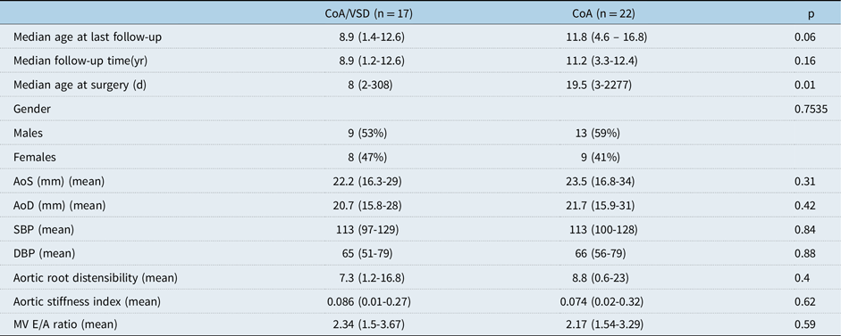
There was no statistically significant difference in the sex distribution between the two groups (p = ns). The group with aortic coarctation and ventricular septal defect had a higher median age at time of surgery (p > 0.01).
Regarding the healthy controls, they were 20 (10 males and 10 females). There were no statistically significant differences between coarctation of the aorta group versus healthy controls and between coarctation of the aorta/ventricular septal defect group and healthy controls regarding sex and age (p = ns). Mean systolic blood pressure was 114 (99–125) mmHg, whereas mean diastolic blood pressure was 67 (55–77) mmHg.
Genetic abnormalities
Interestingly, there were no patients with Turner syndrome in the cohort. This is important as Turner syndrome is associated with aortopathy and altered aortic stiffness. Reference Bons, Van Den Hoven and Malik21 One patient had trisomy 21 and another had a diagnosis of Kabuki syndrome, both with ventricular septal defect.
Cardiac features
In the group with an intact ventricular septum, 10 (45%) had a trileaflet aortic valve and 12 had a bicuspid aortic valve. In the aortic coarctation and ventricular septal defect group, 11 (65%) had a trileaflet aortic valve and six had a bicuspid aortic valve.
Of those with ventricular septal defects: 8/17 (47%) were muscular defects, 8/17 were perimembranous defects and one was a supracristal defect. Of the 17 patients with a combination of aortic coarctation and ventricular septal defect, 6 (35%) underwent concomitant pulmonary artery banding at the time of coarctation repair.
Follow-up
Median follow-up duration was 11.2 years for the aortic coarctation group and 8.9 years for the aortic coarctation and ventricular septal defect group. All patients were alive without heart transplantation at most recent follow-up appointment. Five patients had not had an outpatient examination for more than 4 years and it was assumed that these patients were either lost to follow-up, had moved to the private system, or were being followed up in a local hospital in an outreach clinic. The most recent follow-up in our institute was used for these patients’ measurements. During the follow-up period, five patients underwent balloon angioplasty of residual coarctation; four in the aortic coarctation group and one in the aortic coarctation and ventricular septal defect group.
One patient had undergone further cardiothoracic surgery—a subaortic ridge resection 6.6 years following initial coarctation repair. No patients were receiving cardiac medications at most recent follow-up appointment.
The blood pressure recordings were from a measurement from an upper limb appropriately-sized blood pressure cuff at the most recent clinic appointment. In one case, the patient had had a recent 24-hour blood pressure monitor and therefore the average systolic and average diastolic readings were used for this patient.
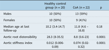
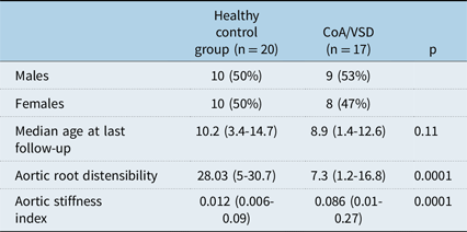
With regard to aortic root distensibility and aortic stiffness index, a statistically significant difference was detected between the healthy control group and simple aortic coarctation patients (in both cases p < 0.0001). The same statistical significance was in comparison with aortic coarctation and ventricular septal defect (in both cases p < 0.0001). See Tables 1 and 2. No differences were found with regard to aortic root distensibility and aortic stiffness index between the two pathological groups (both p = ns). Statistically significant differences were missing even when comparing patients with trileaflet aortic valve to those with a bileaflet valve (p = ns). Furthermore, no statistically significant difference was detected in relation to left ventricular diastolic function expressed by trans-mitral flow E/A ratio (See Table 3). No correlation was detected between the two indexes of aortic distensibility and left ventricular diastolic function (expressed as E/A ratio) in both simple aortic coarctation and aortic coarctation combined with ventricular septal defect groups (r = 0.121, p = ns and r = 0.097, p = ns respectively).
Healthy controls versus simple aortic coarctation (CoA) patients

Healthy controls versus patients with aortic coarctation associated with ventricular septal defect (CoA/VSD)

Normotensive patients with aortic coarctation associated with ventricular septal defect (COA/VSD) versus normotensive patients with simple aortic coarctation (CoA)

Acronyms: CoA = aortic coarctation; VSD = ventricular septal defect; AoS = aortic diameter at left ventricular systole (from parasternal long axis view); AoD = aortic diameter at left ventricular diastole (from parasternal long axis view); SBP = systolic blood pressure; DBP: diastolic blood pressure.
Discussion
Little research exists comparing patients with isolated aortic coarctation and those with coarctation of the aorta associated with ventricular septal defect.
As to aortic stiffness, measured by means of two different techniques based on echocardiography and blood pressure measurements, a statistically significant difference was detected between both simple aortic coarctation and aortic coarctation/ventricular septal defect patients and their healthy peers. This is not surprising, as there is a significant body of knowledge about altered elastic properties in patients affected by aortic coarctation. This may be due to extension of the ductal tissue into the aortic wall or to cystic medial necrosis. Reference Elzenga, Gittenberger-de Groot and Oppenheimer-Dekker22,Reference Isner, Donaldson, Fulton, Bhan, Payne and Cleveland23 An extension of ductal tissue into the aortic wall exceeding 30% is considered abnormal. Reference Russell, Berry, Watterson, Dhasmana and Wisheart24 Regarding cystic medial necrosis, it is usually defined as depletion and disarray of elastic tissue. In a previous cases series, it was observed in 100% of aortic coarctation specimens. Furthermore, in approx. 70% of the specimens, the extent of cystic medial necrosis, graded semi-quantitatively on a scale from 0 (normal aorta) to 3+, was severe (3+). Reference Isner, Donaldson, Fulton, Bhan, Payne and Cleveland23 In our sample size, both aortic coarctation and aortic coarctation/ventricular septal defect subgroups included patients with bicuspid aortic valve. However, as per a previous report, concomitance of aortic coarctation and bicuspid aortic valve seems not to have influence on arterial stiffness augmentation. Reference Róg, Okólska and Dziedzic-Oleksy7 This is likely due to the small sample size and the very young age at the time of study.
The association between aortic coarctation and ventricular septal defect is well documented, but little research exists about the features distinguishing the cases of “pure” aortic coarctation from those with aortic coarctation/ventricular septal defect. Reference Wollins, Ferencz, Boughman and Loffredo25 Although altered in both cases, we did not find any difference between the two groups in term of aortic stiffness. In other words, the association between aortic coarctation and ventricular septal defect did not exert any detrimental effect on aortic artery rigidity in comparison with isolated aortic coarctation. Ventricular septal defects are common CHDs, Reference Bassareo, Calcaterra, Deidda, Marras and Mercuro26,Reference Bassareo, Marras and Mercuro27 but unlike other congenital cardiac abnormalities such as tetralogy of Fallot or hypoplastic left heart syndrome Reference Niwa28 , they do not seem to represent an additional independent risk factor for increased aortic rigidity.
Again, diastolic function was normal and similar in simple aortic coarctation patients and in those with the association aortic coarctation/ventricular septal defect. It means that the concomitance of a ventricular septal defect did not represent a detrimental factor in worsening left ventricular diastolic function.
Regarding the lack of any significant relationship between the two parameters of aortic distensibility and left ventricular diastolic function expressed as E/A ratio, it should be pointed out that aortic coarctation leads to chronic left ventricular overload and in turn to left ventricular diastolic impairment and heart insufficiency over time. In fact, chronic left ventricular overload triggers cardiomyocyte hypertrophy, mismatch between cardiac oxygen request and supply, and apoptosis with replacement fibrosis. All these pathological changes alter the left ventricular viscoelastic properties and compliance thus resulting in diastolic dysfunction. Reference Nishimura and Borlaug29 One of the aims of aortic coarctation surgery is to prevent these complications and, in our patients, it seems to have been reached. In fact, all our patients were surgically treated as infants, whereas left ventricular diastolic dysfunction is more commonly seen in adult aortic coarctation patients. Reference Egbe, Miranda and Connolly30
The present study has its own limitations, namely: 1) the small sample size that needs to be implemented; 2) the fact that left ventricular diastolic function was assessed by pulsed wave Doppler E/A ratio rather than with other more advanced tools (for example by E/e’ ratio on tissue Doppler imaging). Reference Isaaz31 In fact, tissue Doppler imaging proved to be more accurate than conventional Doppler in detecting impaired diastolic function in patients with heart failure and normal ejection fraction; Reference Kasner, Westermann and Steendijk32 3) the intrinsic limitations linked with the retrospective nature of the study (e.g. inferior level of evidence in comparison to prospective studies; the fact that controls may not be representative of the general population and prone to selection bias); 4) some of the findings of this study, in particular the absence of any association with indexes of diastolic dysfunction might not be replicated in a slightly older sample or in patients with secondary hypertension. This is just a hypothesis. Ideally, the study should be repeated enrolling patients undergoing early versus late repair.
In conclusion, in both isolated aortic coarctation and aortic coarctation/ventricular septal defects subgroups, aortic stiffness is increased in comparison with healthy peers. The detected increased aortic rigidity is similar in both subgroups. In this respect, ventricular septal defects did not exert any negative influence on aortic stiffness.