Introduction
Lichenicolous fungi represent a highly specialized group of fungi with life strategies ranging from saprotrophs to aggressive parasites (Lawrey & Diederich Reference Lawrey and Diederich2003). This wide range of nutritional modes further reflects the high phylogenetic diversity found in this group of fungi, recently revealed by several molecular studies (Crespo et al. Reference Crespo, Kauff, Divakar, del Prado, Pérez-Ortega, Amo de Paz, Ferencova, Blanco, Roca-Valiente and Núñez-Zapata2010; Millanes et al. Reference Millanes, Diederich, Ekman and Wedin2011; Diederich et al. Reference Diederich, Ertz, Lawrey, Sikaroodi and Untereiner2013; Ertz et al. Reference Ertz, Lawrey, Common and Diederich2013; Frisch et al. Reference Frisch, Thor, Ertz and Grube2014; Pérez-Ortega et al. Reference Pérez-Ortega, Suija, Crespo and de los Ríos2014; Suija et al. Reference Suija, Ertz, Lawrey and Diederich2015). While research on lichenicolous fungi has received increased interest during the past decades, it is clearly geographically biased, with a greater focus concentrated in certain areas. Thus, if we compare polar and subpolar regions of both hemispheres, arctic and subarctic areas have received much more attention (e.g. Zhurbenko & Laursen Reference Zhurbenko and Laursen2003; Zhurbenko Reference Zhurbenko2009 a, Reference Zhurbenko b , Reference Zhurbenko2010) than their equivalents in the Southern Hemisphere (Lawrey & Diederich Reference Lawrey and Diederich2003; Hawksworth & Iturriaga Reference Hawksworth and Iturriaga2006; Etayo & Sancho Reference Etayo and Sancho2008). One aspect of lichenicolous infections that has received little attention is the characterization of the host-parasite interfaces (de los Ríos & Grube Reference de los Ríos and Grube2000; Grube & de los Ríos Reference Grube and de los Ríos2001; Lawrey & Diederich Reference Lawrey and Diederich2003). This is probably due to the need for more sophisticated techniques than light microscopy to identify and ultrastructurally characterize the interactions between lichenicolous fungi and components of the lichen host. During our study on the phylogeography of the lichen-forming fungus Mastodia tessellata (Hook. f. & Harv.) Hook. f. & Harv., a lichenicolous fungus was discovered that grew abundantly on the surface of this species in samples collected in Antarctica and Tierra del Fuego. Morphological and anatomical characters, such as the perithecioid ascomata, the presence of periphyses and bitunicate 8-spored asci with 3-septate hyaline ascospores, led us to consider its affinities with the genus Pseudostigmidium Etayo, recently described from southern South America (Etayo & Sancho Reference Etayo and Sancho2008) and with Stigmidium Trevis. However, the presence of some characters deviating from these two genera led us to describe a new genus.
Here we describe the new genus Austrostigmidium, with the new species A. mastodiae, and discuss its possible affinities with apparently closely related genera. Furthermore, we shed light on its phylogenetic relationships by means of nuLSU and nuSSU markers, demonstrating its adscription to the Teratosphaeriaceae (Capnodiales, Dothideomycetes). Finally, using transmission electron and fluorescence microscopies, we show that the new taxon interacts preferentially with the photobiont of Mastodia tessellata and discuss possible implications of these interactions for the lichen symbiosis.
Material and Methods
Morphological studies
Specimens were examined under a Leica S8APO dissecting microscope, and macroscopic photographs were taken with a Leica EC3 image capture system. Hand-cut sections of ascomata were observed using a Zeiss Axioplan 2 microscope fitted with “Nomarski” differential interference contrast (DIC) and photographs were taken with a Zeiss AxioCam digital camera. Microscopic observations and measurements were made on material mounted in water by means of the Zeiss Axiovision 4.8 image analyser system. 10% KOH, Lugol's iodine, without (I) or with (K/I) pretreatment with KOH, lactophenol cotton blue and Blue Cresyl (BCr) were used both for tissue dissociation and examination, and for testing possible colour reactions of ascomatal elements and vegetative hyphae. Measurements are given as the average, followed by its standard deviation and the maximum and minimum values (in parentheses) in each case. The length/breadth ratio gives minimum and maximum values, average and its standard deviation.
The voucher and type specimens are deposited in MA. Author citations follow MycoBank (http://www.mycobank.org/).
Taxon sampling, DNA extraction, PCR amplification, and DNA sequencing
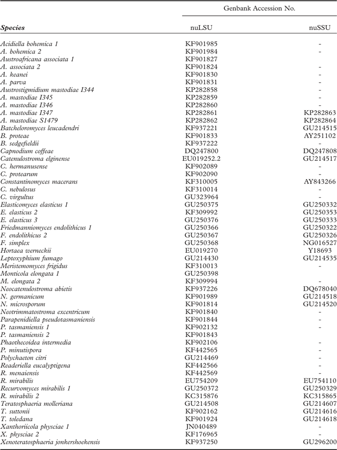
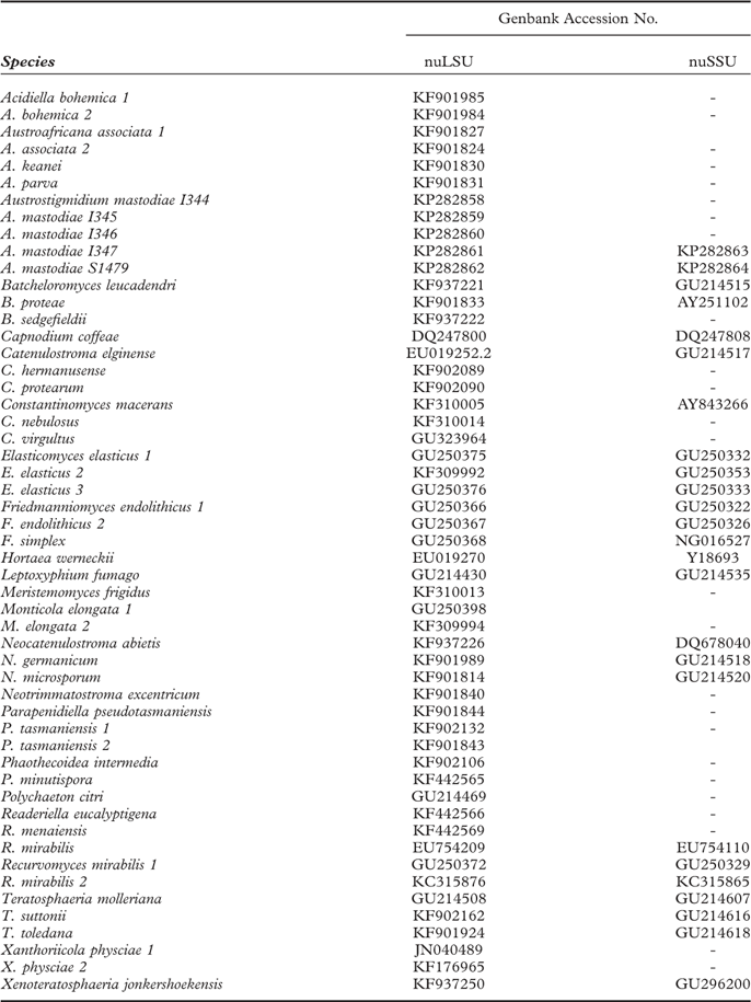
DNA sequences were generated from five specimens from Tierra del Fuego (Chile) and Antarctica populations (Table 1). Due to the tiny size of the fruiting bodies, c. 10–15 pseudothecia were sampled from the same host thallus portion. A sterilized razor blade and an acupuncture needle were used for removing the pseudothecia from thalli and for cleaning their surface. The remnants were then further inspected under both the dissecting and light microscopes in order to avoid contamination by other fungi or algae. Genomic DNA isolation was performed by means of a modified version of the CTAB method (Cubero et al. Reference Cubero, Crespo, Fatehi and Bridge1999). In order to establish the phylogenetic position of the selected taxa we partially amplified the nuLSU and nuSSU rDNA regions, using the primer pairs LR0R (Rehner & Samuels Reference Rehner and Samuels1994) and LR5 (Vilgalys & Hester Reference Vilgalys and Hester1990) for nuLSU, and nssu131-nssu1088 (Kauff & Lutzoni Reference Kauff and Lutzoni2002) for nuSSU. PCR reactions were performed in a total volume of 20 μl, containing 5 μl of template DNA, 2 μl of each primer (10 μM), 10 μl of DNA AmpliTools Fast Master Mix (2×) (Biotools®); final volume was reached by adding distilled water. The PCR cycle for nuLSU was as follows: an initial 5 min heating phase at 95°C, followed by 25 cycles of 28 s at 95°C, 28 s at 52°C and 30 s at 72°C, followed by 10 identical cycles but using 50°C for the annealing step; for nuSSU: initial denaturation for 5 min at 95°C, followed by 10 cycles of 30 s at 95°C, 30 s at 56°C and 30 s at 72°C, followed by an identical 25 cycles but using 52°C for the annealing step; the final elongation for both nuLSU and nuSSU was 15 s at 72°C. The PCR reactions were visualized on 1% agarose gel stained with PRONASAFE nucleic acid stain solution (CONDA Laboratories). PCR products were purified and cleaned using the UltraClean® PCR Clean-Up Kit (MOBIO Laboratories, Inc.). Both complementary DNA strands were sequenced at Macrogen Europe (The Netherlands). Electropherograms were checked and assembled using SeqmanII v.5.07© (Dnastar Inc.).
Specimens used in this study and their accession numbers
Sequence alignment and phylogenetic analysis
We retrieved 48 nuLSU and 23 nuSSU sequences from GenBank corresponding to species in the Teratosphaeriaceae (and 3 outgroup species), based on BLAST searches of the newly generated sequences and previous studies on the family (Ruibal et al. Reference Ruibal, Gueidan, Selbmann, Gorbushina, Crous, Groenewald, Muggia, Grube, Isola and Schoch2009; Egidi et al. Reference Egidi, de Hoog, Isola, Onofri, Quaedvlieg, de Vries, Verkley, Stielow, Zucconi and Selbmann2014). They were aligned together with the new sequences generated in this study (5 nuLSU, 2 nuSSU) (Table 1) using Muscle v3.6 (Edgar Reference Edgar2004) and they were manually examined in Bioedit v.7.0.9 (Hall Reference Hall1999). Gblocks 0.91b (Castresana Reference Castresana2000) was used to remove ambiguously aligned regions, using the least restrictive options for removing gaps and allowing for gaps in less than 50% of the sequences. The combined nuLSU+nuSSU dataset consisted of 1677 bp (722 bp for nuLSU and 955 bp for nuSSU). The software jModelTest v.2.1.4 (Darriba et al. Reference Darriba, Taboada, Doallo and Posada2012) was used to select the best nucleotide substitution model for subsequent analyses using the Bayesian Information Criterion (BIC, Schwarz Reference Schwarz1978). The GTR+G+I model was selected for nuLSU and the K80+I for nuSSU. Partitions were used for the combined nuLSU+nuSSU dataset in both analyses. Capnodium coffeae Pat., Leptoxyphium fumago (Woron.) R. C. Srivast. and Polychaeton citri (Pers.) Lév. were selected as outgroup taxa. Maximum likelihood (ML) analyses were carried out using the online version of RAxML-HPC2 implemented in the CIPRES Science Gateway (Stamatakis Reference Stamatakis2006; Stamatakis et al. Reference Stamatakis, Hoover and Rougemont2008; Miller et al. Reference Miller, Pfeiffer and Schwartz2010). Nodal support was evaluated using 1000 bootstrap pseudoreplicates. Bayesian analyses were those implemented in Mr. Bayes v3.2.2 in the CIPRES Science Gateway (Miller et al. Reference Miller, Pfeiffer and Schwartz2010; Ronquist et al. Reference Ronquist, Teslenko, van der Mark, Ayres, Darling, Höhna, Larget, Liu, Suchard and Huelsenbeck2012). Two parallel, simultaneous runs with four-chain runs were performed over 10 M generations starting with a random tree. Sampling was performed after every 100th step; the first 25% of saved data was discarded as ‘burn-in’ and the 50% majority-rule consensus tree and posterior probabilities (PP) were calculated from the rest of the trees. Convergence of chains and ESS values were checked in Tracer v.1.6 (available at http://tree.bio.ed.ac.uk/software/tracer/). Phylogenetic trees were visualized in FigTree v. 1.3.1 and Adobe Illustrator CS2® was used for artwork. Bootstrap support values ≥70 and Bayesian posterior probabilities ≥0·95 were regarded as significantly supported. The combined nuLSU+nuSSU ML tree was chosen as the working phylogenetic hypothesis.
Transmission electron microscopy
Small fragments of Mastodia tessellata thalli-dwelling Austrostigmidium pseudothecia were fixed in 3% glutaraldehyde and 1% osmium tetroxide, dehydrated in a graded ethanol series, and embedded in Spurr's resin following the protocol described in de los Ríos & Ascaso (Reference de los Ríos and Ascaso2002). Ultrathin sections were post-stained with lead citrate (Reynolds Reference Reynolds1963) and observed in a Zeiss EM910 transmission electron microscope (TEM).
Results
Taxonomy
Austrostigmidium Pérez-Ortega & Garrido-Benavent gen. nov.
MycoBank No.: MB811127
Vegetative hyphae present, hyaline to dark brown; pseudothecia sessile to semi-immersed, globose to subglobose; pseudothecial wall brown, paraplectenchymatous, ostiolate; periphyses abundant; short pendant filaments (pseudoparaphyses) present; asci clavate to subcylindrical, fissitunicate, ocular chamber present, I–, KI–; ascospores hyaline, not ornamented, 3-septate. Pycnidia sessile or with one third immersed in the host thallus, conical to subglobose; conidiogenous cells more or less ampulliform; conidiospores hyaline, simple, bacilliform, straight to curved.
Type species: Austrostigmidium mastodiae Pérez-Ortega & Garrido-Benavent.
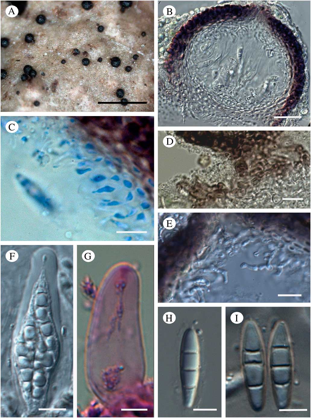
(Figs 1 & 2)
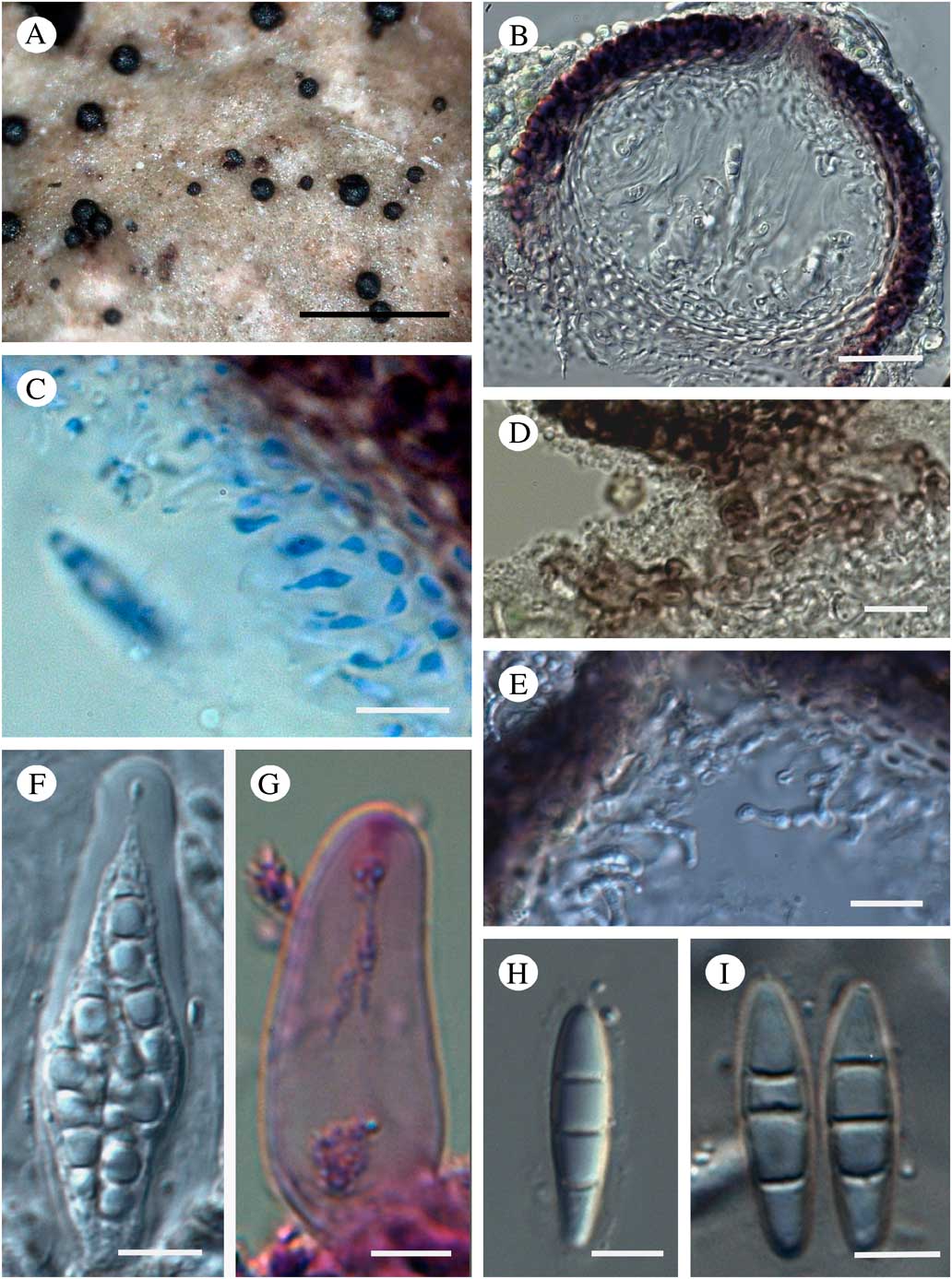
Austrostigmidium mastodiae, habitus and anatomical characters. A, pseudothecia on the surface of Mastodia tessellata; B, longitudinal section of a pseudothecium (DIC); C, pendant filaments (pseudoparaphyses) in lactophenol cotton blue; D, detail of brown vegetative hyphae below the pseudothecium; E, periphyses (DIC); F, mature ascus showing ascospores (DIC); G, ascus tip in BCr showing ocular chamber and the stained apical apparatus; H, young ascospore with an apparent halo (DIC); I, mature ascospores (DIC). Scales: A = 500 μm; B = 25 μm; C, D & E = 10 μm; F, G, H & I = 5 μm. In colour online.
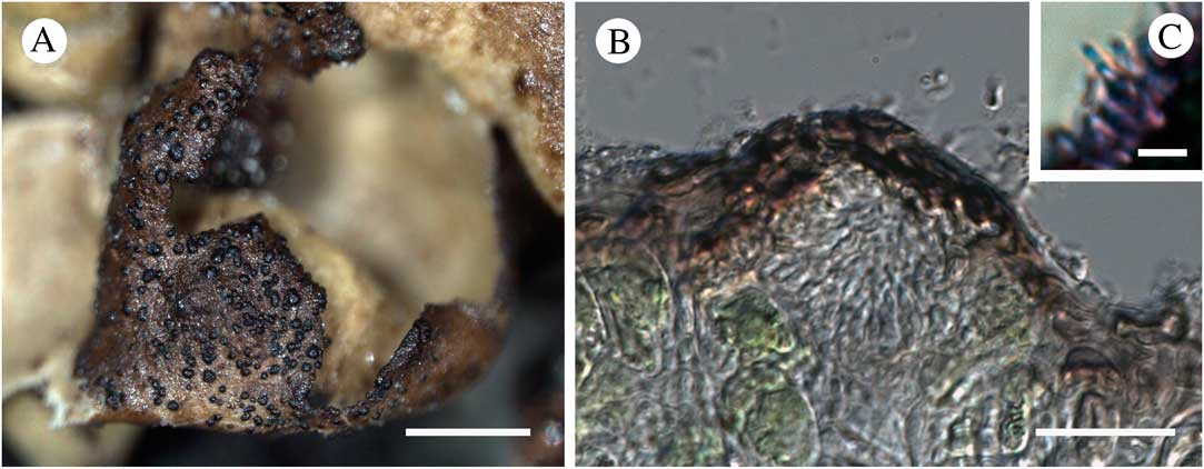
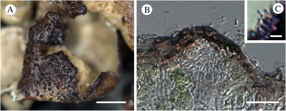
Austrostigmidium mastodiae, pycnidia and conidiospores. A, pycnidia on Mastodia tessellata; B, longitudinal section of a pycnidium; C, conidiogenous cells and conidium in BCr. Scales: A = 500 μm; B = 25 μm; C = 5 μm. In colour online.
Etymology. The generic name refers to its geographical origin (australis, southern) and Stigmidium, which the new taxon resembles anatomically.
Austrostigmidium mastodiae Pérez-Ortega & Garrido-Benavent sp. nov.
MycoBank No.: MB811128
Vegetative hyphae hyaline to dark brown, torulose; pseudothecia globose to subglobose, (58–)95·5±18·9(–130); pseudothecial wall brown, paraplectenchymatous, composed of 4–9 layers of polygonal cells; periphyses abundant; short pendant filaments (pseudoparaphyses) composed of two cells; hymenial gel I–, KI–; asci clavate to subcylindrical, fissitunicate, ocular chamber present (30·5–)39·6±4·8(–48·0)×(8·0–)11·1±1·6(–14·4) μm, 8-spored, I–, KI–; ascospores hyaline, not ornamented, 3-septate, halonate when young, (14·5–)16·7±1·3(–20·0)×(3·5–)4·1±0·5(–5·0) μm. Pycnidia present, conical to subglobose, (34–)48±10·1(–65·0)×(32·0–)36·7±3·9(–43·0) μm; conidiogenous cells more or less ampulliform, simple, hyaline, (3·5–)5·1±0·8(–7·0)×(2·5–)2·8±0·25(–3·00) μm; conidiospores hyaline, simple, bacilliform, straight to curved, (2·5–)3·4±0·6 (–5·0)×(1·0–)1·8±0·2(–1·5) μm.
Type: Antarctica, South Shetlands, Livingston Island, Glaciar Rocoso, 3 m a.s.l., 62° 42·824′S, 60°24·548′ W, 31 January 2014, Asunción de los Ríos (MA–Lich 18215).
(Figs 1 & 2)
Vegetative hyphae usually hyaline to light brown, torulose, usually immersed in host thallus, rarely observed on thallus surface, then dark brown, 2–3 μm wide, I–, KI–, BCr+ violet.
Pseudothecia globose to subglobose, black, usually with glossy appearance, (58·0–)95·5±18·9(–130·0) μm diam. (n = 36), sessile on thallus host, or with the base slightly immersed; ostiolate, ostiolum (12·0–)15·5±2·2(–18·0) μm (n = 12), centred. Pseudothecial wall brown, K– (turning dark blackish brown), (10–)14±3·8(–23·0) μm, usually paler in the lower third, even hyaline in the basal part in contact with the host thallus, paraplectenchymatous, composed of several layers of cells (4–9), polygonal (2·0–)3·8± 1·4(–8·0)×(1·0–)2·3±0· 5(–3·4) μm (n = 28), with thin cell wall, inner wall layers hyaline. Periphyses abundant around ostiolum, hyaline, simple or slightly branched towards the apex, septate with rounded apex, (10·5–)14·3±3·0(–21·5)×(1·5–)1·7±0·28(–2·50)μm (n = 16). Short pendant filaments (pseudo-paraphyses) composed of two cells found in all specimens (9·0–)12·3±2·7(–15·5)×(1·5–)1·6±0·21(–2·50) μm (n = 13), but some specimens show longer filaments up to 20 μm. Other interascal filaments not observed. Hymenial gel I–, KI–. Asci clavate to subcylindrical, fissitunicate, ocular chamber present at least in young asci, no external gelatine observed, (30·5–)39·6±4·76(–48·0)×(8·0–) 11·1±1·65(–14·40) μm (n = 23), 8-spored, I–, KI–, wall BCr–, epiplasm BCr+ violet, apical apparatus revealed with BCr in form of a±light purple multilayered area stained above the ocular chamber. Ascospores hyaline, not ornamented, 3-septate (with a fourth septum in some mature ascospores), sometimes slightly constricted at the septum, apex blunted, halonate when young (1–4 μm), (14·5–)16·7±1·3(–20·0)×(3·5–)4·1±0·5(–5·0)μm [length/breadth ratio = (3·2–) 4·2±0·6(–5·8) (n = 86)].
Pycnidia present, sessile or with one third immersed in the host thallus, black, usually with a glossy appearance, conical to subglobose, dispersed or aggregated in groups, (34–)48±10·09(–65)×(32–)36·75±3·88(–43) μm (n = 24); conidiomatal wall brown, K– (turning dark blackish brown), (6–)8±4·6(–13·0) μm, paler or usually in the basal part, paraplectenchymatous, composed of several layers of cells (2–5), rectangular, similar in size to those in ascomata wall. Conidia cells more or less ampulliform (upper third narrower), simple, hyaline, (3·5–)5·06±0·8(–7·0)×(2·5–)2·76±0·25(–3·0) μm (n = 30). Conidia hyaline, simple, bacilliform, straight to curved, (2·5–)3·34±0·64(–5·0)×(1·0–)1·37±0·21(–1·5) μm.
Etymology. The epithet mastodiae refers to the host genus.
Other material studied. Austrostigmidium mastodiae: Antarctica: South Shetlands: Livingston Island, Punta Hanna, seashore, 0–5 m a.s.l., 62°39′S, 60°37′W, 12 ii 2012, C. Laguna Fiol s/n (MA–Lich 18202); King George Island (Isla 25 de Mayo), Potter Peninsula, Peñon VI, 0–5 m a.s.l., 62.261203°S, 58.618951°W, 22 xii 2009, F. Fernández-Mendoza & S. Domaschke (MA–Lich 18203); King George Island (Isla 25 de Mayo), Potter Peninsula, Stranger Point, 0–5 m a.s.l., 62.261502°S, 58.617572°W, 22 xii 2009 F. Fernández-Mendoza & S. Domaschke (MA–Lich 18206); Avian Island (Adelaide Island), seashore, 0–5 m a.s.l., 67°46′S, 68°53′W, 27 i 2007, J. C. García Galindo & J. Romagni (MA–Lich 18210); Rongé Island, seashore, 0–5 m a.s.l., 64°43′S, 60°54′W, 25 i 2007, J. C. García Galindo & J. Romagni (MA–Lich 18211); Antarctic Peninsula, Cierva Cove, seashore, 0–5 m a.s.l., 64°09′S, 60°57′W, 26 i 2007, J. C. García Galindo & J. Romagni (MA–Lich 18212); Yalour Island, seashore, 0–5 m a.s.l., 65°15′S, 64°11′W, 26 i 2007, J. C. García Galindo & J. Romagni (MA–Lich 18209).—Chile: Región de Magallanes y Antártica Chilena: Tierra del Fuego, Isla Basket, seashore, 0–2 m a.s.l., 54°42′13"S, 71°34′53"W, 2009, S. Pérez-Ortega 3179bis (MA–Lich 18204); ibid., S. Pérez-Ortega 3242bis (MA–Lich 18213); Beagle Channel, Isla Chair, Bahía Darwin, seashore, 0–5 m a.s.l., 54°53′59′′S, 70°00′48′′W, 2009, S. Pérez-Ortega 3227 (MA–Lich 18207); Peninsula Brunswick, cerca del cabo Froward, Bahía de San Nicolás, 0–5 m a.s.l., 53°47′7·4"S, 70°58′38"W, 18 xii 2009, U. Søchting (MA–Lich 18208).
Stigmidium acetabuli Calat. & Triebel: Spain: Aragón: Teruel, Tramacastilla, road Tramacastilla-Villar del Cobo, Quercus faginea forest, 30TXK163732, 1460 m a.s.l., on Pleurosticta acetabulum, 2010, S. Pérez-Ortega 1539 (MA-Lich 18216).
Pseudostigmidium nephromiarium (Linds.) Etayo: Chile: Región de Magallanes y Antártica Chilena: Isla Navarino, subida al cerro Bandera 250 m a.s.l., 54°57′10′′S, 67°38′33′′W, 19 i 2008, S. Pérez-Ortega (MA-Lich 18217).
Phylogenetic relationships
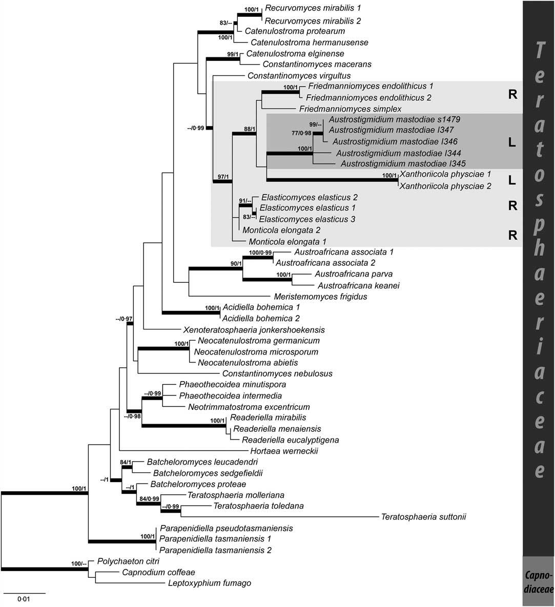
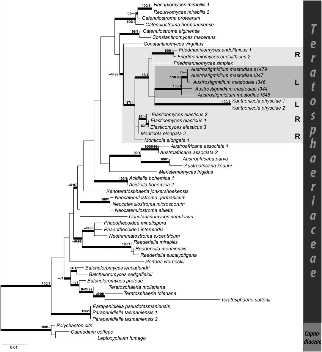
A total of 7 rDNA sequences were generated (5 nuLSU and 2 nuSSU, Table 1). The final alignment consisted of 53 sequences and 1677 characters (722 corresponding to nuLSU, and 955 to nuSSU). Bayesian and ML analyses resulted in similar and congruent topologies, so only the best tree obtained in the ML search is shown in Fig. 3 with posterior probabilities added to supported branches. The five specimens of Austrostigmidium mastodiae formed a single well-supported clade both by posterior probability (PP = 1) and bootstrap (B = 100%). Within this clade, three specimens (I346, I347, S1479) appear together within a well-supported group (PP = 1, B = 100%), whereas the relationships of this group with the two remaining specimens (I344 and I345) remain unclear. The former three specimens were all collected in Tierra del Fuego, and the latter two were collected in Antarctica. Austrostigmidium mastodiae formed a supported clade together with specimens of Xanthoriicola physciae (Kalchbr.) D. Hawksw. and species of the genus Friedmanniomyces Onofri (F. endolithicus Onofri and F. simplex Selbmann et al.) (PP = 1, B = 88%). However, phylogenetic relationships among these three groups remain unresolved. This clade, composed of Austrostigmidium, Xanthoriicola and Friedmanniomyces, constitutes the sister clade to a group composed of Elasticomyces elasticus Zucconi & Selbmann and Monticola elongata Selbmann & Egidi (PP = 0·99, B = 97%).
Maximum likelihood phylogenetic tree obtained in the RAxML analysis (Bayesian analysis recovered a similar topology). Bold branches depict either bootstrap (B) or/and posterior probability (PP) support, figures are given above the branches (B/PP). Austrostigmidium mastodiae is highlighted in dark grey and the group to which it belongs in light grey. R = rock-inhabiting fungi, L = lichenicolous fungi.
Host-parasite interface
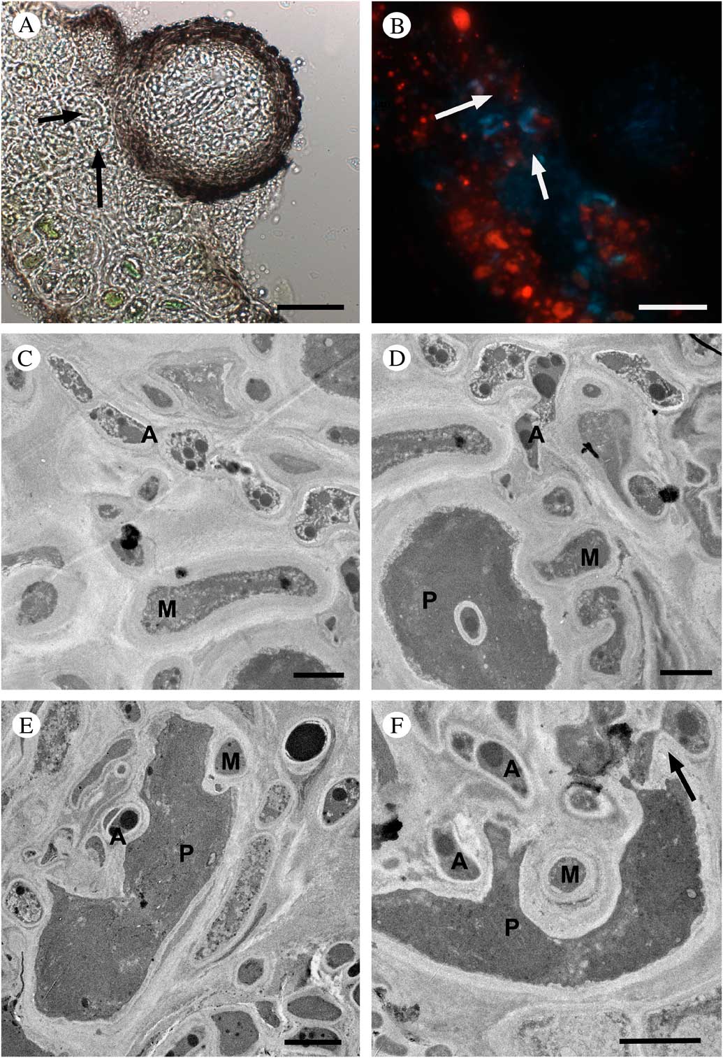
Austrostigmidium mastodiae shows close interactions with the thallus of Mastodia tessellata, especially with the photobiont partner. Vegetative hyphae of A. mastodiae arise from the base of the pseudothecium and penetrate into the host thallus (Fig. 1D). We often observed the decrease of living photobiont cells beneath the pseudothecia, this being particularly visible under the fluorescence microscope. Figures 4A and B show the same thallus section with the surface of a pseudothecium, using transmitted light (Fig. 4A) and fluorescence (Fig. 4B) microscopy. In Figure 4A, it is possible to observe photobiont cells with a healthy appearance only in the lower part of the host thallus. These cells showed a chlorophyll autofluorescence signal (red colour) by fluorescence microscopy (Fig. 2B). The autofluorescence signal was much lower, when observable, in damaged algal cells situated beneath the pseudothecium (arrows in Fig. 4A & B), highlighting the lack of viability of these photobiont cells. The host-parasite interface of this lichenicolous infection has been characterized by means of transmission electron microscopy (TEM). Host fungal and lichenicolous hyphae were clearly distinctive using TEM (Fig. 4C). Mastodia hyphae (M) usually show longer and much larger cells than Austrostigmidium (A), which usually also has more torulose hyphae (Fig. 4C). Cell walls are also thicker in M. tessellata hyphae (0·7–0·8 μm vs 0·3–0·4 μm). On the other hand, Austrostigmidium hyphae showed a large number of electron-dense osmophylic globules in the cytoplasm.
Austrostigmidium mastodiae, anatomical and ultrastructural interactions. A, light microscopy image of a section of Mastodia thallus with an Austrostigmidium pseudothecium, arrows point to collapsed photobiont cells; B, same section in fluorescence microscopy, arrows point to the same collapsed photobiont cells showing faint fluorescence signal (red); C, TEM image showing an A. mastodiae hypha (A) with numerous osmophylic globules and M. tessellata hyphae (M); D, TEM image showing M. tessellata hypha (M) interacting with a Prasiola borealis cell (P), Austrostigmidium hyphae (A) are situated near the photobiont cell but do not interact; E, TEM image showing fungal hyphae (M and A) interacting with a photobiont cell (P) and producing the invagination of the cell; F, TEM image showing penetrations of the cell wall produced by Austrostigmidium (A) and M. tessellata (M) and the Austrostigmidium intracellular haustorium into the collapsed photobiont cell (arrow). Scales: A & B = 25 μm; C, D, E & F = 2 μm.
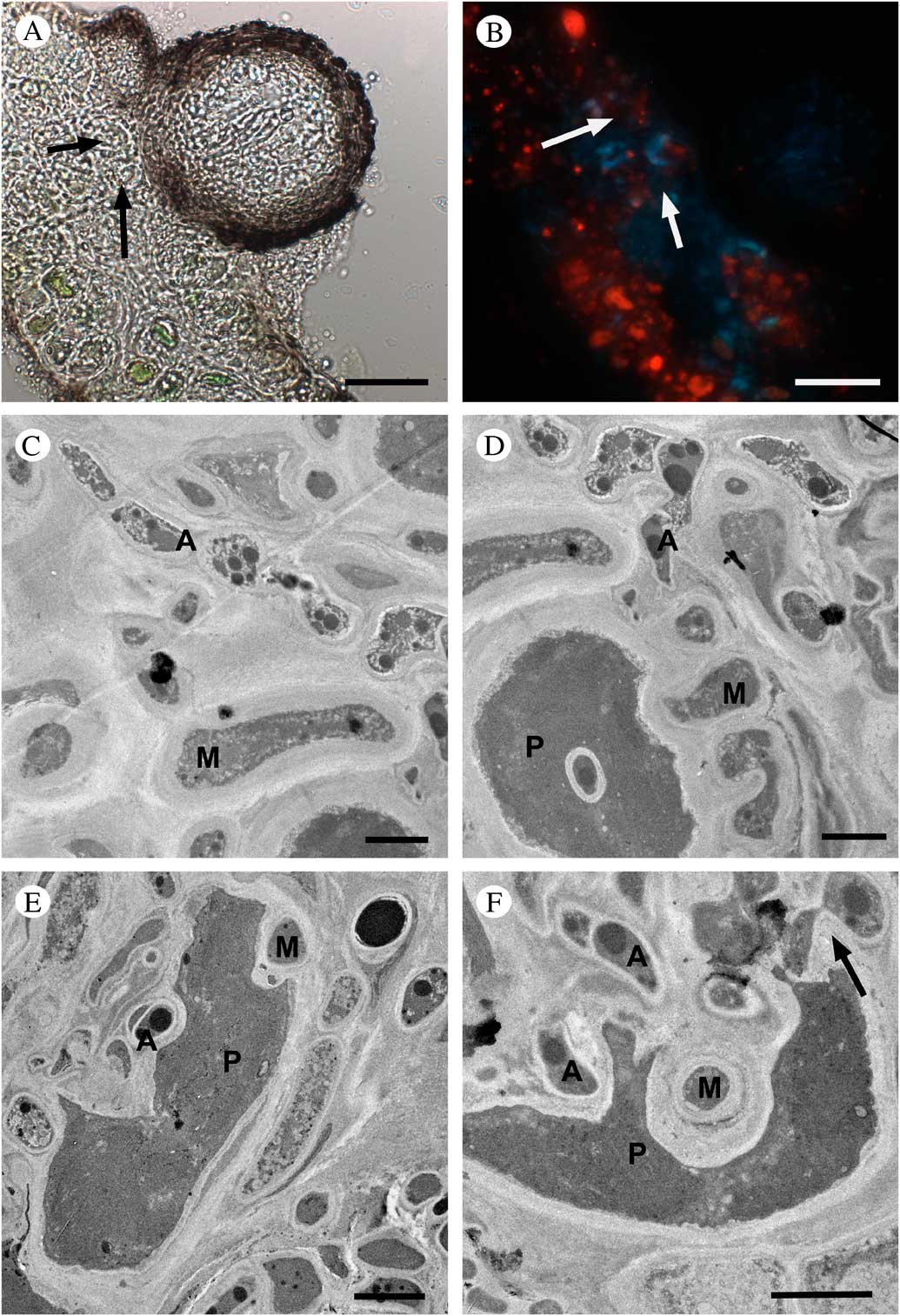
Our TEM study reveals frequent interactions of Austrostigmidium hyphae with the photobiont cells of M. tessellata, but not with the mycobiont hyphae. In Figures 4D–F it is possible to observe the sequence of interactions between the photobiont cells and Austrostigmidium hyphae. In Figure 4D it is shown how Mastodia hyphae interact with a Prasiola borealis cell forming the typical complex haustorial system with multiple lobes, as described in Pérez-Ortega et al. (Reference Pérez-Ortega, de los Ríos, Crespo and Sancho2010). It is also possible to observe Austrostigmidium hyphae near the photobiont cell but still without any visible interaction. In a second stage (Fig. 4E), Austrostigmidium hyphae clearly interact with the P. borealis cell and push the cell wall producing an invagination. Finally, in Fig. 4F it is possible to observe a completely degraded photobiont cell with both Mastodia and Austrostigmidium hyphae attacking it and inducing the collapse of photobiont cells. Mastodia and Austrostigmidium hyphae penetrated the host cell wall but it is even possible to observe intracellular haustoria by an Austrostigmidium hypha (arrow).
Discussion
The new genus Austrostigmidium is described for material of a pseudothecioid species with 3-septate ascospores growing on Mastodia tessellata. A new genus is proposed as the new species does not fit any available genus. Austrostigmidium shows affinities with the recently described Pseudostigmidium Etayo (Etayo & Sancho Reference Etayo and Sancho2008). This genus, described from southern South America, occurs on epiphytic macrolichens from the Peltigerales and is characterized by its conical to subconical ascomata, the lack of hamathecial filaments, presence of periphyses and 3-septate ascospores. These characters are shared with Austrostigmidium. The main difference between the two genera relies on the structure of the ascomatal wall. In Pseudostigmidium, the wall is composed of a network of more or less radial torulose hyphae (Etayo & Sancho Reference Etayo and Sancho2008), whereas Austrostigmidium has a paraplectenchymatous wall composed of polygonal cells. Only P. biseptatum Etayo shows a different paraplectenchymatous wall structure. This species is also deviant regarding ascospore septation, showing 2-septate ascospores, and shape of the ascomata, subglobose in P. biseptatum and conical in other species of the genus. Therefore we consider that A. mastodiae does not belong to Pseudostigmidium s. str., but its relationships to P. biseptatum need to be studied further.
Austrostigmidium also shows close affinities with Stigmidium Trevis. This genus is divided into several groups and may be polyphyletic (Roux & Triebel Reference Roux and Triebel1994; Calatayud & Triebel Reference Calatayud and Triebel2003). Stigmidium and Austrostigmidium share the presence of hyaline to dark brown vegetative hyphae, paraplectenchymatous wall of the pseudothecia, bitunicate asci with an ocular chamber and an apical apparatus revealed by BCr. Stigmidium shows a range of suprahymenial elements, from short to long pseudoparaphyses (Roux & Triebel Reference Roux and Triebel1994; Calatayud & Triebel Reference Calatayud and Triebel2003). In Austrostigmidium, most of the specimens studied showed short pseudoparaphyses of the type-a (composed of two cells of unequal length) of Roux & Triebel (Reference Roux and Triebel1994). This type of pseudoparaphyse appears in Stigmidium s. str. (Roux & Triebel Reference Roux and Triebel1994). Long suprahymenial filaments were found in one specimen which are more similar to those of type-b sensu Roux & Triebel (Reference Roux and Triebel1994) and present in the Stigmidium placynthii group (Roux & Triebel Reference Roux and Triebel1994). The presence of both types of suprahymenial filaments in the same species is intriguing and deserves further research in order to find out whether this anomaly is due to the abnormal expansion of the periphyses towards lower areas of the pseudothecium cavity. Other ascomatal characters in these specimens do not differ from the rest.
Thus, the main differences between Stigmidium and Pseudostigmidium rely on ascospore characters. Stigmidium always has 1-septate ascospores, although species of the Stigmidium psorae group (Calatayud & Triebel Reference Calatayud and Triebel2003) may have 1–3–5-septate ascospores, but only when ascospores are old, and then they become brown. In Austrostigmidium, ascospores are always 3-septate, with a fourth septum appearing in very mature ascospores, but never turning brown. Furthermore, Austrostigmidium has ascospores with thicker cell walls than Stigmidium.
Other genera showing similarities with Austrostigmidium are Epibryon Döbbeler, Sphaerellothecium Zopf. and Clauzadella Nav.–Ros. & Cl. Roux. Epibryon is a genus of predominantly bryophilous species with I+, KI+ blue hymenial gel, lacking periphyses and an ocular chamber in the asci, and most of the species show setae in the ascomata (Döbbeler 1985). It has been recently adscribed within the Chaetothyriales in the family Epibryaceae (Gueidan et al. Reference Gueidan, Aptroot, da Silva Cáceres, Badali and Stenroos2014). Sphaerellothecium differs by a typical superficial network of brown vegetative hyphae and the presence of interascal paraphysoids (Roux & Triebel Reference Roux and Triebel1994). As in Austrostigmidium, Sphaerellothecium may show 3-septate ascospores in some species, but they tend to turn brown when mature. Finally, Clauzadella also shows 3-septate ascospores; it is characterized by the presence of large pseudothecia (0·2–0·3 mm) with walls tinged by a characteristic violet pigment (Navarro-Rosinés & Roux Reference Navarro-Rosinés and Roux1996).
Molecular analyses revealed the phylogenetic adscription of the new genus Austrostigmidium to the family Teratosphaeriaceae (Capnodiales), more precisely to the clade Teratosphaeriaceae I (according to Egidi et al. Reference Egidi, de Hoog, Isola, Onofri, Quaedvlieg, de Vries, Verkley, Stielow, Zucconi and Selbmann2014), as the family Teratosphaeriaceae has been shown to be polyphyletic by Ruibal et al. (Reference Ruibal, Gueidan, Selbmann, Gorbushina, Crous, Groenewald, Muggia, Grube, Isola and Schoch2009). Several anatomical characters in Austrostigmidium support its inclusion within Teratosphaeriaceae, such as the presence of ostiolate pseudothecia with a periphysate canal, pseudoparaphyses, 8-spored bitunicate asci with multilayered endotunica and the presence of a mucoid sheath in the ascospores (Crous et al. Reference Crous, Braun and Groenewald2007). Other characters common within the family, such as the presence of stroma or the 2-septate ascospores turning brown when mature, are absent in Austrostigmidium. The clade containing the new genus also comprises the lichenicolous genus Xanthoriicola, a hyphomycetous lichenicolous fungus with broad cupulate enteroblastic conidiogenous cells generating dark brown, simple and warted conidia which grow on the apothecia of Xanthoria parietina (L.) Beltr. (Ruibal et al. Reference Ruibal, Millanes and Hawksworth2011) and Friedmanniomyces, a hyphomycetous rock-inhabiting endemic genus from Antarctica with pale to dark brown hyphae, having simple to multicellular conidia (Selbmann et al. Reference Selbmann, De Hoog, Mazzaglia, Friedmann and Onofri2005). The sister clade to the group Austrostigmidium-Friedmanniomyces-Xanthoriicola consists of two rock-inhabiting genera, Elasticomyces Zucconi & Selbmann described from Antarctica and Monticola Selbmann & Egidi described from Italy. Basal to these two clades is Constantinomyces Egidi & Onofri, another recently described genus of rock-inhabiting fungi including species from the Iberian Peninsula and Mallorca (Egidi et al. Reference Egidi, de Hoog, Isola, Onofri, Quaedvlieg, de Vries, Verkley, Stielow, Zucconi and Selbmann2014).
Rock-inhabiting fungi are slow-growing microorganisms associated with natural rocky substrata (Egidi et al. Reference Egidi, de Hoog, Isola, Onofri, Quaedvlieg, de Vries, Verkley, Stielow, Zucconi and Selbmann2014). These organisms show physiological and physical characters related to stress tolerance, such as the presence of melanin-like compounds in their cell walls, lack of morphologically differentiated sexual phases and the production of few metabolites and morphological structures for survival (Egidi et al. Reference Egidi, de Hoog, Isola, Onofri, Quaedvlieg, de Vries, Verkley, Stielow, Zucconi and Selbmann2014; Sterflinger Reference Sterflinger2006). It has been pointed out that rock-inhabiting fungi may be the probable ancestors of the lichenized order Verrucariales (Gueidan et al. Reference Gueidan, Villaseñor, De Hoog, Gorbushina, Untereiner and Lutzoni2008). Now with the presence of two lichenicolous fungi, Austrostigmidium and Xanthoriicola, in a clade of predominantly rock-inhabiting fungi, it is clear that this group of fungi can also be the ancestor of some groups of lichenicolous fungi. This switch of lifestyle is not surprising; on the one hand it has been shown that some rock-inhabiting fungi are able to form associations with lichen photobionts, at least in culture (Gorbushina et al. Reference Gorbushina, Beck and Schulte2005), and on the other hand, Selbmann et al. (Reference Selbmann, Grube, Onofri, Isola and Zucconi2013) have recently shown that lichens constitute a very valuable niche for black meristematic fungi in Antarctica, where epilithic conditions are very often prohibitive for life. In this study, the species Friedmanniomyces endolithicus was isolated from Lecidea cancriformis C. W. Dodge & G. E. Baker, and Elasticomyces elasticus was isolated from Lecanora fuscobrunnea C. W. Dodge & G. E. Baker, Lecanora sp. and Usnea antarctica Du Rietz. Furthermore, a similar switch of lifestyle has already been reported within the recently described order Lichenostigmatales (Ertz et al. Reference Ertz, Lawrey, Common and Diederich2013).
It has been shown how A. mastodiae interacts with its host Mastodia tessellata. These interactions are not morphogenetic, as they do not produce deformations or galls in the host thallus. Austrostigmidium mastodiae does not produce any apparent damage in the host thallus, at least not any observable under the dissecting microscope. Based on this observation, A. mastodiae is a commensalistic species, according to Hawksworth (Reference Hawksworth1982). However, this study using transmission electron microscopy has revealed close interactions between A. mastodiae hyphae and the photobiont cells of M. tessellata, those of the alga Prasiola borealis (Pérez-Ortega et al. Reference Pérez-Ortega, de los Ríos, Crespo and Sancho2010; Moniz et al. Reference Moniz, Rindi, Novis, Broady and Guiry2012). These interactions result in the total collapse of photobiont cells, once the lichenicolous fungus interacts with them. Whereas M. tessellata produces peg-like haustoria and biotrophic intraparietal penetrations (Pérez-Ortega et al. Reference Pérez-Ortega, de los Ríos, Crespo and Sancho2010), A. mastodiae seems to first push against the cell wall producing invaginations and later it penetrates into the cell wall and plasma membrane. Pérez-Ortega et al. (Reference Pérez-Ortega, de los Ríos, Crespo and Sancho2010) highlighted that the symbiosis between M. tessellata and Prasiola borealis was not similar to other known lichen symbioses. Instead, this case seems to be more a dynamic equilibrium dependent on biotic and abiotic factors. The presence of a further fungus interacting with the photobiont cells appears to collapse the system and lead to the common loss of photobiont cells beneath the pseudothecia of Austrostigmidium. It is likely that the collapse of photobiont cells is dependent on several factors such as the fitness of the lichen symbiosis at the time the infection occurs, and other abiotic factors such as the level of nitrogen and phosphorous available in the habitat (Pérez-Ortega et al. Reference Pérez-Ortega, de los Ríos, Crespo and Sancho2010). A higher incidence of Austrostigmidium pseudothecia has been observed in those localities close to penguin colonies.
Interactions between lichenicolous fungi and the photobiont cells of their hosts are common (Rambold & Triebel Reference Rambold and Triebel1992; de los Ríos & Grube Reference de los Ríos and Grube2000; de los Ríos et al. Reference de los Ríos, Ascaso and Grube2002). One of the best illustrated examples is how the lichenicolous fungus Zwackhiomyces coepulonus (Norman) Grube & R. Sant. attacks and kills the photobiont cells of its host Xanthoria parietina by means of intracellular haustoria (Grube & Hafellner Reference Grube and Hafellner1990; de los Ríos et al. Reference de los Ríos, Ascaso and Grube2002). Infection by Austrostigmidium mastodiae does not seem as harmful for the host as cases where the lichenicolous fungus directly interacts with the mycobiont cells (de los Ríos & Grube Reference de los Ríos and Grube2000; de los Ríos et al. Reference de los Ríos, Ascaso and Grube2002; Grube & de los Ríos 2002). Lawrey & Diederich (Reference Lawrey and Diederich2003) raised the hypothesis that “highly stable and specialized biotrophic interactions will exhibit modes of nutrient acquisition involving only one lichen biont and little host damage”. Thus, considering the observations made in this study, A. mastodiae can be considered a highly stable and specialized biotrophic association.
This work was funded by the grants CTM2012-38222-C02-02 and FPU AP2012-3556. The authors would like to thank Virginia Souza for technical assistance and UTM (BAE Juan Carlos I) for their logistical support during Antarctic sample collection. We also thank Fernando Fernández-Mendoza (Universität Graz), Clara Laguna (UCM, Madrid), Juan Carlos Galindo (UCA, Cádiz), Ulrik Søchting (University of Copenhagen) and Joanne Romagni (St. Thommas University) for contributing with collections. We are obliged to Leopoldo García Sancho (UCM, Madrid) for his support during the collection of samples in Tierra del Fuego by the first author. We also would like to thank Paul Diederich and an anonymous reviewer for their suggestions and comments on the original manuscript.