INTRODUCTION
An infectious disease of cattle producing fever, haemoglobinuria, anaemia and fatalities, has probably haunted farmers since the beginning of livestock production in warm regions of the Old World. This disease, popularly known as ‘redwater fever’, ‘cattle tick fever’, ‘Spanish fever’ or ‘Texas cattle fever’, was later introduced into the New World by early settlers with imported cattle. The first documented reports date from around 1810 in North America and 1870 in Australia (Clark, Reference Clark1951; Angus, Reference Angus1996). By the beginning of the 20th century, pioneering research studies had already shown that: (i) the disease is caused by an intra-erythrocytic microorganism transmitted through tick bites; (ii) fever and decrease of haematocrit are its main clinical manifestations; (iii) the severity of these symptoms as well as the speed of recovery and mortality rates are inversely related to age; (iv) when naïve animals are inoculated with Babesia-infected blood, the ensuing disease is less severe than when animals naturally acquire the infection through tick bites; and (v) cattle inoculated with infected blood develop a protective immunity upon recovery (Babes, Reference Babes1888; Smith and Kilborne, Reference Smith and Kilborne1893; Connaway and Francis, Reference Connaway and Francis1899; Guthrie, Reference Guthrie1905). This highly valuable information provided the foundations for the development of vaccines for ‘redwater’. The present review will examine our current situation with respect to the control of this disease, especially in the field of vaccination, and will comment on interesting perspectives that have arisen out of progress in cell and molecular biology.
THE AETIOLOGICAL AGENTS OF BOVINE BABESIOSIS AND THEIR HOSTS
Victor Babes was the first to observe a microorganism, which he first thought to be a bacterium, inside bovine erythrocytes and to correlate this observation with the appearance of haemoglobinuria in Rumanian cattle (Babes, Reference Babes1888). Further research showed that this microorganism was in fact a protozoan that was transmitted by ticks and that similar entities could be found in other domestic animals, such as sheep, dog and horse (Smith and Kilborne, Reference Smith and Kilborne1893; Schnittger et al. Reference Schnittger, Rodriguez, Florin-Christensen and Morrison2012). The genus, that later received the name of Babesia, has continued to expand with the progressive incorporation of over 100 species infecting eutherian mammals, marsupials and even birds. Babesia parasites can have devastating consequences on some domestic animals and humans, yet commonly cause asymptomatic infections and remain unnoticed in wildlife. Due to the growing array of previously unrecognized species, it has been proposed that possibly any existing vertebrate species may turn into a Babesia carrier host as long as there is a transmitting tick vector available (Uilenberg, Reference Uilenberg2006; Schnittger et al. Reference Schnittger, Rodriguez, Florin-Christensen and Morrison2012).
With the establishment of the genus Babesia, the name ‘bovine babesiosis’ has been introduced to describe Babesia infections of cattle. Among the several Babesia species shown to infect bovines, Babesia bovis and Babesia bigemina have the highest impact on cattle health. The zoonotic Babesia divergens, although geographically more restricted, is also important as a human pathogen, apart from its impact on bovines (Zintl et al. Reference Zintl, Mulcahy, Skerrett, Taylor and Gray2003; Uilenberg, Reference Uilenberg2006; Schnittger et al. Reference Schnittger, Rodriguez, Florin-Christensen and Morrison2012). From an economic point of view, bovine babesiosis is considered the most important arthropod-transmitted disease of cattle (Bock et al. Reference Bock, Jackson, de Vos and Jorgensen2004). In general, the financial costs of this disease are connected to mortalities, abortions, decrease in meat and milk productivity, control measures and loss of productive potential of endemic areas; while its local impact depends on regional factors, such as the type of livestock involved and the availability and cost-effect ratio of control measures (Uilenberg, Reference Uilenberg1995).
Babesia spp. are unicellular eukaryotes which are taxonomically classified with Theileria spp. and Cytauxzoon spp. as Piroplasmorida (Apicomplexa). Piroplasmorida have been defined as piriform, round, or rod-shaped parasites that lack conoids and flagellae in all stages; without oocysts; and with sexual stages associated with the formation of a large axopodium-like ‘Strahlen’ (Adl et al. Reference Adl, Simpson, Farmer, Andersen, Anderson, Barta, Bowser, Brugerolle, Fensome, Fredericq, James, Karpov, Kugrens, Krug, Lane, Lewis, Lodge, Lynn, Mann, Mc Court, Mendoza, Moestrup, Mozley-Standridge, Nerad, Shearer, Smirnov, Spiegel and Taylor2005, Reference Adl, Simpson, Lane, Lukeš, Bass, Bowser, Brown, Burki, Dunthorn, Hampl, Heiss, Hoppenrath, Lara, Le Gall, Lynn, McManus, Mitchell, Mozley-Stanridge, Parfrey, Pawlowski, Rueckert, Shadwick, Schoch, Smirnov and Spiegel2012). Babesia bovis, B. bigemina and B. divergens are ‘true babesia’, in contrast to other species with unclear taxonomy placement, such as Babesia microti, and fall within the Babesia sensu strictu clade (corresponding to Clade VI in Schnittger et al. Reference Schnittger, Rodriguez, Florin-Christensen and Morrison2012). Their life cycle involves an invertebrate host, an Ixodidae tick, where sexual reproduction takes place, and a vertebrate host, where the parasite undergoes asexual reproduction exclusively within erythrocytes. In addition, true babesia are characterized by transovarial transmission in the tick (Florin-Christensen and Schnittger, Reference Florin-Christensen and Schnittger2009).
Transmission of the sporozoites, the infectious parasite stage, takes place through tick bites during blood meals. Iatrogenic and transplacental transmission has been described for related parasites, but these routes have not yet been investigated for bovine babesias (Chhabra et al. Reference Chhabra, Ranjan, Uppal and Singla2012; Joseph et al. Reference Joseph, Purtill, Wong, Munoz, Teal, Madison-Antenucci, Horowitz, Aguero-Rosenfeld, Moore, Abramowsky and Wormser2012; Beard et al. Reference Beard, Pelzel, Rush, Wright, Galgut, Hennager, King and Traub-Dargatz2013). Geographic distribution of bovine Babesia spp. strictly follows that of their tick vectors, which is mainly influenced by temperature and humidity (de Waal and Combrick, Reference de Waal and Combrink2006). Babesia bovis and B. bigemina are transmitted by Rhipicephalus microplus, Rhipicephalus annulatus and Rhipicephalus geigyi ticks, present in vast tropical and temperate regions around the world. In addition, B. bigemina can also be transmitted by Rhipicephalus decoloratus and Rhipicephalus evertsi, further extending its distribution into the African continent, making it the most widespread bovine Babesia species (Bock et al. Reference Bock, Jackson, de Vos and Jorgensen2004; Rodriguez et al. Reference Rodriguez, Schnittger, Tomazic, Florin-Christensen, Castillo and Harris2013a). Babesia divergens, on the other hand, is transmitted mainly by Ixodes ricinus, which only thrives in moisture-saturated microhabitats but can tolerate a larger range of temperatures. It has been reported in several European countries, as well as northern Africa (Zintl et al. Reference Zintl, Mulcahy, Skerrett, Taylor and Gray2003; de Waal and Combrick, Reference de Waal and Combrink2006).
Bos taurus (European cattle) and Bos indicus (zebu cattle), and lately Bubalus bubalis (water buffalo) as well, have been described as hosts for B. bovis and B. bigemina, but clinical cases of the disease do not normally occur in zebu cattle or water buffalo, which could be a result of long co-evolutionary adaptations of these cattle types with Rhipicephalus ticks and Babesia parasites (Bock et al. Reference Bock, Jackson, de Vos and Jorgensen2004; Ferreri et al. Reference Ferreri, Benitez, Dominguez, Rodriguez, Asenzo, Mesplet, Florin-Christensen and Schnittger2008). In addition, B. bovis and B. bigemina-infected deer have been detected by PCR-based and serological diagnostic tests, implying that these animals might act as parasite carriers, which would have important epidemiological consequences (Cantu et al. Reference Cantu, Ortega-S, Mosqueda, Garcia-Vazquez, Henke and George2007; Ramos et al. Reference Ramos, Cooper and Holman2010; da Silveira et al. Reference da Silveira, Rabelo and Ribeiro2011; Holman et al. Reference Holman, Carroll, Pugh and Davis2011). However, attempts to experimentally infect deer with B. bovis have so far been unsuccessful (G. A. Scoles, personal communication), thus the role of deer in Babesia spp. transmission remains uncertain. Babesia bovis and B. bigemina have also been detected in horse and pampas deer, but further studies are needed to evaluate the significance of these findings (Criado-Fornelio et al. Reference Criado-Fornelio, Buling, Asenzo, Benitez, Florin-Christensen, Gonzalez-Oliva, Henriques, Silva, Alongi, Agnone, Torina and Madruga2009; Silveira et al. Reference Silveira, Rabelo, Lacerda, Borges, Tomás, Pellegrin, Tomich and Ribeiro2013).
On the other hand, B. divergens infections produce clinical disease in European cattle, as well as in immunocompromised humans and other primates (Zintl et al. Reference Zintl, Mulcahy, Skerrett, Taylor and Gray2003). They have also been detected in cottontail rabbits and cervids, but at least some of the latter infections are probably due to the closely related species Babesia capreoli, which poses no threat to cattle or humans (Malandrin et al. Reference Malandrin, Jouglin, Sun, Brisseau and Chauvin2010; Schnittger et al. Reference Schnittger, Rodriguez, Florin-Christensen and Morrison2012).
In B. taurus, the aetiology of bovine babesiosis varies according to the infecting species, parasite strain and the age of the animal. Clinical cases are typically observed in adults, while animals younger than 9–10 months usually remain asymptomatic. Due to the accumulation of infected erythrocytes in lung and brain capillaries, acute babesiosis caused by B. bovis is frequently severe and characterized by hypotension, respiratory stress syndrome and neurological symptoms. Consequently, B. bovis infections normally show low levels of parasitaemia. On the other hand, babesiosis caused by B. bigemina and B. divergens results in high parasitaemias, and pathogenicity is mainly associated with massive erythrocyte destruction, leading to severe anaemia. Haemoglobinuria is observed at the peak of the haemolytic crisis in B. bigemina and B. divergens infections, and is also observed in the latter stages of babesiosis caused by B. bovis, hence the disease is commonly called ‘redwater’. Fever is frequent in all three infections, and can lead to abortions in pregnant cattle or temporary reduction of fertility in bulls (Zintl et al. Reference Zintl, Mulcahy, Skerrett, Taylor and Gray2003; Bock et al. Reference Bock, Jackson, de Vos and Jorgensen2004). Clinical cases resulting from B. divergens infections in humans resemble the symptoms of the acute bovine disease, including haemoglobinuria (Gray, Reference Gray2006).
VACCINES: WHERE WE ARE NOW
Why and when to vaccinate
In areas where there is a continuous inoculation of cattle with Babesia spp. by infected ticks, calves are likely to be in contact with the parasite during the first 10–12 months of life, when typically they do not show any clinical manifestations. Babesia parasites are able to establish persistent infections in these animals that thus develop into parasite carriers with strong acquired immunity and resistance to disease. Mahoney (Reference Mahoney1974) described this situation as enzootic stability and stated that if 75% of the animals in a herd have been exposed to B. bovis before 9 months of age, as indicated by specific antibody titres, the chance of observing clinical cases is very low. Although natural endemic stability is in principle an ideal condition where no control measures are needed, this situation is rare and, when it does exist, it can be easily broken by variations in climate, host genotypes and management strategies. In addition, artificially maintaining endemic stability by yearly manipulating transmission rates in each herd is highly impractical. Importantly, it has been argued that the threshold of 75% exposed cattle as a means to predict the appearance of clinical cases should be taken with caution when extrapolated to different regions and host-tick-pathogen systems from those on which the model was based (Jonsson et al. Reference Jonsson, Bock, Jorgensen, Morton and Stear2012).
In regions of enzootic instability, or when cattle are relocated from tick-free to tick-infested regions, prophylactic immunization has proved an effective method to prevent the occurrence of babesiosis outbreaks (Bock et al. Reference Bock, Jackson, de Vos and Jorgensen2004).
Currently available vaccines
The development of vaccines against bovine babesiosis was prompted by early observations indicating that cows that recovered from natural Babesia spp. infections developed long-lasting immunity; and inoculation of their blood into susceptible cattle resulted in a less virulent form of the disease. Thus, the first vaccine formulations consisted of blood from donor bovines that had recovered from infection (Connaway and Francis, Reference Connaway and Francis1899; de Waal and Combrink, Reference de Waal and Combrink2006). A breakthrough in the development of bovine babesiosis vaccines was achieved by Australian researchers, who observed that rapid successive blood passages of B. bovis between splenectomized calves resulted in progressive virulence decrease, with diminished post-vaccination changes in body temperature and haematocrit (Callow, Reference Callow1979). Later, attenuation of B. bigemina was also achieved, but in this case the procedure involved slow successive passages among spleen-intact calves (Dalgliesh et al. Reference Dalgliesh, Callow, Mellors and McGregor1981). Attenuation also leads to the waning of nervous symptoms in the case of B. bovis and is sometimes, but not always, associated with a loss of tick transmissibility (Timms et al. Reference Timms, Stewart and De Vos1990; Mangold et al. Reference Mangold, Aguirre, Cafrune, de Echaide and Guglielmone1993; Mafra et al. Reference Mafra, Patarroyo and Silva1994). Since the spleen is important in the trapping and destruction of infected erythrocytes, the use of splenectomized bovines yields adequately high parasitaemias in the case of B. bovis (Bock et al. Reference Bock, Jackson, de Vos and Jorgensen2004). Current vaccines against B. bovis and B. bigemina are based on these attenuation procedures.
The mechanisms underlying attenuation are still unknown. It has been hypothesized that during the attenuation process, parasites lose their capacity to express certain virulence-mediating genes, and/or that a subpopulation of parasites with a mild pathogenicity phenotype, present in the initial pathogenic field isolate, is selected. The latter hypothesis is supported by studies carried out in B. bovis using molecular markers and whole genome comparisons between parental virulent strains and resulting attenuated ‘daughter’ strains that showed that the genetic complexity of the population is reduced upon attenuation (Carson et al. Reference Carson, Timms, Cowman and Stewart1990; Lau et al. Reference Lau, Kalyanaraman, Echaide, Palmer, Bock, Pedroni, Rameshkumar, Ferreira, Fletcher and McElwain2011; Baravalle et al. Reference Baravalle, Thompson, Valentini, Ferreira, Torioni de Echaide, Florin-Christensen and Echaide2012; Mazuz et al. Reference Mazuz, Molad, Fish, Leibovitz, Wolkomirsky, Fleiderovitz and Shkap2012; Combrink et al. Reference Combrink, Troskie, Pienaar, Latif and Mans2014). However, the attenuation scenario is likely more complex than just a selection procedure since, on one hand, an attenuated strain can be composed of virulent and avirulent subpopulations, as was shown in B. bovis clones derived from the Australian Ka vaccine strain; and, on the other, an avirulent clone can reverse its phenotype to a virulent one upon passage through a spleen-intact bovine (Timms et al. Reference Timms, Stewart and De Vos1990). In any case, research efforts are still needed to unravel the host-pathogen interactions that yield a less virulent parasite population upon blood passages.
In order to identify possible genes involved in the pathogenic/attenuated phenotypes, comparison of transcription patterns before and after attenuation of B. bovis has been recently carried out using genome sequencing or microarray technology. It has been found that the pattern of transcription of protease genes in B. bovis blood-stage parasites did not show differences when a pathogenic parental strain was compared with its attenuated derivative (Mesplet et al. Reference Mesplet, Palmer, Pedroni, Echaide, Florin-Christensen, Schnittger and Lau2011). In another study, a number of genes were shown to be differentially expressed when the whole transcriptome of a virulent and its attenuated derivative strain were compared, but further studies will be necessary to demonstrate whether any of these have a functional role in virulence and/or attenuation (Pedroni et al. Reference Pedroni, Sondgeroth, Gallego-Lopez, Echaide and Lau2013). Understanding gene regulation events responsible for turning genes on or off during attenuation can open the way to new therapeutic and preventive approaches. Nucleosome rearrangements have been shown to be involved in the regulation of the expression of a family of variable antigens in B. bovis, and complex machinery for histone-based epigenetic regulation is encoded in the genome of this parasite (Dominguez et al. Reference Dominguez, Ferreri and Schnittger2011; Huang et al. Reference Huang, Xiao and Allred2013). Future studies are needed to unravel whether these or other types of gene regulation mechanisms are involved in attenuation.
It has also been hypothesized that lipid composition could be a virulence determinant since lipids extracted from virulent B. bovis merozoites elicited a stronger pro-inflammatory response in mice and bovines than those of an attenuated strain, possibly allowing immune evasion of the latter (Gimenez et al. Reference Gimenez, Magalhaes, Belaunzarán, Poncini, Lammel, Gonzalex-Cappa, Bozza and Isola2010, Reference Gimenez, Belaunzarán, Poncini, Blanco, Echaide, Zamorano, Lammel, González Cappa and Isola2013).
In the case of B. divergens, effective live vaccines were produced either in splenectomized calves or intact gerbils (Meriones unguiculatus). Virulent parasite field isolates were used in these formulations, and thus treatment with babesiacides was needed to avoid clinical manifestations after vaccination. Commercial production of the live vaccine against B. divergens was discontinued over 10 years ago in Ireland, and there is no ‘web-available’ information about current production in other countries (Zintl et al. Reference Zintl, Mulcahy, Skerrett, Taylor and Gray2003; Bock et al. Reference Bock, Jackson, de Vos and Jorgensen2004). Most attempts to attenuate B. divergens by different methods, including bovine passages, were unsuccessful, with the exception of in vitro cultivation that yielded a decrease in virulence, but this methodology was not applied to vaccine formulation (Winger et al. Reference Winger, Canning and Culverhouse1989; Zintl et al. Reference Zintl, Mulcahy, Skerrett, Taylor and Gray2003).
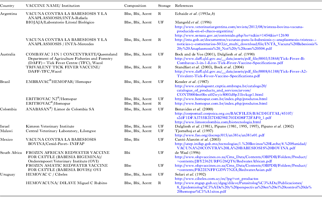
Table 1 presents information on known live vaccines against B. bovis and B. bigemina currently commercialized around the world. Additionally, vaccine production in Cuba and Zimbabwe, as well as an experimental B. bigemina vaccine based on an Uzbekistan attenuated B. bigemina strain, have also been reported (Shkap et al. Reference Shkap, de Vos, Zweygarth and Jongejan2007a, Reference Shkap, Rasulov, Abdurasulov, Fish, Leibovitz, Krigel, Molad, Mazuz and Savitskyb).
Commercially available vaccines against bovine babesiosis. Bbo, Bbi and Acent: bovine erythrocytes infected with Babesia bovis, B. bigemina or Anaplasma centrale, respectively; UF: ultra-frozen; R: refrigerated. With the exception of in vitro produced Bbo and Bbi-infected erythrocytes used in the vaccines of INTA-Rafaela and Laboratorio Litoral Biologico, Argentina, the rest of the vaccines use infected erythrocytes obtained from splenectomized calves

Live vaccines against B. bovis and B. bigemina can be prepared typically as a bivalent formula that contains around 107 erythrocytes infected with each of these parasites, although a reduced dose of 2·5×106 infected erythrocytes has also been reported as effective for protection against B. bigemina in Australia (Standfast et al. Reference Standfast, Bock, Wiecek, Devos, Jorgensen and Kingston2003). Often, a trivalent formula is commercialized that also contains 107 erythrocytes infected with the rickettsia Anaplasma centrale providing cross-protection against Anaplasma marginale, another intra-erythrocytic tick-borne pathogen causing a related disease with wide distribution in tropical and temperate regions (OIE, 2010). In most cases for B. bovis and B. bigemina and always for A. centrale, infected erythrocytes are obtained from ad hoc infected splenectomized calves. To this aim, donor animals, free of Babesia spp. and other blood-transmissible pathogens, are inoculated with suitable attenuated strains and monitored, blood is collected from the jugular vein during the acute phase of the reaction, and parasitaemia is determined to standardize the number of infected erythrocytes per vaccine dose (Bock et al. Reference Bock, Jackson, de Vos and Jorgensen2004; de Waal and Combrink, Reference de Waal and Combrink2006). In vitro production of Babesia parasites (see section on ‘In vitro culture’) has opened additional possibilities for vaccine development. Indeed, in vitro cultured B. bovis and B. bigemina have been successfully used for vaccination purposes in Argentina for almost 2 decades, and have also been reported to be effective in Mexico (Mangold et al. Reference Mangold, Vanzini, Echaide, de Eschaide, Volpogni and Guglielmone1996; Cantó Alarcón et al. Reference Cantó Alarcón, Martínez, Rojas Ramírez, Ramos Aragón, Mosqueda Gualito, Vega y Murguía and Figueroa Millán2003; Shkap et al. Reference Shkap, de Vos, Zweygarth and Jongejan2007a). In vitro vaccines allow more controlled and standardized conditions, and have reduced risks of pathogen co-transmission, which can be further decreased by gamma irradiation of the serum used for parasite cultivation (Rojas et al. Reference Rojas, Figueroa, Alvarado, Mejia, Mosqueda, Falcon, Vega and Alvarez2006; Shkap et al. Reference Shkap, de Vos, Zweygarth and Jongejan2007a). Interestingly, it was shown that older B. taurus bulls pre-immunized with culture-derived parasites showed good tolerance when later inoculated with a live B. bovis/B. bigemina vaccine, while a significant number of non pre-immunized animals showed clinical signs of B. bigemina infection (Shkap et al. Reference Shkap, Leibovitz, Krigel, Hammerschlag, Marcovics, Fish, Molad, Savitsky and Mazuz2005). As drawbacks, in vitro cultivation is labour-intensive, requires permanent supply of erythrocytes and serum from a suitable donor animal, as well as adequate laboratory equipment and trained personnel.
Importantly, standardized conditions need to be followed to maintain the attenuated state and immunogenicity of vaccinal parasites, including monitoring the number of in vitro or in vivo passages, and carrying out periodic inoculation of naïve cattle in the case of in vitro vaccines (Echaide, Reference Echaide2008). Additionally, thorough testing of vaccine donors and codes of good manufacturing techniques are necessary (OIE, 2010). Several vaccines produced around the world are based on the Australian vaccinal strains, while some countries, such as Argentina, South Africa, Mexico and Uzbekistan use their own locally generated attenuated strains.
Live vaccines have limited shelf-life, lasting between 4 and 7 days at 4 °C (Bock et al. Reference Bock, Jackson, de Vos and Jorgensen2004). Ultra-freezing of infected erythrocytes in liquid nitrogen allows long-term storage, and vaccines can then be prepared upon demand, resulting in increased cost-efficiency. Importantly, the safety and protective efficacy of frozen vaccines can be tested prior to their release, while testing of chilled vaccines can only be done in retrospect, although the high cost of these tests hampers their routine application (Bock et al. Reference Bock, Jackson, de Vos and Jorgensen2004; de Waal and Combrick, Reference de Waal and Combrink2006).
It has been shown that when glycerol is used as cryoprotectant, ultra-frozen B. bovis and B. bigemina parasites can keep their viability for up to 24 h upon thawing, although inoculation within 8 h is recommended (Mangold et al. Reference Mangold, Aguirre and Guglielmone1990; Bock et al. Reference Bock, Jackson, de Vos and Jorgensen2004; de Waal and Combrink, Reference de Waal and Combrink2006). Dimethylsulphoxide is used as a cryoprotectant in some vaccine formulations but due to its higher toxicity and lower post-thaw storage life it has lesser acceptance than glycerol (Bock et al. Reference Bock, Jackson, de Vos and Jorgensen2004; de Waal and Combrink, Reference de Waal and Combrink2006). In the case of B. divergens, attempts to recover significant numbers of viable parasites after freezing and thawing have so far failed (de Waal and Combrink, Reference de Waal and Combrink2006).
Live Babesia vaccines are recommended to be applied to 4- to 10-month-old calves that generally show good tolerance, though a transient clinical response to vaccination can sometimes take place (Fish et al. Reference Fish, Leibovich, Krigel, McElwain and Shkap2008). Adult animals, on the other hand, can develop acute babesiosis upon vaccination, for which daily monitoring for up to 21 days is suggested and babesiacide treatment is often needed (Shkap et al. Reference Shkap, Leibovitz, Krigel, Hammerschlag, Marcovics, Fish, Molad, Savitsky and Mazuz2005; de Waal and Combrink, Reference de Waal and Combrink2006). Protective immunity develops 3–4 weeks after vaccination and normally lasts at least 4 years (Bock and de Vos, Reference Bock and de Vos2001; OIE, 2010). It was observed in South Africa that, after chemosterilization of infections, sterile immunity to B. bigemina lasted for only 16 months, without further boosting of immunity from tick-acquired infections, while immunity to B. bovis lasted for over 3 years (De Waal, Reference de Waal1996). Thus, complete tick control after vaccination is discouraged, so that natural infections through tick bites can aid in the acquisition of a long-term protected status (Bock et al. Reference Bock, Jackson, de Vos and Jorgensen2004; de Waal and Combrink, Reference de Waal and Combrink2006).
Opposite to what was observed with B. divergens live vaccines that produced sterile immunity, B. bovis and B. bigemina live vaccines do not eliminate the parasites but rather produce disease-resistant carrier animals that may act as reservoirs for tick transmission (Zintl et al. Reference Zintl, Mulcahy, Skerrett, Taylor and Gray2003; Bock et al. Reference Bock, Jackson, de Vos and Jorgensen2004). Additionally, live vaccine production can be cumbersome, expensive and unattractive for private industry undertakings; current vaccines around the world are mostly produced with government support. Donor bovines should be kept free of other pathogens, such as bovine leukaemia virus, and vaccines need to undergo strict quality controls to avoid pathogen dissemination at vaccination (Bock et al. Reference Bock, Jackson, de Vos and Jorgensen2004; Shkap et al. Reference Shkap, de Vos, Zweygarth and Jongejan2007a). Notably, donors should be Babesia spp.-free, which can be particularly challenging when vaccines are produced in tick-endemic regions. In order to ensure parasite viability, a strict cold chain needs to be sustained during preparation, maintenance and transportation to the end user. As mentioned above, refrigerated vaccines have a short shelf life, and in the case of frozen vaccines, the timing of application is critical, since parasites rapidly die after thawing (Shkap et al. Reference Shkap, de Vos, Zweygarth and Jongejan2007a). Adverse reactions to vaccination with live parasites, including abortions in pregnant cattle have been reported in highly susceptible cattle, such as high-yielding dairy cows. Additionally, vaccine failures can take place due to incorrect handling or storage, administration of chemotherapeutics immediately before or during vaccination, stress or concomitant infections, pathogenicity reversion of the attenuated vaccine strains, and/or changes in the parasite population that can lead to lack of protection (Bock et al. Reference Bock, de Vos, Kingston, Shiels and Dalgliesh1992; Bock and de Vos, Reference Bock and de Vos2001; de Waal and Combrink, Reference de Waal and Combrink2006).
Thus, in spite of the general efficiency of current vaccines in preventing clinical cases of bovine babesiosis, there is considerable interest in the development of improved subunit vaccines that provide protection against disease and are safer, and easier to handle and produce. The following sections present our current knowledge about bovine Babesia spp. biology as well as technological achievements in the manipulation of these parasites that can be exploited for the rational design of next-generation vaccines.
PUZZLE PIECES FOR NOVEL VACCINES
In vitro culture
In vitro cultivation of bovine Babesia spp. allows vaccine preparations, cheap maintenance of field strains for antigen characterization, drug testing, seroneutralization assays, production of transgenic variants, morphological studies and invasion assays (Müller and Hemphill, Reference Müller and Hemphill2013). Achievement of continuous in vitro cultivation of B. bovis, B. bigemina and B. divergens was reported in the 1980s, and the same methods are still in practice. Culture reagents generally consist of bovine erythrocytes, buffered culture medium and adult bovine serum (reviewed in Schuster, Reference Schuster2002). Other sources of nutrients were also at least partially useful, such as foetal or calf bovine serum and lipid-enriched serum albumin (Grande et al. Reference Grande, Precigout, Ancelin, Moubri, Carcy, Lemesre, Vial and Gorenflot1997; Jackson et al. Reference Jackson, Waldron, Weier, Nicoll and Cooke2001; Schuster, Reference Schuster2002; Zintl et al. Reference Zintl, Westbrook, Mulcahy, Skerrett and Gray2002a; Sánchez et al. Reference Sánchez, Campos and Oliva2009; Sun et al. Reference Sun, Moreau, Chauvin and Malandrin2011). Inclusion of lipid-enriched serum albumin instead of bovine serum in in vitro cultures destined for live vaccine production could decrease the risk of pathogen co-transmission. Interestingly, B. bovis has been shown to be able to grow in serum-free medium, a condition suggested being particularly adequate for drug testing (Bork et al. Reference Bork, Okamura, Matsuo, Kumar, Yokoyama and Igarashi2005). Human, sheep and rodent erythrocytes, as well as sheep serum, can also be used for long-term cultivation of B. divergens (Ben Musa and Phillips, Reference Ben Musa and Phillips1991; Chauvin et al. Reference Chauvin, Valentin, Malandrin and L'Hostis2002; Zintl et al. Reference Zintl, Westbrook, Mulcahy, Skerrett and Gray2002a). On the other hand, invasion of horse and donkey erythrocytes by B. divergens, or of human, horse, pig or goat erythrocytes by B. bovis, has been reported to take place, but this is not usually followed by sustained growth (Gaffar et al. Reference Gaffar, Franssen and de Vries2003; Zintl et al. Reference Zintl, Mulcahy, Skerrett, Taylor and Gray2003).
Low oxygen (2 or 5%) atmosphere has been recommended for Babesia cultivation in cases of low parasitaemia, for example when starting cultures out of carrier animals, or during parasite cloning (Rodriguez et al. Reference Rodriguez, Buening, Green and Carson1983). However, it has been recently shown for the related piroplasmid Theileria equi that addition of L-cysteine replaced the need for a low oxygen atmosphere when establishing in vitro cultures out of field isolates (Zweygarth and Josemans, Reference Zweygarth and Josemans2014). It remains to be tested whether this observation that simplifies laboratory requirements for parasite cultivation, applies also to Babesia spp.
Infected erythrocyte percentages are most commonly monitored by microscopic observation of Giemsa-stained culture smears (Zweygarth et al. Reference Zweygarth, Van Niekerk, Just and De Waal1995). While simple and of low-cost, this method is time-consuming and, since infected erythrocytes do not distribute uniformly in smears, a large number need to be examined to obtain reproducible estimates. During their growth, Babesia parasites consume haemoglobin-bound oxygen leading to colour changes in the medium from brilliant red to almost black (Zweygarth et al. Reference Zweygarth, Van Niekerk, Just and De Waal1995; Schuster, Reference Schuster2002). Spectrophotometric measurement of these changes has been described by Malandrin et al. (Reference Malandrin, Marchand and Chauvin2004) as a fast and reliable method to evaluate in vitro growth of B. divergens. Other available quantitative techniques to monitor growth and/or viability of Babesia spp. include measurements of [3H] hypoxanthine or [3H] thymidine incorporation into DNA, detection of fluorescent nuclear and/or vital dyes by epifluorescence microscopy or flow cytometry, and quantitative PCR (Goff and Yunker, Reference Goff and Yunker1986; Wyatt et al. Reference Wyatt, Goff and Davis1991; Davis et al. Reference Davis, Wyatt, Hamilton and Goff1992; Brasseur et al. Reference Brasseur, Lecoublet, Kapel, Favennec and Ballet1998; Jackson et al. Reference Jackson, Waldron, Weier, Nicoll and Cooke2001; Buling et al. Reference Buling, Criado-Fornelio, Asenzo, Benitez, Barba-Carretero and Florin-Christensen2007; Criado-Fornelio et al. Reference Criado-Fornelio, Buling, Asenzo, Benitez, Florin-Christensen, Gonzalez-Oliva, Henriques, Silva, Alongi, Agnone, Torina and Madruga2009; Fletcher et al. Reference Fletcher, Wigg, Rolls and de Vos2009; Müller and Hemphill, Reference Müller and Hemphill2013). Quantification of parasite-specific mRNA levels by qRT-PCR, or by reporter gene expression in transgenic parasites has also been used for growth/vitality measurements in other apicomplexan organisms (Müller and Hemphill, Reference Müller and Hemphill2013). The use of transgenic B. bovis parasites in growth experiments is now an accessible possibility, given the recent developments in this area discussed in the section on ‘Transfection as a tool for Babesia vaccine development’.
Bovine-pathogen interactions
Interactions at the cellular level
Since all apicomplexan protozoa are obligate intracellular parasites at some point during their life cycle, efficient invasion of host cells is critical for their survival. Thus, the understanding of mechanisms and roles of molecules involved in this event can greatly contribute to the development of vaccines against members of this group of medical and veterinary importance. In the case of Babesia parasites, in addition to relevant specific research that has been carried out, knowledge about conserved cell invasion mechanisms among apicomplexans can be extrapolated from studies on Plasmodium falciparum and Toxoplasma gondii (Yokoyama et al. Reference Yokoyama, Okamura and Igarashi2006; Sun et al. Reference Sun, Moreau, Chauvin and Malandrin2011; Lobo et al. Reference Lobo, Rodriguez and Cursino-Santos2012). Several parasite proteins likely involved in the interactions between bovine Babesia parasites and host erythrocytes have been analysed as potential vaccine candidates and will be introduced in this section.
During the Babesia life cycle in the tick, sporozoites, the vertebrate-infective life stage, are generated by sporogony in the salivary glands and are later injected with an aliquot of tick saliva into the bloodstream of a suitable host during a bloodmeal. Sporozoites invade erythrocytes, transform into haemoglobin-feeding trophozoites, asexually divide into typically two pear-shaped merozoites, and eventually lyse their host cells and invade new ones, repeating this asexual propagation cycle (Kakoma and Mehlhorn, Reference Kakoma, Mehlhorn, Kreier and Baker1994).
Movement of babesias between erythrocytes takes place by gliding, the typical motility style of apicomplexans, as recently demonstrated for B. bovis by time-lapse video microscopy (Asada et al. Reference Asada, Goto, Yahata, Yokoyama, Kawai, Inoue, Kaneko and Kawazu2012a). Gliding involves a microtubule network that, as in other Alveolata, is part of an intricate cellular wrapping, the pellicle, which lies underneath the parasite plasma membrane (Lew et al. Reference Lew, Dluzewski, Johnson and Pinder2002; Klinger et al. Reference Klinger, Nisbet, Ouologuem, Roos and Dacks2013). Interaction between host and parasite molecules is essential for invasion (Yokoyama et al. Reference Yokoyama, Okamura and Igarashi2006). It has been proposed that Babesia parasites bind to erythrocyte sialic acid residues present in glycolipids and/or glycoproteins, such as glycophorins A and B (Kania et al. Reference Kania, Allred and Barbet1995; Zintl et al. Reference Zintl, Westbrook, Skerrett, Gray and Mulcahy2002b; Gaffar et al. Reference Gaffar, Franssen and de Vries2003; Lobo, Reference Lobo2005; Cursino-Santos et al. Reference Cursino-Santos, Halverson, Rodriguez, Narla and Lobo2014).
On the parasite side, glycosylphosphatidylinositol (GPI)-anchored proteins have been implicated in erythrocyte recognition and attachment, as strongly suggested by erythrocyte binding assays, seroneutralization and/or enzymatic cleavage experiments (Hines et al. Reference Hines, Palmer, Jasmer, McGuire and McElwain1992; Suarez et al. Reference Suarez, Florin-Christensen, Hines, Palmer, Brown and McElwain2000; Mosqueda et al. Reference Mosqueda, McElwain, Stiller and Palmer2002a, Reference Mosqueda, McElwain and Palmerb; Wilkowsky et al. Reference Wilkowsky, Farber, Echaide, Torioni de Echaide, Zamorano, Dominguez, Suarez and Florin-Christensen2003; Delbecq et al. Reference Delbecq, Auguin, Yang, Löhr, Arold, Schetters, Précigout, Gorenflot and Roumestand2008; Dominguez et al. Reference Dominguez, Echaide, Echaide, Mosqueda, Cetrá, Suarez and Florin-Christensen2010; Rodriguez et al. Reference Rodriguez, Florin-Christensen, Flores, Echaide, Suarez and Schnittger2014). Prominent members of this family include the Merozoite Surface Antigen (MSA) 1 and 2 antigens of B. bovis, Bd37 of B. divergens and gp45/55 of B. bigemina. Bioinformatic analysis predicted several previously unrecognized GPI-anchored proteins in B. bovis (Rodriguez et al. Reference Rodriguez, Florin-Christensen, Flores, Echaide, Suarez and Schnittger2014). Importantly, due to their surface localization, GPI-anchored proteins are accessible by antibodies and other effector molecules of the host immune system, and considered major vaccine candidates for Babesia spp. and several other pathogenic protozoa. Free GPIs, i.e. GPI molecules not linked to proteins, that are abundantly present in B. bovis membranes and are likely components of the parasite glycocalyx, might also be involved in these first interactions with host erythrocytes (Rodriguez et al. Reference Rodriguez, Couto, Echaide, Schnittger and Florin-Christensen2010).
A high polymorphism and immunogenicity of some GPI-anchored proteins might be connected with host immune evasion mechanisms which might preclude their use in vaccination formulations (Carcy et al. Reference Carcy, Précigout, Schetters and Gorenflot2006). However, B. bovis MSA-2a1 and 2b, which display moderate to high overall polymorphism, contain neutralization-sensitive B-cell epitopes that are conserved among geographically distant isolates (Dominguez et al. Reference Dominguez, Echaide, Echaide, Mosqueda, Cetrá, Suarez and Florin-Christensen2010). It has been also shown that a recombinant form of B. divergens Bd37 provided protection against challenge with different parasite isolates that presented polymorphic versions of this antigen, in an experimental model (Hadj-Kaddour et al. Reference Hadj-Kaddour, Carcy, Vallet, Randazzo, Delbecq, Kleuskens, Schetters, Gorenflot and Precigout2007; see section on ‘I n vivo tested non-live vaccines’). On the other hand, immunogenic GPI-anchored proteins can also be highly conserved. This is the case of B. bovis MSA-2c, which has been used successfully for the development of diagnostic tests and has also been proposed as a vaccine candidate (Wilkowsky et al. Reference Wilkowsky, Farber, Echaide, Torioni de Echaide, Zamorano, Dominguez, Suarez and Florin-Christensen2003; Kim et al. Reference Kim, Blanco, Alhassan, Iseki, Yokoyama, Xuan and Igarashi2008; Alvarez et al. Reference Alvarez, Lopez, Rojas, Borgonio, Sanchez, Castañeda, Vargas and Figueroa2010; Dominguez et al. Reference Dominguez, Echaide, de Echaide, Wilkowsky, Zabal, Mosqueda, Schnittger and Florin-Christensen2012).
On the surface of P. falciparum, ribosome phosphoprotein P0, a conserved neutral protein also found in other eukaryotes, is expressed. It has been shown that this surface protein contains erythrocyte binding domains participating in initial recognition and/or attachment events, and Rajeshwari et al. (Reference Rajeshwari, Patel, Nambeesan, Mehta, Sehgal, Chakraborty and Sharma2004) demonstrated that a P. falciparum P0 peptide has been protective against challenge in a mice model (Arevalo-Pinzon et al. Reference Arevalo-Pinzon, Curtidor, Reyes, Pinto, Vizcaíno, Patarroyo and Patarroyo2010). Babesia P0 proteins show a high degree of inter-species cross-reactivity, and a Brazilian study showed that most cattle naturally infected with B. bovis or B. bigemina displayed antibodies against this protein (Ramos et al. Reference Ramos, Araújo, Souza, Oliveira, Elisei, Soares, Sacco, Rosinha and Alves2009). Based on these observations and the finding that antibodies against B. gibsoni P0 inhibited in vitro growth of B. bovis, P0 has been proposed as a vaccine antigen against bovine babesiosis or even against babesiosis in general (Brown et al. Reference Brown, Norimine, Goff, Suarez and McElwain2006b; Terkawi et al. Reference Terkawi, Jia, Gabriel, Goo, Nishikawa, Yokoyama, Igarashi, Fujisaki and Xuan2007; Ramos et al. Reference Ramos, Araújo, Souza, Oliveira, Elisei, Soares, Sacco, Rosinha and Alves2009).
After the initial contact between parasite and host membranes, babesias re-orient to a perpendicular angle with respect to the erythrocyte, and an apicomplexan-exclusive event takes place: the sequential secretion of proteins contained in apical organelles – micronemes and rhoptries – which are essential for the onset and establishment of parasite internalization (Dubremetz et al. Reference Dubremetz, Garcia-Reguet, Conseil and Fourmaux1998). Parasite and host membranes then form a tight connection starting at the parasite apical pole that proceeds towards the posterior end as the parasite invades the host cell (‘moving junction’). Current studies on Toxoplasma and Plasmodium spp. are deciphering which parasite and host molecules participate in the formation of the moving junction and how they interact with each other. Microneme proteins (MICs) are secreted to the parasite surface in a calcium-dependent fashion and, through their interaction with host receptors, prompt rhoptry protein secretion (Gubbels and Duraisingh, Reference Gubbels and Duraisingh2012). Proteins from the duct-like neck portion of the club-shaped rhoptries (RONs) insert into the host cell membrane forming complexes that interact with MICs present on the parasite surface, such as AMA-1. Another MIC, MIC2 or TRAP in T. gondii or P. falciparum, respectively, binds directly to host membrane aldolase. MICs in turn are connected to the actin-myosin engine located at the submembrane of the parasite. The membrane complexes serve as levers that aid the actin-myosin engine in propelling the parasite into a parasitophorous vacuole (Besteiro et al. Reference Besteiro, Dubremetz and Lebrun2011). During invasion, proteins contained in the bulb portion of the rhoptries (ROPs) are secreted into the host cell to likely fulfil different functional roles, such as the formation of the parasitophorous vacuole, the modulation of protein function through phosphorylation, and hydrolytic processes. Interestingly, while RONs are conserved among apicomplexans, ROPs have been shown to be mostly genus specific, which might be related to specific functional roles according to particular host cell environments encountered by different parasites (Bradley and Sibley, Reference Bradley and Sibley2007; Besteiro et al. Reference Besteiro, Dubremetz and Lebrun2011). Noteworthy, the composition and roles of apicomplexan micronemes and rhoptries seem to be evolutionarily related with ciliate organelles of the endosomal system (early/recycling endosomes, secretory lysosomes) and/or trichocysts, which are involved in membrane trafficking, extracellular digestion and defence mechanisms (Gubbels and Duraisingh, Reference Gubbels and Duraisingh2012; Klinger et al. Reference Klinger, Nisbet, Ouologuem, Roos and Dacks2013). Intramembrane serine rhomboid proteases are thought to take part in the cleavage of MICs upon internalization, destroying the junction between parasite and host membranes, and releasing the parasite into the parasitophorous vacuole (Li et al. Reference Li, Child and Bogyo2012). In Babesia, different from what happens in Toxoplasma and Plasmodium zoites, the parasitophorous vacuole around the parasite disintegrates soon after invasion, and the parasite's membrane remains in direct contact with the erythrocyte cytoplasm at this life-cycle stage (Rudzinska et al. Reference Rudzinska, Trager, Lewengrub and Gubert1976; Asada et al. Reference Asada, Goto, Yahata, Yokoyama, Kawai, Inoue, Kaneko and Kawazu2012a).
Several bovine Babesia spp. MICs and rhoptry proteins have been described and characterized as potential vaccine candidates. MICs include AMA-1 in B. bovis, B. bigemina and B. divergens, as well as two TRAP proteins and one MIC-1-like protein in B. bovis (Gaffar et al. Reference Gaffar, Yatsuda, Franssen and de Vries2004a, Reference Gaffar, Yatsuda, Franssen and de Vriesb; Montero et al. Reference Montero, Rodriguez, Oksov and Lobo2009; Torina et al. Reference Torina, Agnone, Sireci, Mosqueda, Blanda, Albanese, La Farina, Cerrone, Cusumano and Caracappa2010; Silva et al. Reference Silva, Ueti, Norimine, Florin-Christensen, Bastos, Goff, Brown, Oliva and Suarez2010a; Salama et al. Reference Salama, Terkawi, Kawai, Aboulaila, Nayel, Mousa, Zaghawa, Yokoyama and Igarashi2013; Terkawi et al. Reference Terkawi, Ratthanophart, Salama, Aboulaila, Asada, Ueno, Alhasan, Guswanto, Masatani, Yokoyama, Nishikawa, Xuan and Igarashi2013). All described B. bovis MICs have been shown to be neutralization-sensitive. AMA-1 and both TRAP proteins from this parasite are released to culture supernatants, suggesting that they suffer proteolytic cleavage as has been shown for their Plasmodium and Toxoplasma counterparts (Gaffar et al. Reference Gaffar, Yatsuda, Franssen and de Vries2004a, Reference Gaffar, Yatsuda, Franssen and de Vriesb). Accordingly, a family of seven functional rhomboid serine proteases, likely involved in MIC cleavage, is transcribed in B. bovis merozoites (Mesplet et al. Reference Mesplet, Palmer, Pedroni, Echaide, Florin-Christensen, Schnittger and Lau2011).
The rhoptry-associated protein-1 (RAP-1) gene family is ubiquitous among Babesia and was the first group of rhoptry proteins described for these parasites. All RAP-1 members share several well-defined molecular features, including the presence of a signal peptide, strict conservation of four cysteine residues, and various conserved motifs close to the amino termini (Suarez et al. Reference Suarez, Laughery, Bastos, Johnson, Norimine, Asenzo, Brown, Florin-Christensen and Goff2011). While the B. bovis genome contains only two identical and contiguous rap-1 gene copies, the B. bigemina locus is rather complex, displaying a tandem array of three different types of genes: rap-1a, rap-1b and rap-1c (Suarez et al. Reference Suarez, Palmer, Hötzel and McElwain1998, Reference Suarez, Palmer, Florin-Christensen, Hines, Hotzel and McElwain2003). B. bigemina rap-1 genes are transcribed at different rates, and exclusively rap-1a is expressed as protein. Thus, their expression appears to be under tight regulation (Suarez et al. Reference Suarez, Palmer, Florin-Christensen, Hines, Hotzel and McElwain2003). Babesia bovis and B. bigemina RAP-1 proteins elicit strong cellular and humoral immune responses in bovines (Brown et al. Reference Brown, McElwain, Palmer, Chantler and Estes1999a; Norimine et al. Reference Norimine, Suarez, McElwain, Florin-Christensen and Brown2002, Reference Norimine, Mosqueda, Suarez, Palmer, McElwain, Mbassa and Brown2003). In addition, anti-B. bovis RAP-1 antibodies were shown to inhibit erythrocyte invasion (Mosqueda et al. Reference Mosqueda, McElwain, Stiller and Palmer2002a; Yokoyama et al. 2002). The high immunogenicity and conservation of B. bovis and B. bigemina RAP-1 proteins have been exploited for the development of diagnostic tests (Goff et al. Reference Goff, Molloy, Johnson, Suarez, Pino, Rhalem, Sahibi, Ceci, Carelli, Adams, McGuire, Knowles and McElwain2006a, Reference Goff, Johnson, Molloy, Jorgensen, Waldron, Figueroa, Matthee, Adams, McGuire, Pino, Mosqueda, Palmer, Suarez, Knowles and McElwain2008). Recently, two additional highly conserved rhoptry antigens were identified in B. bovis: a subdominant and neutralization sensitive RAP-1-related antigen (RRA), and BboRhop145, the homologue of P. falciparum Rhop68, which appears to be exclusively expressed in intracellular parasites, but not in free merozoites (Baravalle et al. Reference Baravalle, Thompson, de Echaide, Palacios, Valentini, Suárez, Christensen and Echaide2010; Suarez et al. Reference Suarez, Laughery, Bastos, Johnson, Norimine, Asenzo, Brown, Florin-Christensen and Goff2011). In B. divergens, a rap-1 gene has also been described and several isolated rap-1 sequences were later deposited in GenBank, but no further characterization of the product of this gene has yet been reported (Skuce et al. Reference Skuce, Mallon and Taylor1996; GenBank Accession numbers HQ538419 to HQ538430).
Dense granules, another type of secretory organelle present in apicomplexans, are constitutively released immediately after invasion and throughout the parasite life cycle, and are believed to be responsible for host cell modifications (Gubbels and Duraisingh, Reference Gubbels and Duraisingh2012). In Babesia, spherical bodies are considered to be their homologous organelles. Four different B. bovis spherical body proteins (SBP1 to 4) have so far been described and shown to localize either in the erythrocyte cytoplasm or on the cytoplasmic side of the erythrocyte membrane (Hines et al. Reference Hines, Palmer, Jasmer, Goff and McElwain1995; Dowling et al. Reference Dowling, Perryman and Jasmer1996; Ruef et al. Reference Ruef, Dowling, Conley, Perryman, Brown, Jasmer and Rice-Ficht2000a; Terkawi et al. Reference Terkawi, Seuseu, Eko-Wibowo, Huyen, Minoda, AbouLaila, Kawai, Yokoyama, Xuan and Igarashi2011a). Interestingly, SBP2 has been shown to be consistently and significantly up-regulated in attenuated strains compared with their virulent parental strains (Pedroni et al. Reference Pedroni, Sondgeroth, Gallego-Lopez, Echaide and Lau2013). In addition, recombinant SBP4 was the most reliable diagnostic antigen of those tested in detection of B. bovis by indirect ELISA (Terkawi et al. Reference Terkawi, Huyen, Wibowo, Seuseu, Aboulaila, Ueno, Goo, Yokoyama, Xuan and Igarashi2011b). Importantly, the future elucidation of the complete protein composition of rhoptries, micronemes and spherical bodies of different Babesia species is likely to be highly rewarding in the discovery of new therapeutic targets.
Similar to observations for P. falciparum, B. bovis-infected erythrocytes become highly adhesive towards different cell types including endothelial cells, and this property is directly connected with the appearance of protruding ridges, composed of parasite exported proteins, at the erythrocyte membrane (Aikawa et al. Reference Aikawa, Rabbege, Uni, Ristic and Miller1985; Gohil et al. Reference Gohil, Kats, Sturm and Cooke2010). This property, which has not been observed in the case of B. bigemina or B. divergens, results in the sequestration of B. bovis-infected erythrocytes on the endothelium of capillaries, evading main circulation and allowing the parasite to escape destruction in the spleen. Cytoadherence of B. bovis-infected erythrocytes leads to obstruction of brain capillaries, resulting in the neurological symptoms observed during acute B. bovis infections; this is significantly reduced upon parasite attenuation (Sondgeroth et al. Reference Sondgeroth, McElwain, Allen, Chen and Lau2013).
One of the proteins strongly suggested to participate in cytoadhesion is VESA1, a heterodimer formed by subunits 1a and 1b, localized in the ridges of B. bovis-infected erythrocytes, and encoded by a multigenic gene family of over 100 polymorphic ves1α and β genes (O'Connor and Allred, Reference O´Connor and Allred2000; Brayton et al. Reference Brayton, Lau, Herndon, Hannick, Kappmeyer, Berens, Bidwell, Brown, Crabtree, Fadrosh, Feldblum, Forberger, Haas, Howell, Khouri, Koo, Mann, Norimine, Paulsen, Radune, Ren, Smith, Suarez, White, Wortman, Knowles, McElwain and Nene2007). Interestingly, VESA1 undergoes antigenic variation during host infection and this phenomenon could allow the parasite to keep VESA1-related cytoadherence in the phase of an active immune response targeted against this protein, by expressing new versions of this ligand (O'Connor and Allred, Reference O´Connor and Allred2000). Expression of VESA1 variants is under tight regulation and involves creation of mosaic ves genes and chromatin structural remodelling (Allred and Al-Khedery, Reference Allred and Al-Khedery2006; Huang et al. Reference Huang, Xiao and Allred2013). In spite of its high degree of polymorphism, exploitation of VESA1 has been proposed for future therapeutic interventions due to its potential relevance in B. bovis pathogenicity, by (i) creating small molecule mimics of key erythrocyte membrane regions involved in cytoadherence for the development of immunotherapeutics against cerebral babesiosis; (ii) identifying and targeting enzymes involved in the formation of mosaic ves genes; or (iii) inhibiting the assembly of VESA1a and 1b subunits into a functional holoprotein (Allred and Al-Khedery, Reference Allred and Al-Khedery2006). Notably, ves-1 genes have recently been shown to be also abundantly present in the genomes of B. bigemina and B. divergens, and their products, as in the case of B. bovis, are likely involved in antigenic variation. Additionally, a family of ves-like genes encodes abundantly expressed VESA-2 proteins in these two parasites, which are probably secreted into the erythrocyte cytoplasm, with still unknown functions (Jackson et al. Reference Jackson, Otto, Darby, Ramaprasad, Xia, Echaide, Farber, Gahlot, Gamble, Gupta, Gupta, Jackson, Malandrin, Malas, Moussa, Nair, Reid, Sanders, Sharma, Tracey, Quail, Weir, Wastling, Hall, Willadsen, Lingelbach, Shiels, Tait, Berriman, Allred and Pain2014).
After lysis of an infected erythrocyte, the released merozoites rapidly glide away until they find another suitable erythrocyte to invade (Asada et al. Reference Asada, Goto, Yahata, Yokoyama, Kawai, Inoue, Kaneko and Kawazu2012a). The molecules and mechanisms involved in erythrocyte lysis are essentially unknown, but homologues of P. falciparum cysteine protease falcipain-2, which were described in B. bovis and B. bigemina, are likely to participate in this process (Mesplet et al. Reference Mesplet, Echaide, Dominguez, Mosqueda, Suarez, Schnittger and Florin-Christensen2010; Martins et al. Reference Martins, do Rosário and Domingos2012). Indeed, it has been proposed that falcipain-2 degradation of the erythrocyte cytoskeleton proteins ankyrin and protein 4·1 destabilizes the erythrocyte membrane and facilitates parasite release (Dhawan et al. Reference Dhawan, Dua, Chishti and Hanspal2003). Correspondingly, falcipain-2 is secreted to the erythrocyte cytoplasm, a characteristic also observed in the case of its Babesia homologues (Mesplet et al. Reference Mesplet, Echaide, Dominguez, Mosqueda, Suarez, Schnittger and Florin-Christensen2010). Subtisilin-like protein 1 (SUB1), a serine protease, is secreted from dense granules by intracellular P. falciparum merozoites in a Ca-2+ dependent fashion and is also believed to be critical for the egress progress (Agarwal et al. Reference Agarwal, Singh, Garg, Chitnis and Singh2013). Importantly, a homologue of this protein was identified in B. divergens dense granules (Montero et al. Reference Montero, Gonzalez, Rodriguez, Oksov, Blackman and Lobo2006).
In addition to the possible involvement of some proteases in the egress from host cells, the members of the complex parasite degradome, which in B. bovis is composed of 66 functional cysteine, serine, aspartic, threonine and metallo-proteases, must certainly participate in essential events for the parasite life cycle, including haemoglobin degradation as a nutrient source (Mesplet et al. Reference Mesplet, Palmer, Pedroni, Echaide, Florin-Christensen, Schnittger and Lau2011).
Fatty acid salvage is an essential mechanism in apicomplexans Toxoplasma or Plasmodium but particularly so in piroplasmids as they have completely lost the ability for de novo fatty acid synthesis (Mazumdar and Striepen, Reference Mazumdar and Striepen2007; Caballero et al. Reference Caballero, Pedroni, Palmer, Suarez, Davitt and Lau2012). In Babesia, the uptake of exogenous fatty acids and their use in the biosynthesis of phosphatidylcholine and other complex lipids that form part of their membranes, including GPIs, has been demonstrated (Hines et al. Reference Hines, McElwain, Buening and Palmer1989; Carcy et al. Reference Carcy, Precigout, Valentin, Gorenflot and Schrevel1995; Florin-Christensen et al. Reference Florin-Christensen, Suarez, Florin-Christensen, Hines, McElwain and Palmer2000; Rodriguez et al. Reference Rodriguez, Couto, Echaide, Schnittger and Florin-Christensen2010). In P. falciparum, an acyl-CoA synthetase (PfACS) is thought to participate in fatty acid salvage. It is released to the erythrocyte cytoplasm and interacts with the cytoskeletal protein ankyrin bringing it closer to the erythrocyte exterior where exogenous fatty acids are available (Téllez et al. Reference Téllez, Matesanz and Alcina2003). Interestingly, a possible PfACS homologue was identified in B. bovis and shown to be recognized by memory CD4+ T cells of immune cattle, indicating its possible involvement in the development of protective immunity (Norimine et al. Reference Norimine, Ruef, Palmer, Knowles, Herndon, Rice-Ficht and Brown2006).
Babesia undergo a number of changes in their environment during their life cycle, including temperature shifts when they pass from a poikilothermic to a homoeothermic host, or during the onset of fever as a response to infection. These changes create stressful conditions that the parasite needs to overcome in order to survive. Particularly important is to ensure correct protein folding, and to reverse protein misfolding, since proteins tend to denature under stress. This task is carried out mainly by heat-shock proteins, a large family of highly conserved molecular chaperones, many of which are stress-inducible. In P. falciparum, heat-shock proteins are also thought to participate in export of parasite proteins to the erythrocyte, protein trafficking between parasite organelles, and regulation of parasite infectivity and pathogenesis, and are thus considered attractive therapeutic targets for malaria (Shonhai, Reference Shonhai2010).
Heat shock proteins of 20 and 70 kDa (Hsp-20 and Hsp-70) have been described in B. bovis, B. bigemina and B. divergens (Carcy et al. Reference Carcy, Précigout, Valentin, Gorenflot, Reese and Schrével1991; Ruef et al. Reference Ruef, Ward, Oxner, Conley, Brown and Rice-Ficht2000b; Brown et al. Reference Brown, Ruef, Norimine, Kegerreis, Suarez, Conley, Stich, Carson and Rice-Ficht2001; Montero et al. Reference Montero, Rodriguez, Gonzalez and Lobo2008; AbouLaila et al. Reference AbouLaila, Terkawi, Seuseu, Ota, de Macedo, Yokoyama, Xuan and Igarashi2012), as well as a heat shock protein of 90 kDa (Hsp-90) in B. bovis (Ruef et al. Reference Ruef, Ward, Oxner, Conley, Brown and Rice-Ficht2000b). Notably, B. divergens Hsp-20 and Hsp-70 were shown to be up-regulated during stressful conditions, such as nutrient depletion or temperature increase (Carcy et al. Reference Carcy, Précigout, Valentin, Gorenflot, Reese and Schrével1991; Montero et al. Reference Montero, Rodriguez, Gonzalez and Lobo2008). Given the significant roles of Hsps for parasite survival, they have been proposed and some have been tested as vaccine candidates, as will be mentioned below. Importantly, B. bovis Hsp-20 contains strain-conserved T helper cell epitopes (Brown et al. Reference Brown, Ruef, Norimine, Kegerreis, Suarez, Conley, Stich, Carson and Rice-Ficht2001; Norimine et al. Reference Norimine, Mosqueda, Palmer, Lewin and Brown2004) and antibodies against B. divergens Hsp-20 neutralize invasion (Carcy et al. Reference Carcy, Précigout, Valentin, Gorenflot, Reese and Schrével1991). On the other hand, some Hsps show a high degree of sequence conservation between host and parasite and thus are not suitable as vaccine candidates.
Interactions at the systemic level: immune response
Current knowledge on immunity against Babesia species pathogenic for bovines is mainly based on B. bovis, while relatively few studies have been carried out for B. bigemina and B. divergens. Based on these studies the following main features of immunity against Babesia infections of cattle have been recognized (Rodriguez et al. Reference Rodriguez, Schnittger, Tomazic, Florin-Christensen, Castillo and Harris2013a).
First, contrary to the expected, an inverse age resistance is observed, meaning that after an infection, calves up to 9 months of age rarely show any clinical signs of disease regardless of the Babesia spp. involved or the immune status of the dams. Protection is likely based on cellular innate immune mechanisms rather than on transfer of colostral antibodies (Riek, Reference Riek1966; Zintl et al. Reference Zintl, Gray, Skerrett and Mulcahy2005).
Second, splenectomy results in severe clinical disease and death of calves and also adult animals. This observation emphasizes the importance of the spleen in the process of filtration of infected erythrocytes and the protective cellular immune mechanisms situated in this organ (Montealegre et al. Reference Montealegre, Levy, Ristic and James1985; Wright and Goodger, Reference Wright, Goodger and Ristic1988; Homer et al. Reference Homer, Aguilar-Delfin, Telford, Krause and Persing2000). Adoptive transfer of antisera has been reported to protect diseased animals (Mahoney, Reference Mahoney and Morrison1986). Humoral immunity is considered of importance for protection of adult animals while innate cellular immunity is sufficient for protection of calves (Brown and Palmer, Reference Brown and Palmer1999; Zintl et al. Reference Zintl, Mulcahy, Skerrett, Taylor and Gray2003; Brown et al. Reference Brown, Norimine, Knowles and Goff2006a).
Third, a concomitant immunity is developed by animals that recover from infection. These carrier animals are protected from disease but not from infection as they bear extremely low numbers of parasites in the blood. Concomitant immunity lasts for a number of years with B. bovis and for a few months in B. bigemina. However, in the case of B. divergens a sterile immunity may develop (Zintl et al. Reference Zintl, Mulcahy, Skerrett, Taylor and Gray2003). Protection after infection can be broken down by stress factors such as starvation or concurrent disease, and clinical signs may reappear, while repeated infections result in permanent immunity. It has been also reported that infection with B. bigemina can lead to a degree of cross-protection against subsequent B. bovis infection (Wright et al. Reference Wright, Goodger, Leatch, Aylward, Rode-Bramanism and Waltisbuhlm1987).
As mentioned above, in calves an innate immune response commonly leads to protection after infection. In contrast, an efficient innate immunity does not develop in adult animals and this age group depends on the development of an adaptive immunity for protection. Many studies have therefore focused on the elucidation of innate immune response mechanisms in calves and contrasted them with the corresponding non-protective response in adult animals. Innate immune recognition of pathogens is mediated via PRRs (pattern-recognition receptors) which recognize PAMPs (pathogen-associated molecular patterns). Molecules that are strongly implicated as PAMPs in B. bovis infection are unmethylated CpG DNA and GPI (Brown and Corral, Reference Brown and Corral2002). The former has been shown to stimulate macrophages, dendritic cells (DC) and B cells and is recognized by Toll-like receptor-(TLR)-9, while the latter, either as free GPI, or as GPI-anchor of surface antigens, has been described as a potent stimulator of macrophages in the closely related P. falciparum, and is likely to play a critical role in B. bovis-infection (Krishnegowda et al. Reference Krishnegowda, Hajjar, Zhu, Douglass, Uematsu, Akira, Woods and Gowda2005; Carcy et al. Reference Carcy, Précigout, Schetters and Gorenflot2006; Uematsu and Akira, Reference Uematsu, Akira, Bauer and Hartmann2008; Rodriguez et al. Reference Rodriguez, Couto, Echaide, Schnittger and Florin-Christensen2010, Reference Rodriguez, Florin-Christensen, Flores, Echaide, Suarez and Schnittger2014). Babesia bovis-lipid extracts stimulated TLR-2-mediated activation of macrophages likely provoked by remains of membrane-derived GPI (Shoda et al. Reference Shoda, Palmer, Florin-Christensen, Florin-Christensen, Godson and Brown2000; Gimenez et al. Reference Gimenez, Magalhaes, Belaunzarán, Poncini, Lammel, Gonzalex-Cappa, Bozza and Isola2010; Rodriguez et al. Reference Rodriguez, Couto, Echaide, Schnittger and Florin-Christensen2010). TLR-2 has been described as a principal PRR of GPI in protozoans. The similar severe immunopathological consequences of cerebral malaria and B. bovis-infection are thought to be caused by overproduction of cytokines TNF-α and IFN-γ, and infected erythrocyte adherence in vascular capillaries (Krause et al. Reference Krause, Daily, Telford, Vannier, Lantos and Spielman2007). In the case of P. falciparum evidence has been presented that parasite-derived GPI stimulates production of these cytokines on the cellular level and it is likely that GPI plays a similar role in the immunopathology of B. bovis-infection.
As splenectomy breaks immune protection in calves and adult animals, cells of the innate immune system in this organ seems to be of central importance for the development and maintenance of an efficient innate and adaptive immune response, respectively. Consequently, it has been shown that after B. bovis-infection natural killer (NK) cells, DC, and monocytes/macrophages (Mo/Ma) accumulate in the marginal zones of the spleen (Schneider et al. Reference Schneider, Yan, Bastos, Johnson, Gavin, Allen, Barrington, Herrmann-Hoesing, Knowles and Goff2011). Subsequent crosstalk between NK cells and DC lead to the formation of a NK/DC cellular synapse and secretion of IFN-γ by NK cells. This cytokine is crucial for the full activation of Mo/Ma effector cells in the presence of parasitic compounds (infected erythrocytes, merozoites, PAMPs) causing production of TNF-α, Il-12, IL-18 and IL-1β (Goff et al. Reference Goff, Storset, Johnson and Brown2006b, Reference Goff, Bastos, Brown, Johnson and Schneider2010; Bastos et al. Reference Bastos, Johnson, Mwangi, Brown and Goff2008).
The presence of IFN-γ and TNF-α, the latter as an autocrine amplifier, promotes a type 1 cellular response upregulating nitric oxide synthase (iNOS) with subsequent production of nitric oxide (NO) by Mo/Ma (Goff et al. Reference Goff, Johnson, Horn, Barrington and Knowles2003). It has been shown that NO is produced in babesiacidal concentrations inhibiting parasite growth in vitro and its synthesis corresponds with the appearance of B. bovis crisis forms in vivo indicative of host recovery (Goff et al. Reference Goff, Johnson, Parish, Barrington, Tuo and Valdez2001; Brown et al. Reference Brown, Norimine, Knowles and Goff2006a). However, NO has been shown to be only partially responsible for parasite growth inhibition and the contribution of additional factors has been postulated (Shoda et al. Reference Shoda, Palmer, Florin-Christensen, Florin-Christensen, Godson and Brown2000). Such an additional factor may be reactive oxygen species (ROS) as they have shown to be produced in vivo and to be effective in vitro against B. bovis (Johnson et al. Reference Johnson, Cluff, Goff and Wyatt1996; Court et al. Reference Court, Jackson and Lee2001). In contrast, B. divergens seems to be resistant to oxidative radicals, and this mechanism may rather contribute to the observed immunopathological side effects of infection (Court et al. Reference Court, Jackson and Lee2001; Zintl et al. Reference Zintl, Gray, Skerrett and Mulcahy2005). Cytokines IL-10 and TGF-β are paramount in regulating this immune response. IL-10 controls NO production by down-regulation of TNF-α while TGF-β inhibits secretion of IFN-γ and TNF-α. This is analogous to murine malaria, where it has been reported that an early increased level of TGF-β correlates with failure to control parasitaemia while at a later time it prevents inflammatory pathology (Goff et al. Reference Goff, Bastos, Brown, Johnson and Schneider2010).
Studies revealed that the inefficient innate immune response in adults corresponds with important differences to the protective innate response in calves (Goff et al. Reference Goff, Johnson, Parish, Barrington, Tuo and Valdez2001, Reference Goff, Johnson, Parish, Barrington, Elsasser, Davis and Valdez2002a Reference Goff, Johnson, Tuo, Valdez, Parish, Barrington and Davisb, Reference Goff, Bastos, Brown, Johnson and Schneider2010). In contrast to calves, iNOS expression and NO production are not observed in adult animals and this observation corresponded with a 3-day delay in production of IL-12 and IFN-γ cytokine post-infection. It is thought that in contrast to the situation in calves, the delayed IFN-γ and IL-12 secretion prevents the development of an efficient innate immune response since it does not procure a time window before down-regulation of the response by cytokines IL-10 and TGF-β via IFN-γ and TNF-α occurs, as described above (Brown et al. Reference Brown, Norimine, Knowles and Goff2006a; Goff et al. Reference Goff, Bastos, Brown, Johnson and Schneider2010).
Accordingly, upon first pathogen exposure, adult animals readily succumb to the infection. Exclusively adult animals that have either developed an adaptive immunity recovering from B. bovis-infection or retained an adaptive immunity developed during first pathogen exposure as calves are solidly immune. To protect adult animals against death after natural infection, this adaptive immunity is generated in farm management either by vaccination with attenuated live parasites or by an ‘infection and treatment’ procedure (de Waal and Combrink, Reference de Waal and Combrink2006).
Importantly, also in adult animals the interaction of innate immune cells is a prerequisite for the mounting of an adaptive immune response. Accordingly, an innate cellular response by NK and DC crosstalk and synapse formation leads to activation and migration of immature DC (iDC) to sites where infected erythrocytes and merozoites enter the spleen (spleen sinus). Presumably iDC process parasite antigen and subsequently migrate as mature DC (mDC) to CD4+ T lymphocyte-rich domains as antigen-presenting cells (APC) during a primary infection (Schneider et al. Reference Schneider, Yan, Bastos, Johnson, Gavin, Allen, Barrington, Herrmann-Hoesing, Knowles and Goff2011). These primed CD4+ T lymphocytes develop into antigen-specific effector CD4+ and memory T cells, the latter of which play a pivotal role during secondary pathogen exposure. Rapid activation of CD4+ T cells, followed by polarization into Th1 (no IL-4 secretion) or Th0 cells (IL-4 secretion) and production of IFN-γ result eventually in cellular response as has been described above and a humoral response (Estes and Brown Reference Estes and Brown2002; Brown et al. Reference Brown, Norimine, Knowles and Goff2006a).
The humoral response results in generation of IgG1 and IgG2 antibodies. These antibody subclasses have opsonizing (IgG2) and complement-fixing (IgG1 and IgG2) activities (McGuire et al. Reference McGuire, Musoke and Kurtti1979). Parasite invasion of erythrocyte can be neutralized with antibodies specific for parasite surface antigens in vitro (Brown and Palmer, Reference Brown and Palmer1999; Florin-Christensen et al. Reference Florin-Christensen, Schnittger, Dominguez, Mesplet, Rodriguez, Ferreri, Asenzo, Wilkowsky, Farber, Echaide and Suarez2007). Importantly, passive immunization experiments have shown that mixtures of IgG1 and IgG2 antibodies are protective against homologous strain challenge (Mahoney, Reference Mahoney and Morrison1986). However, passive immunization experiments with B. divergens have provided inconclusive results and the protective importance of antibodies have been questioned for this parasite infection (Zintl et al. Reference Zintl, Mulcahy, Skerrett, Taylor and Gray2003). In addition to the age factor, the aetiology of the disease may vary with the immunological status of the host, concurrent infections with other pathogens and genetic factors (Bock et al. Reference Bock, de Vos, Kingston and McLellan1997, Reference Bock, Jackson, de Vos and Jorgensen2004; Zintl et al. Reference Zintl, Mulcahy, Skerrett, Taylor and Gray2003; Benavides and Sacco, Reference Benavides and Sacco2007).
Tick-pathogen interactions
Successive stages of Babesia life cycle take place in different compartments of Ixodidae ticks, and thus, parasites need to cross multiple cellular barriers in their migration through tick tissues and cavities, during which they undergo several metamorphic changes (Kakoma and Mehlhorn, Reference Kakoma, Mehlhorn, Kreier and Baker1994; Florin-Christensen and Schnittger, Reference Florin-Christensen and Schnittger2009). Following uptake of intra-erythrocytic parasites by a tick during a bloodmeal on an infected host, unknown mechanisms elicit gametogenesis, leading to the appearance of elongated gametes (ray bodies) that fuse and yield diploid zygotes. This process can be artificially elicited in B. bigemina (Mosqueda et al. Reference Mosqueda, Falcon, Alvarez, Ramos, Oropeza-Hernandez and Figueroa2004a; Bastos et al. Reference Bastos, Suarez, Laughery, Johnson, Ueti and Knowles2013).
Zygotes adhere to and invade midgut epithelial cells and eventually transform into kinetes, which are released into the tick haemocoel and invade multiple tissues, such as ovary epithelial cells and salivary glands. Interestingly, B. bigemina kinetes can be transiently grown on a cell line from the tick Ixodes scapularis, which is not a vector for this parasite, indicating that at least some tick cell invasion steps take place with low host specificity (Ribeiro et al. Reference Ribeiro, Bastos, Vasconcelos and Passos2009). This system might allow performing interesting tick-pathogen interaction studies, and, if optimized, might provide an alternative to Babesia in vitro cultivation in the absence of vertebrate host donors (Ribeiro et al. Reference Ribeiro, Bastos, Vasconcelos and Passos2009). Following the invasion of the granular acini of salivary glands, the parasites undergo sporogony, and replicate to form sporozoite colonies. Finally, sporozoites are injected into a vertebrate host together with the tick saliva during tick feeding, where they invade erythrocytes and replicate asexually, as described above.
Antigens expressed on the surface of the sporozoite are particularly interesting as vaccine targets, as well as those expressed on other stages of parasite development in the tick, which could also be exploited for blocking transmission. Only a few studies have dealt with these stages, probably due to inherent technical difficulties. Among them, B. bigemina Hsp-20 and RAP-1a were shown to be expressed in tick-isolated sporozoites, sexual stages and kinetes (Mosqueda et al. Reference Mosqueda, Ramos, Falcon, Alvarez, Aragon and Figueroa2004b; Vichido et al. Reference Vichido, Falcon, Ramos, Alvarez, Figueroa, Norimine, Brown, Castro and Mosqueda2008). In B. bovis, RAP-1, MSA-1, MSA-2a, 2b and 2c are expressed in sporozoites (Mosqueda et al. Reference Mosqueda, McElwain, Stiller and Palmer2002a, Reference Mosqueda, McElwain and Palmerb). In addition, a protein with similarities with p67, a Theileria parva partially protective antigen against East coast fever, was shown to be expressed in kinetes, as well as in intra-erythrocytic forms (Freeman et al. Reference Freeman, Kappmeyer, Ueti, McElwain, Baszler, Echaide, Nene and Knowles2010).
In Plasmodium, sexual stage-specific surface proteins, termed CCp (Limulus coagulation factor C domain proteins), are released during gametocyte emergence within the mosquito midgut, and are probably involved in gametocyte or ookinete development. CCp knock-out (KO) inhibits sporozoite formation, making them attractive candidates for transmission blocking vaccines (Pradel et al. Reference Pradel, Hayton, Aravind, Iyer, Abrahamsen, Bonawitz, Mejia and Templeton2004). Genes encoding three members of this family (CCp1, CCp2 and CCp3) have been identified in B. divergens, B. bovis and B. bigemina. Expression of the three CCps was demonstrated in B. bigemina temperature-induced gametocytes (Bastos et al. Reference Bastos, Suarez, Laughery, Johnson, Ueti and Knowles2013). In the case of B. divergens, gametocyte specificity of the identified ccp genes was shown by immunological cross-reactivity of anti-peptide antibodies with Plasmodium berghei gametocytes (Becker et al. Reference Becker, Malandrin, Depoix, Larcher, David, Chauvin, Bischoff and Bonnet2010). On the other hand, no CCp expression could be confirmed in B. bovis tick gut stages (Bastos et al. Reference Bastos, Suarez, Laughery, Johnson, Ueti and Knowles2013). In both B. bigemina and B. divergens, CCp proteins and/or transcripts were also found in intra-erythrocytic stages, and thus it has been speculated that parasites might be already committed to the sexual pathway in the vertebrate host. However, since gametocytes can morphologically not be distinguished, additional evidence is needed to verify this theory (Becker et al. Reference Becker, Malandrin, Depoix, Larcher, David, Chauvin, Bischoff and Bonnet2010; Bastos et al. Reference Bastos, Suarez, Laughery, Johnson, Ueti and Knowles2013).
Additionally, a putative tick-stage family of proteins containing sexual stage antigen s48/45 323 domains, and related to Pfs230, was found in B. bovis. Pfs230 is localized on the P. falciparum gamete surface, and antibodies against it can block transmission within mosquitoes (Williamson et al. Reference Williamson, Keister, Muratova and Kaslow1995). One of the B. bovis identified proteins, Bbo-6cys-E, was shown to be expressed in intra-erythrocytic stages and to contain neutralization-sensitive epitopes (Silva et al. Reference Silva, Ueti, Norimine, Florin-Christensen, Bastos, Goff, Brown, Oliva and Suarez2010b). It remains to be determined whether this or other members of the 6-cys family are also expressed in B. bovis tick stages and if they can be used for transmission-blocking strategies.
In addition to Babesia spp. antigens expressed in tick stages, information on tick proteins that could take part in host-pathogen interactions is relevant for the rational design of control measures. Using an overlay assay coupled to proteomics, interaction between R. microplus tick gut mitochondrial porin VDAC, a possible apoptosis inducer, and B. bigemina sexual stage protein extracts was demonstrated, although the biological significance of this finding is as yet unknown (Rodríguez-Hernández et al. Reference Rodríguez-Hernández, Mosqueda, Alvarez-Sánchez, Neri, Mendoza-Hernández and Camacho-Nuez2012). In a different approach, changes in gene expression patterns upon Babesia spp. infection of Rhipicephalus spp. ticks have been analysed in larvae as well as adult guts and ovaries by proteomics and transcriptomics, using cDNA subtraction libraries and microarrays (Rachinsky et al. Reference Rachinsky, Guerrero and Scoles2007, Reference Rachinsky, Guerrero and Scoles2008; Zivkovic et al. Reference Zivkovic, Torina, Mitra, Alongi, Scimeca, Kocan, Galindo, Almazán, Blouin, Villar, Nijhof, Mani, La Barbera, Caracappa, Jongejan and de la Fuente2010; Antunes et al. Reference Antunes, Galindo, Almazán, Rudenko, Golovchenko, Grubhoffer, Shkap, do Rosário, de la Fuente and Domingos2012; Heekin et al. Reference Heekin, Guerrero, Bendele, Saldivar, Scoles, Gondro, Nene, Djikeng and Brayton2012, Reference Heekin, Guerrero, Bendele, Saldivar, Scoles, Dowd, Gondro, Nene, Djikeng and Brayton2013). Proteins with differential expression in infected and non-infected ticks might participate in vector-parasite interactions and it can be hypothesized that abrogating their expression will block Babesia spp. transmission. Indeed, the load of B. bigemina parasites in R. microplus and R. annulatus ticks was significantly decreased upon RNAi silencing of different genes that are overexpressed in response to infection with this pathogen, such as subolesin, TROSPA and serum amyloid A (Merino et al. Reference Merino, Almazán, Canales, Villar, Moreno-Cid, Galindo and de la Fuente2011; Antunes et al. Reference Antunes, Galindo, Almazán, Rudenko, Golovchenko, Grubhoffer, Shkap, do Rosário, de la Fuente and Domingos2012).
Genome sequencing and derived studies
Pathogen genome sequencing significantly contributes to vaccine and drug development through different approaches, including (i) global comparisons between the genomes of organisms that share particular characteristics such as invasion or transmission mechanisms or life cycle, or between strains with different phenotypes; (ii) homology searches of specific candidate target proteins; (iii) exploration of the presence/absence of particular metabolic or regulatory pathways that can constitute therapeutic targets; (iv) global searches for sets of antigens with desired characteristics using bioinformatics prediction tools; or (v) facilitation of transcriptomics and proteomics expression analysis.
The first annotated Babesia genome corresponds to an American virulent isolate (T2Bo) of B. bovis, and was released in 2007 (Brayton et al. Reference Brayton, Lau, Herndon, Hannick, Kappmeyer, Berens, Bidwell, Brown, Crabtree, Fadrosh, Feldblum, Forberger, Haas, Howell, Khouri, Koo, Mann, Norimine, Paulsen, Radune, Ren, Smith, Suarez, White, Wortman, Knowles, McElwain and Nene2007). Shortly before, an EST database of an Israeli isolate of B. bovis was produced and shared (de Vries et al. Reference de Vries, Corton, Harris, Cornelissen and Berriman2006). Genomic data of an Australian isolate of B. bigemina has been available for downloading and searches for some time (http://www.sanger.ac.uk/resources/downloads/protozoa/babesia-bigemina.html), and recently, new genomic data was produced and deposited in public databases for Mexican, Argentine and Puerto Rico B. bigemina isolates, a Mexican B. bovis clone and two French B. divergens isolates (Jackson et al. Reference Jackson, Otto, Darby, Ramaprasad, Xia, Echaide, Farber, Gahlot, Gamble, Gupta, Gupta, Jackson, Malandrin, Malas, Moussa, Nair, Reid, Sanders, Sharma, Tracey, Quail, Weir, Wastling, Hall, Willadsen, Lingelbach, Shiels, Tait, Berriman, Allred and Pain2014).
Babesia bovis has approximately 3700 genes distributed in four nuclear chromosomes. In addition, it also contains one linear mitochondrial chromosome and a circular apicoplast chromosome (Brayton et al. Reference Brayton, Lau, Herndon, Hannick, Kappmeyer, Berens, Bidwell, Brown, Crabtree, Fadrosh, Feldblum, Forberger, Haas, Howell, Khouri, Koo, Mann, Norimine, Paulsen, Radune, Ren, Smith, Suarez, White, Wortman, Knowles, McElwain and Nene2007). This is a relatively small genome, with tightly packed information and more than 70% of nucleotides forming part of genes. The recently sequenced genome of the emergent human pathogen B. microti proved to be even smaller than that of B. bovis (6·5 vs 8·2 Mbp), and has one chromosome less (3 vs 4). However, this is not proportionally reflected in the number of genes (3513 in B. microti). Interestingly, in contrast to B. bovis and other Apicomplexa, the mitochondrial chromosome of B. microti is circular (Cornillot et al. Reference Cornillot, Hadj-Kaddour, Dassouli, Noel, Ranwez, Vacherie, Augagneur, Brès, Duclos, Randazzo, Carcy, Debierre-Grockiego, Delbecq, Moubri-Ménage, Shams-Eldin, Usmani-Brown, Bringaud, Wincker, Vivarès, Schwarz, Schetters, Krause, Gorenflot, Berry, Barbe and BenMamoun2012). A phylogenetic analysis by Schnittger et al. (Reference Schnittger, Rodriguez, Florin-Christensen and Morrison2012) confirmed previous arguments that B. microti neither belongs to the Babesia sensu strictu nor to the Theileria clades, yet comprises a different taxonomic group. This notion was further corroborated by a phylogenetic genome comparison (Cornillot et al. Reference Cornillot, Hadj-Kaddour, Dassouli, Noel, Ranwez, Vacherie, Augagneur, Brès, Duclos, Randazzo, Carcy, Debierre-Grockiego, Delbecq, Moubri-Ménage, Shams-Eldin, Usmani-Brown, Bringaud, Wincker, Vivarès, Schwarz, Schetters, Krause, Gorenflot, Berry, Barbe and BenMamoun2012).
The genomes of piroplasmids Theileria parva, T. annulata, T. orientalis and T. equi have been sequenced. Theileria parasites share with Babesia spp. a sexual stage in a tick, and an intra-erythrocytic merozoite stage in the vertebrate host (in the case of T. parva, T. annulata and T. orientalis a bovine, African buffalo and/or water buffalo host), yet they also exhibit a schizont parasite stage (Gardner et al. Reference Gardner, Bishop, Shah, de Villiers, Carlton, Hall, Ren, Paulsen, Pain, Berriman, Wilson, Sato, Ralph, Mann, Xiong, Shallom, Weidman, Jiang, Lynn, Weaver, Shoaibi, Domingo, Wasawo, Crabtree, Wortman, Haas, Angiuoli, Creasy, Lu, Suh, Silva, Utterback, Feldblyum, Pertea, Allen, Nierman, Taracha, Salzberg, White, Fitzhugh, Morzaria, Venter, Fraser and Nene2005; Pain et al. Reference Pain, Renauld, Berriman, Murphy, Yeats, Weir, Kerhornou, Aslett, Bishop, Bouchier, Cochet, Coulson, Cronin, de Villiers, Fraser, Fosker, Gardner, Goble, Griffiths-Jones, Harris, Katzer, Larke, Lord, Maser, McKellar, Mooney, Morton, Nene, O'Neil, Price, Quail, Rabbinowitsch, Rawlings, Rutter, Saunders, Seeger, Shah, Squares, Squares, Tivey, Walker, Woodward, Dobbelaere, Langsley, Rajandream, McKeever, Shiels, Tait, Barrell and Hall2005; Hayashida et al. Reference Hayashida, Hara, Abe, Yamasaki, Toyoda, Kosuge, Suzuki, Sato, Kawashima, Katayama, Wakaguri, Inoue, Homma, Tada-Umezaki, Yagi, Fujii, Habara, Kanehisa, Watanabe, Ito, Gojobori, Sugawara, Imanishi, Weir, Gardner, Pain, Shiels, Hattori, Nene and Sugimoto2012, Reference Hayashida, Abe, Weir, Nakao, Ito, Kajino, Suzuki, Jongejan, Geysen and Sugimoto2013; Kappmeyer et al. Reference Kappmeyer, Thiagarajan, Herndon, Ramsay, Caler, Djikeng, Gillespie, Lau, Roalson, Silva, Silva, Suarez, Ueti, Nene, Mealey, Knowles and Brayton2012). A few Plasmodium spp. that share with B. bovis, besides an intra-erythrocytic vertebrate stage, some cell invasion mechanisms and a sexual stage in an arthropod, have also been sequenced (Lau, Reference Lau2009). The fully sequenced species P. falciparum also displays significant pathogenicity similarities, including neurological symptoms due to the accumulation of infected erythrocytes in brain capillaries (Krause et al. Reference Krause, Daily, Telford, Vannier, Lantos and Spielman2007).
In silico studies can be facilitated by integrative resources such as EuPathDB, which supports genomic and functional genomic data, data on isolates, and phylogenomics of eukaryotic pathogens, including Plasmodium spp., B. bovis, T. parva and T. annulata, integrating them with powerful search engines and complex analysis tools (Aurrecoechea et al. Reference Aurrecoechea, Barreto, Brestelli, Brunk, Cade, Doherty, Fischer, Gajria, Gao, Gingle, Grant, Harb, Heiges, Hu, Iodice, Kissinger, Kraemer, Li, Pinney, Pitts, Roos, Srinivasamoorthy, Stoeckert, Wang and Warrenfeltz2013). The power of direct application of genome sequencing to vaccine development is illustrated by the fact that all candidate antigens described for B. bovis after 2007 have been identified by bioinformatic searches in the T2Bo genome. Notably, around 60% of B. bovis genes have been annotated as ‘hypothetical proteins’ (Brayton et al. Reference Brayton, Lau, Herndon, Hannick, Kappmeyer, Berens, Bidwell, Brown, Crabtree, Fadrosh, Feldblum, Forberger, Haas, Howell, Khouri, Koo, Mann, Norimine, Paulsen, Radune, Ren, Smith, Suarez, White, Wortman, Knowles, McElwain and Nene2007). Global in silico genome searches promote the discovery of potential vaccine candidates that belong to this predicted protein group and might otherwise have passed unnoticed, because of the inherent limitations of laboratory techniques for antigen discovery. In silico searches have resulted in the description of the B. bovis degradome (protease repertoire), exportome (proteins trafficking to the erythrocyte cytoplasm and/or membrane) and glycosylphosphatidylinositol (GPI)-anchored proteome (Mesplet et al. Reference Mesplet, Palmer, Pedroni, Echaide, Florin-Christensen, Schnittger and Lau2011; Gohil et al. Reference Gohil, Kats, Seemann, Fernandez, Siddiqui and Cooke2013; Rodriguez et al. Reference Rodriguez, Florin-Christensen, Flores, Echaide, Suarez and Schnittger2014). Some of the predicted exported and GPI-anchored proteins were experimentally validated, supporting the robustness of the predictions (Hines et al. Reference Hines, McElwain, Buening and Palmer1989; Gohil et al. Reference Gohil, Kats, Seemann, Fernandez, Siddiqui and Cooke2013; Rodriguez et al. Reference Rodriguez, Florin-Christensen, Flores, Echaide, Suarez and Schnittger2014). The studies noted above have already provided a pool of attractive therapeutic targets that await further in-depth characterization. Given the large array of potential vaccine candidates that these types of studies generate, a research bottleneck is their further characterization and testing in in vitro and in vivo experiments. Recently developed bioinformatics pipelines can aid in the elimination of potential false candidates, reducing the number of proteins that require experimental validation (Goodswen et al. Reference Goodswen, Kennedy and Ellis2013).
However, it is important to note that in silico prediction of proteins also has limitations. First of all, during the genome annotation process, different gene finder programmes give different results, with the greatest number of discrepancies at the gene start. Furthermore, with the currently available software technology it is not possible to confidently predict all the alternatives of differential splicing, nor the different post-translational modifications and/or different protein interactions (Goodswen et al. Reference Goodswen, Kennedy and Ellis2012).
Finally, genome sequencing has also facilitated the study of genomic features associated with attenuation or virulence (see section on ‘Currently available vaccines’), the development of parasite transfection systems (see section on ‘Transfection as a tool for Babesia vaccine development’) and the development and application of molecular typification methods for strain characterization based on micro- and mini-satellites for B. bovis, and multilocus sequence typing for B. bovis and B. bigemina (Perez-Llaneza et al. Reference Perez-Llaneza, Caballero, Baravalle, Mesplet, Mosqueda, Suarez, Echaide, Katzer, Pacheco, Florin-Christensen and Schnittger2010; Simuunza et al. Reference Simuunza, Bilgic, Karagenc, Syakalima, Shiels, Tait and Weir2011; Flores et al. Reference Flores, Minichiello, Araujo, Shkap, Benitez, Echaide, Rolls, Mosqueda, Pacheco, Petterson, Florin-Christensen and Schnittger2013; Guillemi et al. Reference Guillemi, Ruybal, Lia, González, Farber and Wilkowsky2013). High genetic diversity in the B. bovis populations was shown by both methods, while a micro- and mini-satellite approach could demonstrate population substructuring according to geographic location (Perez-Llaneza et al. Reference Perez-Llaneza, Caballero, Baravalle, Mesplet, Mosqueda, Suarez, Echaide, Katzer, Pacheco, Florin-Christensen and Schnittger2010; Simuunza et al. Reference Simuunza, Bilgic, Karagenc, Syakalima, Shiels, Tait and Weir2011). Recently, with a highly sensitive micro- and mini-satellite typing system it was possible to estimate the extensive diversity and substructuring of the B. bovis metapopulation on a global scale (Flores et al. Reference Flores, Minichiello, Araujo, Shkap, Benitez, Echaide, Rolls, Mosqueda, Pacheco, Petterson, Florin-Christensen and Schnittger2013).
Transfection as a tool for Babesia vaccine development
As mentioned before, the availability of the B. bovis genome has greatly facilitated the development of transfection techniques (the incorporation and expression of foreign DNA) in this parasite. The development of the first reported B. bovis stable transfection system required a series of experimental steps, including the identification of functional promoters, methods to transfer DNA into the parasite while maintaining its viability, and the development of a transient transfection system (Suarez and McElwain, Reference Suarez and McElwain2008, Reference Suarez and McElwain2009).
Briefly, transient transfection constructs were initially generated using the promoter and the 3′ region of the rap-1 genes of B. bovis controlling expression of luciferase as a reporter (Suarez et al. Reference Suarez, Palmer, LeRoith, Florin-Christensen, Crabb and McElwain2004). Previous experience collected from other apicomplexan parasites suggested that a B. bovis stable transfection system would require using strong promoters for driving high expression levels of a selectable marker gene. Successful expression of luciferase in B. bovis parasites using this plasmid introduced by classic electroporation of B. bovis-infected erythrocytes was followed by the identification and characterization of stronger promoters, such as the ef-1α promoter, using transient transfection techniques (Suarez et al. Reference Suarez, Norimine, Lacy and Mc Elwain2006). Transient transfection techniques for extra-erythrocytic B. bovis merozoites and infected erythrocytes were also achieved using nucleofection, as an alternative method to electroporation, resulting in higher transfection yields and improved viability of transfected parasites (Suarez et al. Reference Suarez, Lacy, Laughery, Gonzalez and Mc Elwain2007; Suarez and McElwain, Reference Suarez and McElwain2008). Transient transfection experiments demonstrated the feasibility of introduction of foreign DNA in B. bovis parasites and it was thus critical for the further development of a stable transfection technique. Again, genome availability, in addition to previous work performed in Plasmodium spp. helped to identify a target locus for the insertion of an exogenous gene. Analysis of the ef-1α gene locus in the B. bovis genome showed the presence of two identical genes organized in a similar head-to-head arrangement as in Plasmodium parasites. Stable transfection was first carried out using a plasmid designed to target integration of a construct containing the green fluorescent protein gene adjacent to the blasticidin-deaminase gene (gfp-bsd) into the B. bovis elongation factor-1α gene (ef-1α) locus, in a fashion that was similar to a previously described Plasmodium stable transfection system (Fernandez-Becerra et al. Reference Fernandez-Becerra, de Azevedo, Yamamoto and del Portillo2003). In addition, total growth arrest of B. bovis merozoites can be achieved with blasticidin, an effect that is reversed by blasticidin deaminase, the bsd gene product, making the blasticidin/bsd system a suitable selectable marker system for B. bovis transfectants (Suarez and McElwain, Reference Suarez and McElwain2009). The combination of these observations resulted in the first successful development of a stable transfection system in B. bovis. Later on, other stable transfection methods were developed using a construct containing the WR9921/dhfr selection system (Asada et al. Reference Asada, Tanaka, Goto, Yokoyama, Inoue and Kawazu2012b). In these systems, the hdfr gene is under the control of the B. bovis actin promoter, and the exogenous DNA is introduced into B. bovis merozoites using nucleofection. The selectable marker dihydrofolatereductase (dhfr) combined with WR99210 as a selection drug also proved to be effective for the selection of stably transfected parasites (Asada et al. Reference Asada, Tanaka, Goto, Yokoyama, Inoue and Kawazu2012b). Interestingly, this study describes the feasibility of producing stably transfected parasites using either an artificial chromosome strategy based on a circular plasmid containing a B. bovis centromeric region, or by using a homologous recombination approach targeting the thioredoxin peroxidase-1 gene of B. bovis. It was also found that B. bovis parasites transfected with the artificial chromosome approach were able to stably maintain the artificial chromosome without the need for long-term drug selective pressure. Combined, these advances greatly expanded our ability to genetically manipulate B. bovis parasites for a better understanding of the biology of the parasite, and for vaccine production purposes.
While Babesia subunit vaccines might be achievable in the long term, combined use of currently available B. bovis transfection approaches and live vaccine strains might also lead to the rapid development of improved genetically modified live vaccines. Here we will highlight at least three, out of many other possible scenarios, where transfection strategies can directly contribute to the development of improved methods for the prevention of bovine babesiosis, while also impacting positively our fight against ticks and tick-borne diseases.
First, transfection techniques may aid the definition of Babesia virulence factors by gene KO and complementation techniques. Gene KO can contribute to the identification of gene function and regulation, which will improve our understanding of host-parasite interactions and, importantly, can lead to the identification of vaccine antigen candidates. However, phenotype assessments of mutant parasites produced by gene KO require validation by gene complementation, involving re-introducing the wild type copy of the mutated gene back into the functional mutant. This should thus result in the rescue of the wild type phenotype in double sequentially transfected parasites. Achieving this requires the ability to transfect the KO-mutated parasites with the target gene using at least two distinct selectable markers. Thus, combining the power of the B. bovis transfection systems currently available now provides the opportunity to use novel double KO/complementation techniques that could be used for functional gene characterization, identification of B. bovis virulence factors and crucial protective antigens, eventually leading to the design of subunit vaccines.
In addition to the identification of virulence factors as subunit vaccine candidates, transfection-based gene KO methods can also lead to the production of attenuated Babesia strains that are deficient in such virulence factors as alternative and better defined vaccine strains. Organisms that cause persistent infection are able to survive for long periods in a highly hostile milieu containing a barrage of immune effectors in such infected animals. As mentioned before, Babesia parasites, as well as other persistent invading organisms, use several and diverse mechanisms such as capillary sequestration and antigenic variation, in order to avoid the immune responses of the infected hosts. Yet, the notion that subdominant antigens that do not elicit high levels of immune responses are more likely targets of protective immune responses makes intuitive sense as possible candidates for developing vaccines. It is thus possible that low level and limited patterns of expression of such antigens may contribute to their low antigenicity. Additionally, because subdominant antigens such as the RRA (RAP-1 Related Antigen) of B. bovis (Suarez et al. Reference Suarez, Laughery, Bastos, Johnson, Norimine, Asenzo, Brown, Florin-Christensen and Goff2011) usually perform essential functions, they can also be considered as leading vaccine candidates (Brown et al. Reference Brown, Norimine, Goff, Suarez and McElwain2006b; Suarez and Noh, Reference Suarez and Noh2011). Transfection methods may aid not only in the identification and the functional characterization of such subdominant antigens by using, for instance, gene KO and complementation techniques, but also in the production of vaccines based on genetically modified parasites that are able to overexpress subdominant Babesia antigens, or their relevant epitopes, presented to the immune system of the hosts in a highly immunogenic context. Furthermore, overexpression of antigens of interest can also be achieved using the artificial chromosome transfection approach (Asada et al. Reference Asada, Tanaka, Goto, Yokoyama, Inoue and Kawazu2012b) thus avoiding the need for inserting foreign DNA into the parasite genome, which would be an advantage if further modifying the genome of the transfected parasite is difficult or undesired. These novel strategies should likely improve the ability of subdominant antigens to elicit protective immune responses that can block essential functions of the parasites in vaccinated individuals.
Secondly, vaccination with a Babesia attenuated transfected strain in which an easily identifiable molecular marker has been incorporated, could address the inability of current vaccines for discriminating among vaccinated and naturally infected animals in the field (Suarez et al. Reference Suarez, Laughery, Bastos, Johnson, Norimine, Asenzo, Brown, Florin-Christensen and Goff2011, Reference Suarez, Laughery, Schneider, Sondgerot and Mc Elwain2012). It was previously shown that a transfected gfp-bsd gene expressed in the biologically cloned and attenuated Mo7 strain of B. bovis remains genetically stable and, remarkably, could be consistently detected by using nested PCR on total DNA extracted from the blood of experimentally infected animals for at least 10 months. Furthermore, and confirming that transfected parasites are able to cause persistent infection in bovines, transfected parasites expressing the GFP-BSD marker could be isolated in culture for detailed genetic characterization (Suarez et al. Reference Suarez, Laughery, Schneider, Sondgerot and Mc Elwain2012).
The expression and the presence of a molecular marker such as the GFP protein, which can be easily identified and selected using FACS technology, and stably integrated in the genome of transfected parasites, will also facilitate investigations on the trafficking of the parasites in the environment, including tick transmissibility, characterization of sexual stages and recovery of parasites from possible natural reservoirs. Again, parasites could also be labelled with fluorescent or any other kind of molecular tags using the artificial gene approach (Asada, Reference Asada, Tanaka, Goto, Yokoyama, Inoue and Kawazu2012b). This could avoid the need for knocking out or directly modifying genes or non-gene coding regions of the parasite genome.
Thirdly, transfection technologies will allow the development of attenuated parasites that can also function as antigen delivery platforms. Parasites in live vaccines elicited long-term protection in part because they are able to establish persistent infections that are concomitant with strong and continuous stimulation of the immune system and result in elicitation of high levels of antibodies. Importantly, and as mentioned above, transfected B. bovis can also cause mild acute disease and persistent infection in cattle while remaining genetically stable (Suarez et al. Reference Suarez, Laughery, Schneider, Sondgerot and Mc Elwain2012). Furthermore, the transfected strain used in that study was able to elicit strong antibody responses that are undistinguishable from wild type parasites (Suarez, Chun and Laughery, unpublished observations). Overall, experimental infection of bovines with a transfected biologically cloned and attenuated B. bovis Mo7 strain demonstrated that transfection does not appear to cause significant fitness costs to the parasite, and results in a stable construct with the characteristics required of a recombinant attenuated B. bovis vaccine. However, a limitation of the transfection technology described above (Suarez and McElwain, Reference Suarez and McElwain2009; Suarez et al. Reference Suarez, Laughery, Schneider, Sondgerot and Mc Elwain2012) for the development of vaccine delivery platforms is that it allows only the expression of the gfp-bsd gene which is required for the selection of transfected parasites. This limitation was recently addressed by the development of a novel transfection system based on the bidirectional ef-1α promoter controlling independent expression of two genes (Laughery et al. Reference Laughery, Knowles, Schneider, Bastos, McElwain and Suarez2014). In addition, the stable transfection methods described by Asada et al. (Reference Asada, Tanaka, Goto, Yokoyama, Inoue and Kawazu2012b) also allow the expression of at least a second gene in addition to the selectable marker gene, and the potential for expression of foreign genes, without the need for knocking out a parasite gene, can be thus expanded if the artificial chromosomes transfection system is also included. Thus, it would be possible to design vaccines based on transfected parasites that can also deliver heterologous antigens. Consistently, current research efforts are focused on achieving dual vaccines that can protect cattle against clinical babesiosis and interfere with transmission of the parasite by targeting either protective tick antigens and/or B. bovis antigens that are exclusively or differentially expressed in tick stages of the parasite and thus normally ‘invisible’ to the immune system of the bovine host. The inclusion of antigens able to induce protection against vector ticks, such as Bm86 (Willadsen et al. Reference Willadsen, Riding, McKenna, Kemp, Tellam, Nielsen, Lahnstein, Cobon and Gough1989, Reference Willadsen, Bird, Cobon and Hungerford1995; Penichet et al. Reference Penichet, Rodriguez, Castellano, Mandado, Rojas, Rubiera, Sanchez, Lleonart and de la Fuente1994; de la Fuente et al. Reference de la Fuente, Rodríguez, Montero, Redondo, García-García, Méndez, Serrano, Valdés, Enríquez, Canales, Ramos, Boué, Machado and Lleonart1999; Kumar et al. Reference Kumar, Murugan, Ray and Ghosh2012) in a live B. bovis vaccine strain could enhance control not only of B. bovis but also of other haemoparasites transmitted by the same or closely related vectors. Tick-stage antigens expressed by B. bovis remain still mostly undefined, but based on the data so far collected for related parasites such as Plasmodium spp., and the combined application of genomic and transfection approaches, such stage-specific antigens can be first identified and then characterized by using gene KO and complementation techniques. Subsequent expression of tick-stage genes in blood-stage transfected parasites might eventually lead to the development of transmission-blocking vaccines. Other candidate genes that can be expressed by a transfected Babesia vaccine delivery system include genes encoding for protective B. bigemina, Theileria spp. and A. marginale antigens. Overall, some important advantages for the use of such a Babesia antigen delivery system include: (i) expression of the vaccine antigen by a eukaryotic cell; (ii) continuous synthesis and delivery of the antigen by the parasite during persistent infection, which is hypothesized to result in enhanced efficiency in the processing and presentation of the antigens to T- and B-cells in vivo and avoidance of repeated vaccinations needed for maintaining high antibody titres; (iii) reduction of vaccination costs and simplification of practical aspects related to vaccine production and the need for multiple vaccinations of cattle in the field; (iv) reduction in the use of toxic and environmentally unsafe acaricides and babesiacides and decrease in the risk of development of acaricide- or drug-resistant tick and Babesia strains; and (v) inclusion of molecular markers into the vaccine strain allowing identification of vaccinated animals in the field.
It is important to point out that the antibiotics used currently in the selection of transfected parasites (blasticidin and antifolate WR9210) are not applied for the treatment of clinical babesiosis. Thus, the use of transfected parasites in vaccination would not result in the introduction of parasites resistant to critical drugs in cattle herds. In addition, potential concerns of some markets related to the use of genetically modified parasites, might be eased in the future by selecting transfected parasite populations by alternative means. For instance, a FACS-based cloning technique can allow the selection and propagation of a single cell, and derived cultures can then be further screened for the expression of the foreign gene. Selection of transfected parasites can be facilitated by the use of a fluorescent marker such as GFP. In fact, cell-sorting methods, which can be used with or without the requirement of fluorescent parasites, provide the additional advantage of the production of a clonal transfected line (C. Johnson, W. C. Davis, P. Lacy, J. Laughery and C. E. Suarez, unpublished). These alternatives suggest that neither inclusion nor exclusion of antibiotic markers are limitations for the production and use of transfection-based vaccines. Notably, vaccines based on genetically modified P. falciparum sporozoites, attenuated by gene disruption, is a new strategic approach being explored in malaria vaccinology (Arama and Troye-Blomberg, Reference Arama and Troye-Blomberg2014).
Alternative experimental vaccines
As referred to in the section on ‘Vaccines: where we are now’, vaccines based on live parasites are generally effective in conferring immune protection against bovine babesiosis. However, their several drawbacks indicate the need for alternative vaccination approaches. In this section, we will present the available systems for vaccination trials, studies of non-live vaccines that have been tested by vaccination and challenge experiments, and relevant information on immunomodulators and antigen expression systems that bring new perspectives to this field.
In vivo and in vitro systems for vaccine candidate testing
Ideally, and considering the lack of other convenient animals models, bovine babesiosis vaccines should be tested in cattle, using at least two groups (vaccinated and non-vaccinated), each one with a representative number of susceptible animals. A third group of bovines injected only with adjuvant allows analysis as to which effects are specific to the immunogen of choice, and an additional group vaccinated with an already validated live vaccine can serve as a positive control. There is no standardized challenge assay for bovine babesiosis in cattle. Thus, in different published reports on vaccination trials, a needle challenge using 103–109 erythrocytes infected with a pathogenic Babesia strain was applied, typically intravenously, in a timeframe ranging from a couple of weeks to several months, after verifying the onset of an immune reaction to the vaccine by measuring antibody titres (Timms et al. Reference Timms, Dalgliesh, Barry, Dimmock and Rodwell1983, Reference Timms, Stewart, Rodwell and Barry1984; Montenegro-James et al. Reference Montenegro-James, Ristic, Toro Benitez, Leon and Lopez1985, Reference Montenegro-James, Toro Benitez, Leon, Lopez and Risti1987; McElwain et al. Reference McElwain, Perryman, Musoke and McGuire1991; Hines et al. Reference Hines, Palmer, Jasmer, Goff and McElwain1995; Patarroyo et al. Reference Patarroyo, Prates, Tavares, Mafra and Vargas1995; Norimine et al. Reference Norimine, Mosqueda, Suarez, Palmer, McElwain, Mbassa and Brown2003; Hope et al. Reference Hope, Riding, Menzies, Colditz, Reverter and Willadsen2005; Fish et al. Reference Fish, Leibovich, Krigel, McElwain and Shkap2008; Alvarez et al. Reference Alvarez, Lopez, Rojas, Borgonio, Sanchez, Castañeda, Vargas and Figueroa2010). In a single case, a challenge with Babesia-infected ticks was performed under controlled conditions (Machado et al. Reference Machado, McElwain, Pancracio, Freschi and Palmer1999). In order to establish whether an immunogen elicited protection, rectal temperature and haematocrit, appearance of clinical signs (jaundice, haemoglobinuria, nervous symptoms) and development of parasitaemia were monitored and compared in vaccinated and non-vaccinated animals (OIE, 2010). The high cost of these assays, as well as the complications and special facilities required to work with bovines, has made it so far practically impossible to analyse in-depth important vaccine formulation parameters, such as the effect of different immunomodulators or antigen delivery systems, quantities and mode of preparation of immunogens, number of doses and different challenge protocols.
The use of model systems, when available, can be of great help to reduce costs and/or increase the array of variables to be tested. In the case of B. divergens, which can also infect rodents, a gerbil model system has been developed, and applied to vaccine and drug development (Lewis and Williams, Reference Lewis and Williams1979; Liddell et al. Reference Liddell, Lucas and Williams1980; Gray, Reference Gray1983; Hadj-Kaddour et al. Reference Hadj-Kaddour, Carcy, Vallet, Randazzo, Delbecq, Kleuskens, Schetters, Gorenflot and Precigout2007). Gerbils react to high B. divergens inocula with fever, jaundice, anorexia, haemoglobinuria and death within a few days, and are thus a suitable model for the acute phase of babesiosis. In contrast, with a lower B. divergens load, gerbils can recover and develop sterile immunity. Likewise, sheep do not develop acute clinical signs upon B. divergens infection, but establish a persistent low-level parasitaemia and have therefore been proposed as a model system for the chronic phase of the disease (Moreau et al. Reference Moreau, Jouglin, Chauvin and Malandrin2009).
In vaccination trials on gerbils, animals were challenged by intraperitoneal injection of B. divergens infected erythrocytes and the degree of protection evaluated by survival rates, development of parasitaemia and clinical signs, including haematocrit decrease (Precigout et al. Reference Precigout, Gorenflot, Valentin, Bissuel, Carcy, Brasseur, Moreau and Schrevel1991; Carcy et al. Reference Carcy, Precigout, Valentin, Gorenflot and Schrevel1995; Hadj-Kaddour et al. Reference Hadj-Kaddour, Carcy, Vallet, Randazzo, Delbecq, Kleuskens, Schetters, Gorenflot and Precigout2007).
SCID mice were shown to be susceptible to B. bovis infection, after adoptive transfer of bovine erythrocytes. In addition to high levels of parasitaemia, infected mice developed cerebral babesiosis (Tsuji et al. Reference Tsuji, Fujioka, Arai, Taniyama, Ishihara and Aikawa1996). Probably due to its inherent difficulties, this system has not been applied to vaccination trials. Finally, inoculation of mice with Babesia-conserved antigens and monitoring the development of B. microti parasitaemia upon challenge has also been proposed as a model for bovine babesiosis vaccine testing (AbouLaila et al. Reference AbouLaila, Terkawi, Seuseu, Ota, de Macedo, Yokoyama, Xuan and Igarashi2012).
Before a Babesia antigen is tested in a vaccination experiment, a first indication that it might participate in the invasion of erythrocytes and thus constitutes a potential vaccine candidate comes usually from an in vitro neutralization assay. In this type of assay, either merozoites are purified and incubated with a dilution of the test serum prior to the start of in vitro cultures, or, alternatively, the antibody is directly incorporated into the culture medium. A significant decrease in the parasitaemia as compared with controls after 48–96 h of culture is taken as evidence that the targeted antigen was bound by antibodies and invasion of erythrocytes was prevented. This assay has been largely applied to the characterization of bovine Babesia antigens. However, in vitro neutralization tests are not standardized and there is no clear correlation between in vitro neutralization of erythrocyte invasion and the protective capacity of an antigen. Since both humoral and cellular responses have been implicated in the resolution of bovine babesiosis, other indications for inclusion of an antigen in vaccine formulations should be taken from the production of immunoglobulins IgG1 and IgG2, and of the cytokine IFN-γ after vaccination, as well as from the in vitro stimulation of T lymphocyte proliferation when exposed to the antigen (Brown and Palmer, Reference Brown and Palmer1999; Brown et al. Reference Brown, Norimine, Goff, Suarez and McElwain2006b).
In vivo tested non-live vaccines
Mahoney (Reference Mahoney1967) obtained partial protection against homologous challenge using a vaccine based on killed B. bovis parasites. Likewise, a B. divergens vaccine, based on formalin-treated infected erythrocytes, has been shown to generate a consistent immune response and protection against experimental challenge, without causing severe clinical symptoms in calves, and has been successfully used in Austria (Hinaidy, Reference Hinaidy1981; Edelhofer et al. Reference Edelhofer, Kanout, Schuh and Kutzer1998; de Waal and Combrink, Reference de Waal and Combrink2006). These approaches indicated the feasibility of obtaining protection in the absence of circulating parasites, which provides the foundation for the development of subunit vaccines.
The establishment of in vitro cultivation protocols paved the way to analyse the immunological capacity of antigens released by the parasite into the culture medium (often referred to as exoantigens). Interestingly, B. bovis and B. bigemina culture supernatant preparations emulsified in saponin elicited moderate to high protective immunity in cattle, as measured by reduced or absent parasitaemia, and absence of clinical signs upon experimental challenge with homologous and heterologous strains (Timms et al. Reference Timms, Dalgliesh, Barry, Dimmock and Rodwell1983, Reference Timms, Stewart, Rodwell and Barry1984; Montenegro-James et al. Reference Montenegro-James, Ristic, Toro Benitez, Leon and Lopez1985, Reference Montenegro-James, Toro, Leon, Guillen, Lopez and Lopez1992; Patarroyo et al. Reference Patarroyo, Prates, Tavares, Mafra and Vargas1995; Fish et al. Reference Fish, Leibovich, Krigel, McElwain and Shkap2008). Saponin-emulsified B. divergens-culture supernatants also showed protective capacity in vaccination-challenge experiments in gerbils (Winger et al. Reference Winger, Canning and Culverhouse1987; Precigout et al. Reference Precigout, Gorenflot, Valentin, Bissuel, Carcy, Brasseur, Moreau and Schrevel1991). The possibility of long-term storage of lyophilized preparations, the lack of adverse reactions in older cattle, and the absence of the generation of parasite-carrier animals after vaccination speaks in favour of this type of vaccines over those based on live parasites (Montenegro-James et al. Reference Montenegro-James, Toro, Leon, Guillen, Lopez and Lopez1992). However, a total lack of protection by babesial exoantigens upon challenge has also been reported, which might be indicative of difficulties in standardizing these preparations (Echaide et al. Reference Echaide, de Echaide and Guglielmone1993a, Reference Echaide, de Echaide, Mangold and Guglielmoneb).
Notably, B. bovis lipids were also explored as immunogens. Cattle vaccinated with a parasite lipid extract showed an immune response directed to parasite lipids as well as a delayed and decreased parasitaemia when compared with non-vaccinated animals (Goodger et al. Reference Goodger, Commins, Waltisbuhl, Wright and Rode-Bramanis1990).
Given the critical role of rhoptries in erythrocyte invasion, it has been hypothesized that a vaccine based on these organelles may confer immune protection. In fact, for B. bigemina, after three rounds of vaccination with purified rhoptries, emulsified in incomplete Freund´s adjuvant (FA), a significant degree of protection upon challenge was achieved in cattle (Machado et al. Reference Machado, McElwain, Pancracio, Freschi and Palmer1999). Although such a vaccine would be highly expensive to produce, these experiments suggest that it should be possible to identify rhoptry components that mimic the protective effects of the whole organelle. Accordingly, vaccination of cattle with immunopurified B. bigemina native RAP-1a resulted in a significant reduction of parasitaemia, yet this preparation was only partially protective (McElwain et al. Reference McElwain, Perryman, Musoke and McGuire1991). Partial protection and reduction in parasitaemia were obtained with two native purified B. bigemina GPI-anchored proteins of 45 and 55 kDa (McElwain et al. Reference McElwain, Perryman, Musoke and McGuire1991). However, lack of transcription in some strains and strong sequence polymorphism among isolates has so far discouraged further exploration of their usefulness in vaccine formulations (Fisher et al. Reference Fisher, McElwain and Palmer2001).
Recombinant vaccines offer numerous advantages over those based on parasites, parasite fractions or isolated proteins, including low cost, easiness to scale-up, standardization, storage and distribution, and increased safety. Also, as previously discussed, several Babesia vaccine antigens that are functionally and antigenically relevant and thus candidates to be included in subunit vaccines have so far been identified. However, and in part because vaccine formulations, including recombinant and other subunit vaccines, rarely recapitulate the type and intensity of the immune responses achieved by live vaccines that contain such antigens, an ongoing challenge is to find an adequate vaccine formulation that provides the same degree of protection as live vaccines. So far, as outlined in the following, only a few recombinant vaccine tests have been reported, with varying degree of success.
One of the first experimental recombinant vaccines against B. bovis was based on three recombinant subdominant antigens, 12D3, 11C5 and 21B4, which had been identified by systematic testing of the protective capacity of protein fractions obtained by chromatographic separation of parasite extracts. This vaccine led to a significant reduction in parasitaemia upon challenge with a homologous B. bovis strain in cattle (Wright et al. Reference Wright, Casua, Comminsa, Dalrymple, Gale, Goodger, Riddles, Waltisbuhl, Abetz, Berrie, Bowles, Dimmock, Hayes, Kalnins, Leatch, McCrae, Montague, Nisbet, Parrodi, Peters, Scheiwe, Smith, Rode-Bramanis and White1992). No further information was available about this vaccine until in 2005 partial protection was reported using two of these antigens, r12D3 and r11C5, in combination and separately. In this trial, Montanide ISA 50 V, Quil A and DEAE dextran were used as adjuvants and two vaccination doses of each protein were applied. Although animals developed clinical signs of babesiosis and needed treatment, the observed decrease in parasitaemia can be taken as an indication of the potential usefulness of these antigens for subunit vaccine development (Hope et al. Reference Hope, Riding, Menzies, Colditz, Reverter and Willadsen2005).
On the other hand, immunodominant B. bovis antigens such as MSA-1 and RAP-1 failed to confer protection in vaccination and challenge experiments in bovines. In the case of RAP-1, bovines were immunized with four doses of recombinant forms of the whole protein or the N-terminal (NT) region which contains conserved T cell epitopes. Ribi, an oil-in-water emulsion, was used as adjuvant and animals received an injection of human IL-12 immediately after the first dose. All animals tested developed identical clinical signs of babesiosis after challenge with a virulent strain regardless of their previous immunizations. The only difference observed was that the group vaccinated with rNT-RAP-1 showed a significant lesser decrease in haematocrit at days 9 to 11 after challenge (Norimine et al. Reference Norimine, Mosqueda, Suarez, Palmer, McElwain, Mbassa and Brown2003).
In another assay, bovines were immunized with B. bovis rRAP-1 emulsified in complete FA followed by two boosters using incomplete FA. Parasitaemia development and haematocrit decrease were similar in the vaccinated and non-vaccinated group, though in the former, fewer animals needed treatment with babesiacides (Fish et al. Reference Fish, Leibovich, Krigel, McElwain and Shkap2008).
In the case of MSA-1, bovines immunized with four doses of recombinant protein emulsified in saponin did not show significant differences in the appearance of clinical signs upon challenge as compared with non-vaccinated animals (Hines et al. Reference Hines, Palmer, Jasmer, Goff and McElwain1995). More recently, MSA-1 was again tested in combination with 12D3 and MSA-2c applied in two doses using Montanide 75 adjuvant. The observed body temperature increase and haematocrit decrease were not significantly different between vaccinated and non-vaccinated animals upon challenge with a mildly virulent strain (Alvarez et al. Reference Alvarez, Lopez, Rojas, Borgonio, Sanchez, Castañeda, Vargas and Figueroa2010).
Reports of recombinant vaccine formulations against B. bigemina are reduced to one trial in which two calves were inoculated with five doses of rRAP-1a, emulsified in Ribi. After homologous challenge, clear reductions in parasitaemia and haematocrit decreases were observed in both animals, as compared with the values obtained in two calves immunized with an unrelated antigen (Brown et al. Reference Brown, Estes, Chantler, Kegerreis and Suarez1998). In spite of these encouraging results, no further trials using rRAP-1a have so far been reported.
In the case of B. divergens, no reports on vaccine trials in bovines are so far available. However, a significant advance in vaccine development was achieved using the gerbil model. A GPI-anchored protein, Bd37, was identified as part of the protective exoantigen fraction and an anti-Bd37-monoclonal antibody conferred gerbils’ passive protection against challenge (Carcy et al. Reference Carcy, Precigout, Valentin, Gorenflot and Schrevel1995; Delbecq et al. Reference Delbecq, Precigout, Vallet, Carcy, Schetters and Gorenflot2002; Precigout et al. Reference Precigout, Delbecq, Vallet, Carcy, Camillieri, Hadj-Kaddour, Kleuskens, Schetters and Gorenflot2004). Subsequent studies showed that rBd37 emulsified in Quil A saponin was able to confer total protection in gerbils against challenge with homologous or heterologous B. divergens strains in terms of survival rates and absence of clinical signs (Delbecq et al. Reference Delbecq, Hadj-Kaddour, Randazzo, Kleuskens, Schetters, Gorenflot and Précigout2006; Hadj-Kaddour et al. Reference Hadj-Kaddour, Carcy, Vallet, Randazzo, Delbecq, Kleuskens, Schetters, Gorenflot and Precigout2007). The presence of NT hydrophobic moieties in rBd37, absent in the mature protein, were critical to achieve protection (Delbecq et al. Reference Delbecq, Hadj-Kaddour, Randazzo, Kleuskens, Schetters, Gorenflot and Précigout2006). This information should be considered relevant when interpreting results of other vaccination experiments that did not confer the desired protection using truncated versions of antigens lacking the signal peptide hydrophobic region, as in the vaccination trials of Alvarez et al. (Reference Alvarez, Lopez, Rojas, Borgonio, Sanchez, Castañeda, Vargas and Figueroa2010).
All the above-mentioned recombinant proteins used in vaccination studies were obtained in Escherichia coli expression systems, which are simple, economical and yield high levels of protein production, yet do not confer 3D conformations and/or post-translational modifications, which are typically present in native eukaryotic proteins, and may participate in protection. The use of Trypanosoma theileri, a non-pathogenic protozoan that can establish persistent infections in cattle, was recently explored for the heterologous expression of proteins of bovine pathogen origin to better mimic their native counterparts. It has been observed that inoculation of bovines with B. divergens Bd37 expressed in secreted or membrane-bound forms in T. theileri resulted in long-term immunity against this antigen (Mott et al. Reference Mott, Wilson, Fernando, Robinson, MacGregor, Kennedy, Schaap, Matthews and Matthews2011).
Contributing to the array of vaccine formulation options, alternative prokaryotic systems different from E. coli were developed for B. bovis RAP-1, using Mycobacterium bovis and Brucella abortus, which elicited humoral and cellular responses in experimental vaccines in mice (Santangelo et al. Reference Santangelo, McIntosh, Bigi, Armôa, Campos, Ruybal, Dellagostin, McFadden, Mendum, Gicquel, Winter, Farber and Cataldi2007; Sabio et al. Reference Sabio y García, Farber, Carrica, Cravero, Macedo, Bigi, Oliveira, Rossetti and Campos2008). Future vaccines using this approach could serve the double purpose of vaccinating against bovine babesiosis and the carrier agent. Other antigen production systems such as baculovirus, yeast or DNA remain to be explored for bovine babesiosis. A recently developed liposome made from non-purified lipids of egg yolk constitutes an interesting tool for future DNA vaccines, since liposome delivery of plasmid DNA encoding the B. bovis msa-2c gene was significantly more efficient in eliciting a specific antibody response in mice than naked DNA (Rodriguez et al. Reference Rodriguez, Zamorano, Wilkowsky, Torrá, Ferreri, Dominguez and Florin-Christensen2013b).
Vaccine formulations based on different antigen expression systems and immunomodulators can be composed to elicit host immune effectors to achieve desired protection levels. Immunomodulators, like parasite DNA and/or lipids, that have been shown to elicit innate immune responses involved in the resolution of clinical babesiosis, may be included in vaccine formulations as adjuvants (Brown et al. Reference Brown, Estes, Chantler, Kegerreis and Suarez1998, Reference Brown, Suarez, Shoda and Estes1999b; Shoda et al. Reference Shoda, Palmer, Florin-Christensen, Florin-Christensen, Godson and Brown2000, Reference Shoda, Kegerreis, Suarez, Roditi, Corral, Bertot, Norimine and Brown2001; Gimenez et al. Reference Gimenez, Magalhaes, Belaunzarán, Poncini, Lammel, Gonzalex-Cappa, Bozza and Isola2010, Reference Gimenez, Belaunzarán, Poncini, Blanco, Echaide, Zamorano, Lammel, González Cappa and Isola2013). Furthermore, multi-adjuvanted vaccines could simultaneously trigger several signalling pathways, eliciting robust immune protection, as has been proposed for other diseases (Mount et al. Reference Mount, Koernig, Silva, Drane, Maraskovsky and Morelli2013). Importantly, live vaccines can also benefit from the use of immunomodulators. Recently it has been shown that inoculation of bovines with Lactobacillus casei enhanced the immune protective activity of a bivalent live vaccine against B. bovis and B. bigemina needle and tick challenge, probably due to the elicitation of enhanced immune responses mediated by stimulation of Toll-like receptors by L. casei PAMPs (Bautista et al. Reference Bautista, Alvarez, Mosqueda, Falcon, Ramos, Rojas, Figueroa and Ku2008, Reference Bautista, Castañeda, Alvarez, Rojas, Figueroa and Rodriguez2012).
OTHER CONTROL STRATEGIES
Tick control
Acaricides have long been applied by farmers and disease and pest control agencies worldwide in order to reduce the deleterious impact of ticks and tick-borne pathogens on cattle health and productivity. An impressive example of tick control with acaricides is provided by the tick eradication campaign mounted in the southern USA at the beginning of the 20th century. This endeavour, which lasted four decades, resulted in the eradication of the cattle tick and, hence, its transmitted parasites (Clark, Reference Clark1951). Thus, bovine babesiosis was the first arthropod-transmitted disease to be eradicated anywhere. With the exception of a quarantine buffer zone established in the southern border with Mexico, the USA was declared free of bovine babesiosis and substantial savings were achieved by the livestock industry (Graham and Hourrigan, Reference Graham and Hourrigan1977; Bram et al. Reference Bram, George, Reichar and Tabaciinic2002).
In general, the success of tick eradication depends on the degree of isolation of the eradication target area, as the maintenance of a tick-free status is increasingly difficult with an extended boundary to a tick-infested area. The USA shares an extended southern border with tick-infested areas of Mexico that can be occasionally trespassed by tick- and Babesia-infected cattle and, probably, deer. Not surprisingly, babesiosis outbreaks have recently been reported outside the quarantine region, and thus epidemiological surveillance is a permanent concern (Guerrero et al. Reference Guerrero, Bendele, Davey and George2007; Pérez de León et al. Reference Pérez de León, Strickman, Knowles, Fish, Thacker, de la Fuente, Krause, Wikel, Miller, Wagner, Almazán, Hillman, Messenger, Ugstad, Duhaime, Teel, Ortega-Santos, Hewitt, Bowers, Bent, Cochran, McElwain, Scoles, Suarez, Davey, Howell Freeman, Lohmeyer, Li, Guerrero, Kammlah, Phillips and Pound2010; Holman et al. Reference Holman, Carroll, Pugh and Davis2011). In contrast, successful tick eradication has been possible in a few islands where the maintenance of a tick-free status is facilitated in part due to geographic constraints (McCosker, Reference McCosker1993).
Despite their generally powerful action, chemical acaricides bear important drawbacks such as contamination of the environment and animal products with chemical residues (Salas et al. Reference Salas, González, Noa, Pérez, Díaz, Gutiérrez, Zazueta and Osuna2003; Graf et al. Reference Graf, Gogolewski, Leach-Bing, Sabatini, Molento, Bordin and Arantes2004). In addition, new acaricides would be needed to face the progressive emergence of multi-acaricide-resistant tick populations, but the development of new effective synthetic drugs is usually a lengthy and very costly process (Graf et al. Reference Graf, Gogolewski, Leach-Bing, Sabatini, Molento, Bordin and Arantes2004; Riek et al. Reference Riek, Marcondes Klafke, Webster, Dall'Agnol, Scheffer, Araújo Souza, Bamberg Corassini, Vargas, Silveira dos Santos and de Souza Martins2014).
Anti-tick vaccines constitute a more affordable and environmentally friendly alternative (Willadsen, Reference Willadsen2006; de la Fuente, Reference de la Fuente2012). Indeed, recombinant versions of the R. microplus gut protein Bm86, a concealed antigen, elicit humoral responses in cattle that produce significant, although partial, mortality and loss of viability of R. microplus and R. annulatus tick populations feeding on vaccinated animals (Willadsen, Reference Willadsen2006). Reduction in tick-borne diseases has also been reported in Bm86-vaccinated herds, although this is probably just associated with tick number reduction, since Bm86 silencing had no effect on the transovarial transmission of B. bovis (Bastos et al. Reference Bastos, Ueti, Knowles and Scoles2010). Alternative antigens that surpass the efficacy of Bm86 or that can be used in combination with it are actively sought for exploiting the benefits of post-genomic technologies (Willadsen, Reference Willadsen2006; Maritz-Olivier et al. Reference Maritz-Olivier, van Zyl and Stutzer2012; de la Fuente and Merino, Reference de la Fuente and Merino2013). Using antigens involved in pathogen interactions that can at the same time control ticks and block pathogen transmission, or chimeric recombinant constructions of protective antigens of ticks and tick-borne pathogens, it might be possible to achieve a highly desirable aim of combined control of ticks and tick-transmitted pathogens (Merino et al. Reference Merino, Almazán, Canales, Villar, Moreno-Cid, Galindo and de la Fuente2011, Reference Merino, Antunes, Mosqueda, Moreno-Cid, Pérez de la Lastra, Rosario-Cruz, Rodríguez, Domingos and de la Fuente2013; de la Fuente and Merino, Reference de la Fuente and Merino2013; Torina et al. Reference Torina, Moreno-Cid, Blanda, Fernández de Mera, de la Lastra, Scimeca, Blanda, Scariano, Briganò, Disclafani, Piazza, Vicente, Gortázar, Caracappa, Lelli and de la Fuente2014). In addition, antigens that confer cross-protection against different tick species would be particularly attractive (Parizi et al. Reference Parizi, Githaka, Logullo, Konnai, Masuda, Ohashi and da Silva Vaz2012).
Finally, entomopathogenic fungi, pheromone-acaricide impregnated decoys, and herbal essential oils constitute highly interesting alternative possibilities of tick control (Sonenshine et al. Reference Sonenshine, Kocan and de la Fuente2006; Ghosh et al. Reference Ghosh, Azhahianambi and Yadav2007; Posadas and Lecuona, Reference Posadas and Lecuona2009; Ellse and Wall, Reference Ellse and Wall2013).
Chemical control of bovine Babesia parasites
Imidocarb dipropionate is the most widely available babesiacide drug and has dual activity for therapy and prophylaxis against babesiosis (Vial and Gorenflot, Reference Vial and Gorenflot2006). Prophylactic treatment can be of use if the host is guaranteed to contract babesiosis during the protective period, which varies from 3 to 8 weeks for different bovine Babesia species (Zintl et al. Reference Zintl, Mulcahy, Skerrett, Taylor and Gray2003; Bock et al. Reference Bock, Jackson, de Vos and Jorgensen2004). However, the drug can interfere with the development of immunity following live vaccination as it completely eliminates the parasite (de Vos et al. Reference de Vos, Dalgliesh and McGregor1986). Although the accurate mode of action of imidocarb is not clear, two mechanisms have been proposed: (i) interference with polyamine production and/or utilization, and (ii) prevention of the intake of inositol into the parasitized erythrocyte, resulting in starvation of the parasite (Bacchi et al. Reference Bacchi, Nathan, Hutner, Duch and Nichol1981; McHardy et al. Reference McHardy, Woollon, Clampitt, James and Crawley1986). Diminazene aceturate is another babesiacide, which is also used as a trypanocide in tropical areas (Vial and Gorenflot, Reference Vial and Gorenflot2006). This drug protects cattle from re-infestation with B. bovis or B. bigemina for 2 and 4 weeks, respectively (de Vos, Reference de Vos1979; Bock et al. Reference Bock, Jackson, de Vos and Jorgensen2004). In spite of their effectiveness, the above drugs leave residues in the food chain, which led to their withdrawal from some markets (Mdachi et al. Reference Mdachi, Murilla, Omukuba and Cagnolati1995; Zintl et al. Reference Zintl, Mulcahy, Skerrett, Taylor and Gray2003). Furthermore, these drugs are regularly applied in high doses facilitating the development of resistant parasites, which highlights the need for alternative effective compounds (Rodriguez and Trees, Reference Rodriguez and Trees1996; Galay et al. Reference Galay, Maeda, Aung, Umemiya-Shirafuji, Xuan, Igarashi, Tsuji, Tanaka and Fujisaki2011; Wickramasekara Rajapakshage et al. Reference Wickramasekara Rajapakshage, Yamasaki, Hwang, Sasaki, Murakami, Tamura, Lim, Nakamura, Ohta and Takiguchi2012). There are currently no commercial substitutes but a number of novel babesiacide compounds have been reported, some of which have an ample spectrum against apicomplexan pathogens (Mosqueda et al. Reference Mosqueda, Olvera-Ramirez, Aguilar-Tipacamu and Canto2012; Rodriguez et al. Reference Rodriguez, Schnittger, Tomazic, Florin-Christensen, Castillo and Harris2013a; Silva et al. Reference Silva, Domingos, Esteves, Cruz and Suarez2013). The need for new compounds with effective babesiacide activity and low toxicity remains.
Naturally resistant cattle
Zebu cattle (B. indicus) display a markedly higher natural resistance to tick infestation as well as to B. bovis and B. bigemina infection, as compared with European cattle (B. taurus). Thus, replacement of B. taurus by B. indicus cattle and their crosses has been proposed as an economically feasible alternative to tick and tick-borne disease control programmes using acaricides and/or other therapeutic agents (Jonsson, Reference Jonsson2006). On the other hand, since ticks feeding on zebu cattle and their crosses have significantly lower Babesia infection rates than when feeding on B. taurus, it has been postulated that the presence of B. indicus blood might lead to enzootic instability; but this issue remains controversial (Jonsson et al. Reference Jonsson, Bock and Jorgensen2008; Oliveira et al. Reference Oliveira, Oliveira-Sequeira, Regitano, Alencar, Néo, Silva and Oliveira2008). However, since bovine babesiosis outbreaks can occur among herds of B. indicus and its crosses, and these types of cattle are as sensitive to A. marginale infection as B. taurus, application of the trivalent vaccine has been recommended when ticks are present (Jonsson et al. Reference Jonsson, Bock and Jorgensen2008).
Water buffalos are increasingly used as cattle alternative in tick-infested areas (for example, in the Argentine north-eastern regions) both for their high adaptability to poor pastures and floodable lands, as well as their lower susceptibility to tick-borne and other diseases (Ottley, Reference Ottley1984). Control measures including acaricide application and vaccination would be advisable for water buffalos sharing pastures with European cattle in tick-eradication areas, since they can be infected by ticks and act as Babesia parasite carriers (Ferreri et al. Reference Ferreri, Benitez, Dominguez, Rodriguez, Asenzo, Mesplet, Florin-Christensen and Schnittger2008; Benitez et al. Reference Benitez, Cetrá and Florin-Christensen2012).
Interestingly, experimental inoculation of B. taurus with B. bovis disclosed a high degree of variability in the susceptibility of individual animals to infection. This might open the way to the genetic selection of Babesia-resistant B. taurus breeds (Benavides and Sacco, Reference Benavides and Sacco2007).
CONCLUDING REMARKS
Currently available vaccines against bovine babesiosis are based on live attenuated parasites which have remained essentially unchanged for the last 30 years. While not negligible, relatively few improvements, such as a prolonged shelf life by ultra-freezing and/or the avoidance of pathogen contamination by in vitro parasite cultivation, have been adopted by some vaccine-producing centres.
Genome sequencing projects have led to the current post-genomics era which is boosting a considerable amount of knowledge on the parasites, their antigens and their interactions with the host that can be exploited for the development of safer and industry-friendlier subunit vaccines. In addition, transfection methodologies have been established that allow the development of next-generation live vaccines which can be designed to simultaneously evoke an immune response against Babesia spp. and also against ticks and/or other tick-transmitted pathogens.
In the face of growing concerns on acaricide resistance and residues in cow meat and milk resulting from the use of currently available chemicals against ticks and Babesia parasites, research efforts are also devoted to the design of anti-tick vaccines and alternative babesiacides.
In comparison to chemotherapy and acaricides, vaccines are a more sustainable way of disease control. It may therefore be worthwhile to invest in and intensify vaccine trials to test already available vaccine candidates using a variety of formulations in order to overcome this bottleneck of bovine babesiosis vaccine research.
In addition, other current bottlenecks include a paucity of critical human and financial resources devoted towards the development of improved methods for the control of these neglected diseases, which paradoxically cause important economic losses and limit the availability of food in already socially and economically compromised areas worldwide. Furthermore, there is also the clear risk that the geographic habitat of the ticks responsible for the transmission of Babesia may expand as a result of global climatic change and human activity, resulting in the emergence of babesiosis in areas that are currently free of this disease.
Vaccine development poses similar challenges to human and veterinary medicine scientists, who can benefit from each other, especially when tackling similar organisms (McAllister, Reference McAllister2014). Progress made in babesiosis-related research arenas such as malaria, after substantial financial support, can contribute new parasite targets, novel technical approaches, and hope (Crompton et al. Reference Crompton, Pierce and Miller2010; Vaughan and Kappe, Reference Vaughan and Kappe2012; Arama and Troye-Blomberg, Reference Arama and Troye-Blomberg2014).
This review of the abundant body of work and achievements developed through more than a century of intensive research, together with the constant development and application of novel technologies, suggests that, if resources are properly and efficiently allocated, the development of novel and more effective methods of control of babesiosis, including effective and practical vaccines and novel therapeutics, may be achievable goals in a relatively short time.
FINANCIAL STATEMENT
This work was supported by salaries from the National Research Council of Argentina (CONICET) to MFC and LS; the US Department of Agriculture (USDA) to CES; the National Institute of Agricultural Technology of Argentina (INTA) to AER, and the National Agency for the Promotion of Science and Technology, Argentina (ANPCyT) to DAF; and grants from the European Commission (INCO 245145), INTA (PE 1131034), ANPCyT (PICT 2010-0438 and 2011-0114) and USDA (USDA-ARS CRIS Project No. 5348-32000-034-00D).