I. INTRODUCTION
Bacterial cellulose (BC) is a gelatinous membrane biosynthesized by Gluconacetobacter xylinus at the air–liquid interface in a medium. Reference Hu and Catchmark1 It was first reported by Brown in 1886 and then extensively studied in many studies during the period 1950–1980. Reference Lin, Calvar, Catchmark, Liu, Demirci and Cheng2–Reference Keshk5 Nowadays, BC has become one of the promising materials applied in paper, textile, food, and health care industries due to its high chemical purity and ultrafine reticulated structure. Reference Hu, Catchmark and Vogler6–Reference Siró and Plackett8 As an alternative material in the biomedical field, BC owns many advantages, including good biocompatibility, excellent fluid exchange ability, various physical morphologies and satisfactory mechanical properties. Reference Czaja, Krystynowicz, Bielecki and Brown9,Reference Hu and Catchmark10 It has been specifically extended to skin wound care, Reference Czaja, Young, Kawechi and Brown11 blood vessel substitutes, Reference Klemm, Schumann, Udhardt and Marsch12 and cartilage scaffolds. Reference Svensson, Nicklasson, Harrah, Panilaitis, Kaplan, Brittberg and Gatenholm13,Reference Andersson, Stenhamre, Bäckdahl and Gatenholm14 However, porous properties, including inadequate porosity and small pore size of BC constrain its further application from tissue engineering. It has been suggested that the optimal void size for a biomaterial applied in bone tissue engineering should be more than 300 μm to facilitate cell growth and to accommodate vascularization. Reference Karageorgiou and Kaplan15 However, this is not a ubiquitous rule, as cells prefer to stretch themselves along a flat surface instead of along a space, as well as cells do not bridge the voids on the surface of materials if the pore size is excessively large. Reference Lowery, Datta and Rutledge16 As a result, the appropriate pore size of materials should be matched in terms of specific size of cells.
It has been well known that the pore size on the surface of BC is quite small (less than 10 μm), particularly for the compact side of BC (close to the air side), and meanwhile, the irregular pore arrangement on the cross section of BC is another typical porous character. Reference Hu and Catchmark17 Such a porous feature of BC can hardly fit the requirement of ingrowth of human fibroblast cells (HFBCs) as these cells usually exhibit a size from 60 to 150 μm. One study has shown the preliminary evidence of cell ingrowth when the originally unmodified BC was implanted in a murine model. Reference Helenius, Bäckdahl, Bodin, Nannmark, Gatenholm and Risberg18 However, it is required to obtain more evidences to support cell growing inside BC, and, therefore, attempts to improve the porous properties of BC for cell ingrowth are still worth trying.
Acetic acid is a conventional organic acid, and when combining freeze-drying operations, it can be given by a remarkable function of improving porosity and pore size of bioderived materials. A study has shown that the pore size of collagen could vary from 20 to 50 μm via the treatment of different acetic acid concentrations combining freeze-drying processing, Reference Schoof, Apel, Heschel and Rau19 but this method has never been applied to BC. Accordingly, a similar treatment of acetic acid combining freeze-drying operation was utilized in this study to improve the porosity of BC. The porous character of BC was determined by not only density evaluation but also direct porosity measurement. Scanning electron microscopy (SEM) was used to further observe the variation of porous profile of BC surface and cross section, and mechanical parameters and crystallinity of BC were also characterized. Meanwhile, cell culture of HFBCs was performed to assess cell viability on BC, as well as the potential of cell ingrowth.
II. MATERIALS AND METHODS
A. BC production
BC was produced in a glass tray containing a modified HS-medium developed by Hestrin and Schramm (1954) inoculated by Gluconacetobacter xylinus (ATCC 23769). Reference Hu and Catchmark20 One litter of the medium contained 20.0 g of glucose, 5.0 g of yeast extract, 5.0 g of bacterial peptone, 2.7 g of sodium phosphate dibasic, 1.2 g of citric acid, 5.7 g of magnesium sulfate and 1 mL of corn steep liquor. A seven-day culture was performed until a BC pellicle with a thickness of 3–4 mm was obtained. Next, the BC pellicle was purified using 0.1 mol/L sodium hydroxide for 30 min in the 80 °C water bath to lyze the bacterial cells. The purification was repeated at least three times. Deionized (DI) water was then used to remove alkali until the occurrence of neutral pH and no presence of cell debris. Clean purified BC pellicle was finally stored in DI water at 4 °C.
B. Characterization of different surfaces of BC
One small BC pellicle was marked on the top surface to differentiate the bottom surface from it, prior to removing BC pellicle from the culture medium. After the purification and freeze-drying, one piece cut from the freeze-dried BC pellicle was place in liquid nitrogen and then it was quickly broken with a sharp scalpel to obtain the cross section of it. Next, the top surface of another piece of freeze-dried BC pellicle was peeled off by tweezers to obtain the subsurface (below the top surface) of the sample. All samples containing top surface, subsurface, bottom surface, and cross section of the BC pellicle were coated with gold and then were imaged by SEM (FEI Nova NanoSEM450, Hillsboro, OR) operating at 5 kV.
C. Preparation of BC pellicles treated with acetic acid combined with freeze-drying
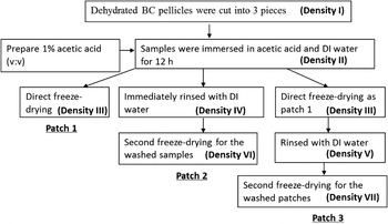
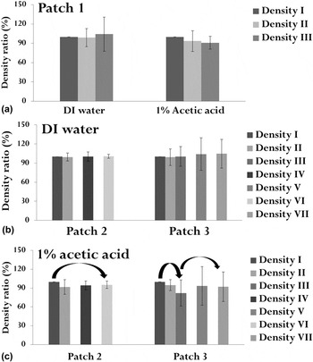
BC pellicle were prepared by 1% of acetic acid (v:v) following the procedure as shown in Fig. 1, where BC samples treated with DI water was used as the control. Purified BC pellicles were freeze-dried and ensured to be dehydrated prior to the first soaking treatment. One dehydrated pellicle was cut into three small pieces of 25.3 × 25.3 mm, labeled as Patches 1–3. BC Patches 1–3 were completely soaked in 1% acetic acid and DI water for 12 h at room temperature. The soaking duration of 12 h and temperature were determined as suggested by most of the analogous Refs. Reference Kato and Gehrke21–Reference Peng and Chen23. Furthermore, our preliminary experiment revealed that longer soaking treatment in the acetic acid solution could probably cause the material to partly degrade, and therefore the processing duration of 12 h was set in this study as the best performance time. BC Patch 1 was directly freeze-dried and the densities of Patch 1 including Densities I, II, III were evaluated based on the measurement of the initial dry weight of Patch 1 and thicknesses at dehydrated (I and III) and hydrated (II) states. To determine the impact of removal of acetic acid on the density of BC, BC Patch 2 was first rinsed by DI water to remove acetic acid prior to freeze-drying and then immediately freeze-dried. The densities of Patch 2 including Densities I, II, IV, VI were evaluated based on the measurement of initial dry weight of Patch 2 and various thicknesses at dehydrated state (I and VI) and hydrated state (II and IV). To investigate the occurrence of density variation of BC during freeze-drying operation or soaking treatment, BC Patch 3 was first freeze-dried after soaking treatment, and then rinsed by DI water followed by an immediate freeze-drying operation. The densities of Patch 3 including Densities I, II, III, V, VII were evaluated based on the measurement of initial dry weight of Patch 3 and various thicknesses at dehydrated states (I, III and VII) and hydrated states (II and V). Freeze-drying operation for all the samples was performed in Freezone 2.5 Freeze Dry system (Laboconco) at −50 °C and a vacuum below 0.133 mBar for 48 h.
Illustration of preparing BC patches treated with 1% acidic acid combined with freeze-drying.

D. Measurement of thicknesses of BC patches and density conversion
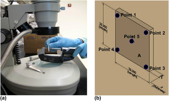
A digimatic point micrometer (Mitutoyo Series 342, Japan) was used to accurately measure the thicknesses (H) of either hydrated or dehydrated patches. Due to the easy-to-compress property of BC, the measurement was performed under an optical microscope (Vista Vision) to ensure adequate and slight contact between the sample surface and the point micrometer stylus [Fig. 2(a)], as well as to avoid the sample compression and the impact of water layer on the outer surface of the sample. Regarding the presence of specific roughness on the BC patch surface, the thickness was recorded as the average of 5 points located on one BC patch (4 at the corners and 1 in the center) as shown in Fig. 2(b). The density and the density variation ratio of one BC patch were calculated based on the average of thicknesses as shown in Eq. (1). Assuming that the original dry weight of one BC sample patch and its surface shape remained nearly unchanged during all the treatments, the material density would be only subjected to the thickness variation. Other than the direct measurement of material porosity, the material density was able to become another appearance parameter evaluating the porosity variation of material when its weight was controlled as a constant.
 $$\rho = {m \over {A \times H}}\quad {\rm{,}}$$
where ρ, A and H are the density, surface area and thickness of the BC patch, and m is the original dry weight of each dehydrated BC Patches 1, 2, 3.
 $${\rm{Density\ ratio}} = {{{\rho _{{\rm{II,III,IV,V,VI,VII}}}}} \over {{\rho _{\rm{I}}}}} \times 100\% \quad {\rm{,}}$$
where ρII,III,IV,V,VI,VII is the Densities II, III, IV, V, VI, VII at dehydrated or hydrated state labeled as shown in Fig. 1, and ρI is the original density of each BC Patches 1, 2, 3 at the dehydrated state labeled as Density I.
Accurate measurement of thickness of BC patch under (a) the optical microscopy and (b) by 5 points illustrated on the BC patch.

E. Measurement of porosity of BC patches
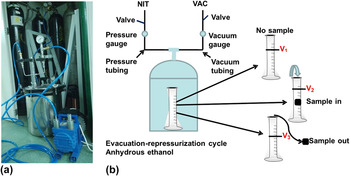
Porosity of BC patches was directly measured by a newly redesigned device [Fig. 3(a)] assembled based on the description provided in the Ref. Reference Nge, Nogi and Yano24. The determination of porosity was performed at a room temperature of around 25 °C in an evacuation–repressurization cycle, as illustrated in Fig. 3(b). The anhydrous ethanol was used as the displacement liquid because BC is highly hydrophilic, while it would not absorb the anhydrous ethanol. The procedure started from a device connection followed by a sample of BC patches immersed in a graduated cylinder containing a known volume of anhydrous ethanol (V 1). The graduated cylinder containing the sample and the anhydrous ethanol was placed in the device, and then the evacuation–repressurization cycle was applied repeatedly for 5 min to force the anhydrous ethanol through the pores of the sample. Due to the volatility of anhydrous ethanol, all operation steps should be carried out as fast as possible other than the strictly controlled evacuation–repressurization cycle of 5 min. Opening the device, the volume of anhydrous ethanol on the graduated cylinder was recorded as V 2, which is the sum of the original volume of anhydrous ethanol and the skeleton of the BC patch. As a result, the volume of the skeleton of the BC patch can be calculated as V 2 − V 1. Next, the sample was removed from the graduated cylinder and then the volume of anhydrous ethanol that remained was recorded as V 3. The void volume of the scaffold, which represents the total volume of the pores of the sample, can be calculated as V 1 − V 3. Thus, the porosity of the BC patch was calculated by Eq. (3).
 $${\rm{Porosity}} = {{({V_1} - {V_3})} \over {({V_2} - {V_1}) + ({V_1} - {V_3})}} = {{({V_1} - {V_3})} \over {({V_2} - {V_3})}}\quad {\rm{.}}$$
(a) The redesigned device of porosity measurement; (b) illustration of porosity measurement of BC patches using an evacuation–repressurization cycle and the anhydrous ethanol as the displacement liquid.

F. Examination of BC patches by SEM
Two samples of BC patches (Density III) modified by DI water and 1% acetic acid were imaged by SEM (HITACHI S-3500N, Japan) at 5 and 20 kV. The dehydrated samples were coated with gold using a vacuum sputter-golder to improve conductivity. The surface and cross section of each sample were both examined.
G. Mechanical properties of BC patches
Mechanical behavior containing tensile strength (stress, MPa) and elongation (strain, %) of freeze-dried BC patches (Density III) were measured by a dynamic mechanical analyzer (DMA Q800, TA Instruments, New Castle, DE) as described in our previous study. Reference Hu and Catchmark10 Young's modulus (GPa) was determined by the gradient in the approximately linear region of stress versus strain graph. Stress is calculated upon the terms of F/A, where F is the loading force and A is the area of the cross section of materials. Strain is calculated upon the terms of ΔL/L, where L is the initial length and ΔL is the difference between the lengths at break and the initial length. All the samples were cut into a size of 20 mm × 3–5 mm (length × width) and the thickness was measured using a caliper prior to loading the samples in DMA. Loading forces were changed at a rate of 0.2 N/min and the temperature was kept at 37 °C.
H. X-ray diffraction analysis of BC patches
To prepare BC samples for x-ray diffraction (XRD), the freeze-dried BC patches (Density III) were pressed by T-Rex system (TRX-1000-D) to make all samples of identical thickness. The compression processing was performed under 500 psi for 2 min, and the resulting samples then were cut to uniform sheets in the size of 2.5 × 2.5 cm. Next, the XRD patterns of the samples were collected on a PANalytical X'Pert Pro MPD θ/θ goniometer (Almelo) with Cu Kα radiation and fixed slit incidence (0.5° divergence, 1.0° anti-scatter, specimen length of 10 mm) and diffracted (0.5° anti-scatter, 0.02 mm nickel filter) optics. Scans were collected at 2° per minute from 5 to 45 degree. OriginPro 8.5.0 SR1 software was used to draw XRD patterns based on the raw data collection.
I. Cell culture of HFBCs
HFBCs, ATCC CCL110, were obtained from the American Type Culture Collection (ATCC). The complete growth medium of HFBCs was composed by Eagle's minimum essential medium (EMEM) (Mediatech Inc., Manassas, VA), 10% of fetal bovine serum (FBS) (ATCC, Manassas, VA), 1% of penicillin/streptomycin (ATCC, Manassas, VA). Starting from thawing the original cell vial, the cell suspension was immediately transferred into 9.0 mL of HFBCs growth medium. After spinning at approximately 125 × g relative centrifugal force (RCF) for 10 min and then discarding the supernatant, the cell pellet was resuspended in the growth medium and then dispensed into a 25 cm2 culture flask. Five milliliters of complete growth medium was added to activate the cells to grow at 37 °C and 5% CO2. When the confluency, used as a measure of the number of the cells by coverage of the flask, reached approximately 80%, the culture medium was removed and then the attached cells were rinsed using phosphate buffered saline of pH 7.4. One milliliter of trypsin–ethylene diamine tetraacetic acid (trypsin-EDTA) solution was added to detach cells at 37 °C. Next, four milliliters of fresh complete growth medium was subsequently added and the cell suspension was then mixed well following centrifugation at 125 × g RCF for 10 min. The resulting cell pellet with fresh medium was transferred to new culture flasks at a ratio of 1:3 to grow. After two-day incubation, the cell detachment and centrifugation were repeated as described above to make the initial cell suspension.
Samples of BC patches (Density III) modified by DI water and 1% acetic acid were autoclaved. Next, the sterile samples were placed to a 16-well nontreated culture plate and sterilized again overnight under UV light. The initial cell suspension (20,000 cells per cubic centimeter) was added directly to the surface of each sample. Prior to observation using confocal microscopy (Olympus Fluoview 300, Japan), samples were stained by two fluorescent dyes, fluorescein di-acetate (FDA) and propidium iodine (PI), prepared as described by Tawakoli et al. Reference Tawakoli, Al-Ahmad, Hoth-Hannig, Hannig and Hannig25 After two-day incubations, the culture medium was removed and the attached cells on the BC patch samples were rinsed with phosphate-buffered saline (PBS). A small drop of FDA as well as PI was added to the samples and the stained samples were then carefully moved to the glass slide under the confocal microscope. FDA indicating green color stained live cells, whereas PI indicating red color stained dead cells.
J. Statistical analysis
All the samples were examined at least three times. The significant differences between sample groups was evaluated using one-way ANOVA (least significant difference) (P < 0.05 confident interval; IBM SPSS Statistical Software; Release 19.0.0).
III. RESULTS AND DISCUSSION
A. Characterization of different surfaces of BC pellicle
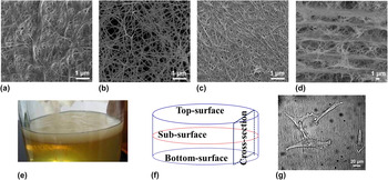
SEM images containing top surface, subsurface, bottom surface, and cross section of BC pellicle are shown in Fig. 4. It is clear that the densest surface was the top surface [Fig. 4(a)]. Less dense surface was the bottom surface [Fig. 4(c)]. The subsurface of BC pellicle exhibits quite a porous structure as compared with the top and bottom surfaces [Fig. 4(b)]. These dense and porous characters of different surfaces of the BC pellicle may be related to the oxygen diffusion impacting the BC production on the different surfaces during cultivation. Cross section of the BC pellicle presents a remarkable layer-by-layer structure with a layer interval size of ∼5 µm. In the light of the HFBC size of ∼100 µm [Fig. 4(g)], the top and bottom surfaces of the BC pellicle might disappoint the cell ingrowth as the pores on their surfaces are too small, while the subsurface and cross-section surface of the BC pellicle might be able to promote cell ingrowth. Given this analysis, if using the BC pellicle as a scaffold in which cells can grow inside, pretreatment to make a cross-section surface or obtain a subsurface by peeling off the top or bottom surface is required.
Porous characters of BC showing (a) top surface; (b) subsurface; (c) bottom surface; (d) cross-section surface; (e) harvested pellicle of BC; (f) schematic graph of different surfaces of BC; and (g) HFBCs under the optical microscopy.

B. Variation of density and porosity of BC patches
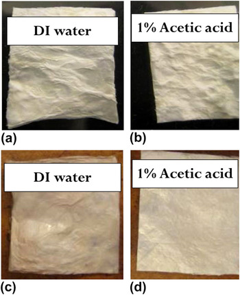
Figure 5 displays the comparison of densities among samples with or without the removal of acetic acid. Due to random growth of fibers inside the BC pellicle network, the same pellicle may be cut into several patches that would probably have different densities. To reduce this error, the density ratio parameter was used to compare densities among different BC patches. The density of the BC patches treated with 1% acetic acid exhibited a slight decrease for almost all samples regardless of the washing operation to remove acetic acid. It is true that no significant difference was found. However, the BC patch treated with acetic acid was indeed observed to show a smoother surface, as well as a visible variation of thickness [Fig. 6(d)]. It is assumed that no significant difference for sample densities might be a result from a small amount of acetic acid left on the samples increasing sample weight so that the density calculated in terms of Eqs. (1) and (2) failed to show an obvious variation. To investigate the effect of the removal of acetic acid on the density of BC patches, two washing operations before freeze-drying (Patch 2) and after freeze-drying (Patch 3) were applied. Due to the large error bars, there is no significant difference among data shown in Fig. 5(c). Regardless of the impact of errors, the density variation from Density I to Density VI of Patch 2 in Fig. 5(c) is smaller than the density variation from Density I to Density III of Patch 3 in Fig. 5(c) based on the average of densities. Meanwhile, the density variation from Density III to Density VII of Patch 3 in Fig. 5(c) shows an increasing trend also based on the average of densities. These variations are related to the impact of removing acetic acid on the density. In addition, the surface profile of the BC patch as shown in Fig. 6 visibly exhibits that the BC surface treated with 1% acetic acid becomes smoother than that treated with DI water, which demonstrates that the density variation should occur after soaking in 1% acetic acid. By combining this visible proof in Fig. 6 with the above analysis of density variations in Fig. 5(c), it is concluded that the density variation of BC patches treated with 1% acetic acid might occur during the process of freeze-drying rather than the treatment of soaking samples in acetic acid.
BC patches at dehydrate and hydrate states showing (a) densities of BC patch 1 soaking in DI water and 1% acetic acid without the removal of acetic acid; (b) various density comparisons between BC patch 2 and patch 3 treated with DI water; (c) various density comparisons between BC patch 2 and patch 3 treated with 1% acetic acid.

Photographs showing the surface profile of dehydrated BC patches before soaking in (a) DI water (Density I) and (b) 1% acetic acid (Density I) and after soaking in (c) DI water (Density III) and (d) 1% acetic acid (Density III).

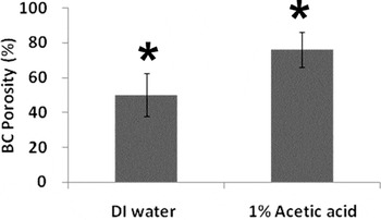
It is well known that the density variation of material generally accompanies the change of porosity if the original dry weight of the BC patch remains unchanged. Although the densities of BC patches as shown in Figs. 5(a)–5(c) did not significantly vary, the result from the direct measurement of BC sample porosity by using the evacuation–repressurization cycle exhibited a significant increase of porosity from 50.3 to 76.43% (P < 0.05, one-way ANOVA; IBM SPSS Statistical Software) as shown in Fig. 7. It can be explained that this improvement of material porosity caused by acetic acid treatment combining freeze-drying operation may be related to the high melting point of acetic acid and the excellent capability of holding water of BC. The formation of pores is subject to the quantity of ice crystals produced by water bound to material, where ice crystals are sublimed completely during freeze-drying and the volume occupied by ice crystals is left entirely to form pores. Reference Schoof, Apel, Heschel and Rau19,Reference Park, Venditti, Jameel and Pawlak26 The amount of water molecules bound to BC can be estimated as described by Peng et al. and Engelund et al. Reference Peng, Gardner and Han27,Reference Engelund, Thygesen, Svensson and Hill28 From Eq. (4), we may infer the number of layers of water molecules around the surface of cellulose fibers. Reference Park, Venditti, Jameel and Pawlak26
 $${\rm{Number\ of\ water\ layer}} = {{W_{\rm{c}} M_{\rm{m}} } \over {M_{{\rm{water}}} }} \times {1 \over S} \times {1 \over {(1 - C)}}\quad {\rm{,}}$$
where W c is the nonfreezing bound water content (0.28 g/g); M m and M water are the molecular weight of the repeating unit (162) and water (18); S is the maximum bonding sites in a monomer (3), and C is the crystallinity of fibers (taken as 44%). One or two layers of water molecules around the cellulose fiber could be estimated from Eq. (4). Moreover, due to higher crystallinity of BC than ordinary plant cellulose and its excellent capability of holding water, C is assumed to be higher and the number of water layers should be greater than 2, suggesting that more water could be bound to BC. On the other hand, acetic acid is a conventional organic acid having a melting point of 16.5 °C. At a low temperature, acetic acid molecules can bind more water molecules through hydrogen bonding and are quickly frozen to ice crystals. Reference Takamuku, Kyoshoin, Noguchi, Kusano and Yamaguchi29 The acetic acid treatment exerted on BC followed by freeze-drying operation thus resulted in more ice crystals, and meanwhile, much void volume was then present when ice crystals were sublimated subsequently. As a result, the porosity of BC was improved.
Porosity variation of BC patches treated with DI water and 1% acetic acid using the evacuation–repressurization cycle, where anhydrous ethanol was used as the displacement liquid. The columns labeled by asterisk (*) indicate that two numerical column data have significant differences (P < 0.05).

C. Analysis of SEM images of BC patches
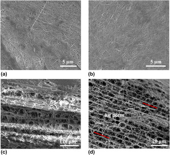
SEM images of the surface and cross section of BC patches (Density III) treated with DI water and 1% acetic acid are shown in Figs. 8(a)–8(d). The porous character on both BC patch surfaces treated with DI water [Figs. 8(a)] and 1% acetic acid [Fig. 8(b)] fails to exhibit a significant difference. Obviously such a physical modification of simple soaking treatment combining freeze-drying operation could hardly improve the porous profile on denser top or bottom surface of BC. However, a significant difference was observed on both cross sections of BC patches treated with DI water [Fig. 8(c)] and 1% acetic acid [Fig. 8(d)]. As shown in Figs. 4(b), 4(d) and 8(c), the inner structure of the BC patch treated with DI water is really porous, while the porous layer-by-layer arrangement on the cross-section surface is not as orderly as the BC patch treated with 1% acetic acid [Fig. 8(d)], and some big pores with approximately 20 μm are only present in Fig. 8(d). These orderly porous structure and big pores would be expected to guide cell ingrowth. It is assumed that this inner porous structure may have resulted from the sublimation of many small ice crystals formed from acetic acid during freeze-drying, which leads to an increase in thickness of the BC patch. The mechanism behind the new porous character of cross section of the BC patch (Density III) treated with acetic acid is consistent with the discussion in the above section. Having a high melting point, acetic acid can bind more water around and inside the BC patch to form more ice crystals during the process of freeze-drying, and thus the porous profile was visibly improved.
SEM images of freeze-dried BC patches (Density III) showing (a) top surface and (b) cross section of the sample treated with DI water; (c) top surface and (d) cross section of the sample treated with 1% acetic acid.

D. Mechanical behavior and XRD analysis of BC patches
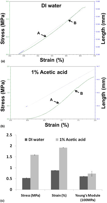
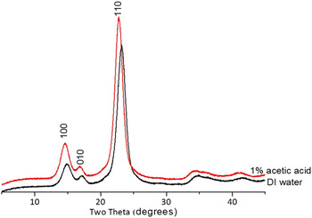
Results from mechanical behavior analysis containing strength (stress at break), elongation (strain at break) and Young's module of BC patches are shown in Fig. 9. BC patches modified with 1% acetic acid present a significant improvement (P < 0.05) for all three mechanical behavior parameters, including stress, strain and Young's module. Both stress and strain of BC patches modified with 1% acetic acid were increased to approximately 300%, while the Young's module (stiffness) had a small significant increase (P < 0.05) as compared with unmodified BC patches [Fig. 9(c)]. This variation probably resulted from the improvement of porosity of the BC patches modified with 1% acetic acid. Although densities between two kinds of BC patches appear to remain unchanged before and after modification, the variation of porosity tells that BC patches modified with 1% acetic acid appear to exhibit a tidier assembly of BC ribbons on the surface and cross section [Figs. 8(b) and 8(d)]. Meanwhile, Fig. 6(d) shows a smoother surface and thicker BC patch after 1% acetic acid modification, and the SEM image in Fig. 8(d) also shows a more orderly inner structure. These proofs demonstrate that BC patches with 1% acetic acid modification might require more force to be broken at a relatively higher elongation, while their stiffness only exhibited a small increase [Figs. 9(a)–9(b)], as the porosity presented a small improvement. The XRD patterns of BC patches as shown in Fig. 10 appear to remain consistent with the mechanical behavior analysis of the BC patches. BC patches modified with or without 1% acetic acid exhibit three typical peaks at roughly 14.5° (100), 16.6° (010), and 22.7° (110) two theta. Reference Gu and Catchmark30 However, it is clear that a higher and wider peak at 22.7° (110) two theta is shown in the XRD pattern of the BC patch modified with 1% acetic acid, suggesting that higher crystalline structure has been achieved and therefore leads to the enhancement of mechanical behavior of BC patches modified with 1% acetic acid.
Mechanical behaviors as shown in stress versus strain graph of BC patches modified by (a) DI water; (b) 1% acetic acid; and (c) comparison of mechanical behaviors containing stress, strain and Young's module of BC patches.

XRD patterns of BC patches modified with DI water and with 1% acetic acid.

These advantages on the mechanical behavior and crystallinity of BC modified by 1% acetic acid were results from the improvement of material porosity in a particular extent. As mentioned in Fig. 7, a small significant enhancement in porosity from 50.3 to 76.43% on average was present, which suggests that such an improvement of material porosity is a benefit for obtaining preferable characteristics of BC. Although higher enhancement of material porosity could result in more porous and compressible BC, we think that too much higher enhancement of porosity might be responsible for the decline of mechanical properties, the imperfection of material structure, and the inadequate surface area offered to cells to attach.
E. Cell culture of HFBCs on BC patches
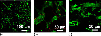
In Fig. 11, it is shown that HFBCs were able to attach and proliferate on BC patches treated with either DI water [Fig. 11(b)] or 1% acetic acid [Fig. 11(c)]. Much better cell viability and density present on tissue culture plate was due to the coating containing cell attachment protein on the surface of the culture plate, as well as relatively flat surface area as compared with the BC surface. The significant differences of cell confluency (viable cell density on a surface) and cell viability between two kinds of samples can be identified in Figs. 11(b)–11(c). From direct observation, it is true that we could hardly find dead cells (red) in Fig. 11(c), while dead cells are present in Fig. 11(b). As we have known, the growth of animal cells on the material is actually quite complex, as various materials present different surface characters, as well as hydrophilic or hydrophobic properties that would affect the attachment and proliferation of cells. The treatment with 1% acetic acid clearly led to the variation of porous profile on the surface of the BC patch as shown in Fig. 6(d). Such a modified surface might provide HFBCs with a flatter substrate to attach, where cell confluency and viability on it were improved. In addition, the orderly porous structure and big pores on the cross section of BC patch treated with 1% acetic acid [Fig. 8(d)] might be also a benefit for guiding cell ingrowth. Further study on a three-dimensional (3D) cell culture is needed to provide more evidence of cell ingrowth on BC treated with 1% acetic acid combining freeze-drying operation.
Cell viability of HFBCs after two-day culture on (a) tissue culture plate; BC patches (Density III) treated with (b) DI water and (c) 1% acetic acid, under the confocal microscope (FDA-PI stain: FDA – live cells and PI – dead cells).

IV. CONCLUSIONS
Acetic acid treatment combining freeze-drying operation was used to improve porous profile of BC including porosity and pore morphology. SEM images of cross section of BC sample treated with 1% acetic acid exhibited more porous layer-by-layer structures, and the number of big pores along the cross section appeared to greatly increase as well. The improvement in porous profile of BC treated with 1% acetic acid could have led to the improvement of porous profile, as well as the enhancement of mechanical properties and crystallinity of BC. The mechanism behind the porosity improvement is described such that acetic acid can bind more water molecules due to the hydrogen bonding between acetic acid and water, and at the same time, BC displays excellent capability of holding water. Furthermore, acetic acid has a high melting point capable of quickly forming more ice crystals with bound water molecules when samples are frozen before freeze-drying. Thus, the sublimation of ice crystals during the process of freeze-drying causes better porous property in BC. Cell culture of HFBCs demonstrated better cell viability on BC treated with 1% acetic acid, where HFBCs preferred to attach and proliferate. This may suggest that the improvement of the porous profile of BC helps to facilitate cell growth on the material, as well as, further induce cells growing inside the BC material.
ACKNOWLEDGMENTS
This work was supported by the Pennsylvania State University Material Research Institute Nano Fabrication Network and the National Science Foundation Cooperative agreement no. 0335765, National Nanotechnology Infrastructure Network, with Cornell University; the National Natural Science Foundation of China, no. 31370978; the Guangdong Natural Science Foundation no. S2013040014248; the Development of Strategic Emerging Industries of the Shenzhen Basic Research project no. JCYJ20130402113127522, no. JCYJ20120831180118537; the Shenzhen Peacock program no. 110811003586331.