INTRODUCTION
Schistosomiasis is a major cause of morbidity especially in sub-Saharan Africa (Chitsulo et al. 2000). The antibody response in schistosomiasis is largely directed against glycans (Cummings and Nyame, 1999; Nyame et al. 2003) produced by the intravascular schistosome worm pairs and by schistosome eggs trapped in host tissues. Both parasite specific and cross-reacting antibodies can be seen in the host, and serology-based diagnostics is useful especially for travellers with early and/or light infection. Parasite-derived glycans are immunogenic and serum antibodies against excretory/secretory products of the schistosome gut epithelium are demonstrable in infected individuals. Other immunogenic glycans, cross-reacting with keyhole limpet haemocyanin (KLH), appear to be excreted via a poorly characterized tubular system (Thors and Linder, 2003). Our knowledge of schistosome glycans is rapidly increasing and several immunogenic glycans, including an immunoregulatory carbohydrate structure, Lewis x (Lex) have been identified (Nyame et al. 2003).
In our studies on the serodiagnostic potential of antibodies to cross-reacting carbohydrate determinants of KLH in schistosomiasis (Thors and Linder, 1998, 2003) we observed that sera from schistosomiasis patients react with carcinomas (unpublished observation). In order to confirm this preliminary finding, we tested sera from experimentally infected mice, before and after infection, for reactivity with a panel of bladder carcinomas. The results of these experiments motivated further exploration of the basis for the putative cross-reactivity between parasites and carcinomas. We therefore decided to test whether monoclonal antibodies against carcinoma antigens would react with schistosomes.
MATERIALS AND METHODS
Immunohistochemistry
For immunohistochemical studies 8-week-old NMRI mice were infected percutaneously with approximately 100 S. mansoni cercariae (Puerto Rican strain) as described previously (Thors and Linder, 1998): At 8–11 weeks post-infection, the mice were killed, intravascular worms recovered by perfusion with RPMI medium and livers were removed for immunohistological staining. Serum specimens were collected and pooled from mice infected for more than 8 weeks. Frozen sections of adult schistosomes and egg granuloma-containing liver tissues were mounted on microscope slides and fixed in acetone at room temperature for 10 min. Paraffin sections of formaldehyde-fixed material were also used for antigen analysis.
Indirect immunofluorescent staining of frozen sections of S. mansoni adult worms and eggs was performed essentially as described previously (Thors and Linder, 1998, 2003). Several monoclonal antibodies against carcinoma-associated antigens were used: mouse anti-Tn (IgG1), recognizing the immunodominant epitope terminal alpha-N-acetylgalactosamine, O-glycosidically linked to protein (GalNAc-alpha-O-Ser/Thr), mouse anti-MUC1 (polymorphic epithelial mucin) (Novocastra Laboratories Ltd), human anti-TF (IgM) (anti-TF1 and anti-TF5) reacting with the antigenic epitope Gal(beta1-3)GalNAc-O-Ser/Thr (Dahlenborg et al. 1997). Anti-immunoglobulin (Ig) conjugates used were goat anti-mouse Ig-FITC (Sigma F-1010) and sheep anti-human Ig-FITC (National Bacteriological Laboratory, Sweden).
Control staining with monoclonal antibodies against the Lewis x (Lex) antigen was performed with anti-human granulocyte associated antigen CD15 (Dako M-0733) (van Dam et al. 1996; Jacobs, Deelder and Van Marck, 1999; Haslam et al. 2000; Wuhrer et al. 2000; Eberl et al. 2001; Huang, Tsai and Khoo, 2001). Human gastric adenocarcinoma cell line AGS (Barranco et al. 1983) was used as control for anti-TF reactivity. The AGS cell line was purchased from American Type Culture Collection (Manassas, VA), and was maintained in RPMI 1640 medium, supplemented with 10% fetal bovine serum, 2 mM L-glutamine, 10 mM HEPES, and 100 IU/ml penicillin and 100 μg/ml streptomycin.
Carcinoma tissue material for indirect immunofluoresence stainings was obtained from a multitissue block with tissue cylinders from urothelial carcinomas from 15 individuals and from 1 urothelial ‘squamous cell’ or ‘keratinizing’ carcinoma of the urinary bladder. This carcinoma was removed by radical cystectomy and was a moderately differentiated (grade II) keratinizing urothelial cell carcinoma that had spread to extra-vesicular tissue representing stage pT4. There was no urothelial component in this tumour. The 15 carcinomas in the multitissue block with 4 mm diameter cylinders were all urothelial malignancies representing low- and high-grade carcinomas. For light microscopic and immunofluorescence studies, the tissues were fixed for a minimum of 24 h in buffered 4% formaldehyde, embedded in paraffin, sectioned and stained with haematoxylin-eosin. The tumours were graded according to the WHO Classification (Eble et al. 2004) and the staging was based on the UICC TNM Classification (Sobin and Wittekind, 2002).
Microscopy was performed using a Leica DMRB fluorescence microscope equipped with filters for FITC fluorescence essentially as described previously (Linder, Thors and Lundin, 1991). For digital photography we used an AxioCam camera (Carl Zeiss, Oberkochen, Germany), Openlab (Improvision, Coventry, UK) and Photoshop 6 (Adobe, San Jose, CA) software for Apple Macintosh G4 computer.
Immunoassay
Anti-TF antibodies were assayed by ELISA using AGP (1 μg/ml) (Sigma A-9791) as antigen, essentially as described (Dahlenborg et al. 1997). Native glycophorin (GP) can be digested by sialidase to express the TF epitope. The resulting AGP was used as antigen to detect anti-TF antibodies (Dahlenborg et al. 1997). Sera from 10 mice were collected at the time of infection with S. mansoni cercariae and subsequently at weekly intervals up to 11 weeks, starting 4 weeks after infection. All sera were tested in ELISA at 1[ratio ]200 dilution. For control purposes, we determined antibodies in serum against GP (Sigma G-7903) (negative control) and keyhole limpet haemocyanin (KLH) (Sigma H-7017) (positive control). The relative anti-TF antibody levels are expressed as arbitrary units (U) using the human monoclonal anti-TF5 antibody as reference (OD value of 2·2. corresponding to 100 U).
RESULTS
Carcinoma seroreactivity associated with schistosomiasis
Pooled sera from S. mansoni-infected mice, but not pre-infection sera, reacted with urothelial carcinomas (Fig. 1).

Fig. 1. Immunohistological staining of paraffin sections of urothelial carcinomas of human bladder with pooled sera from Schistosoma mansoni-infected mice (A, D, G), but not with pooled sera from uninfected mice (B, E, H). The histological appearance of corresponding areas stained with haematoxylin-eosin is seen in C, F and I. Original magnification 400×.
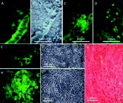
The serum pool from S. mansoni-infected mice reacted with both transitional cell and urothelial cell carcinoma tissues. The reaction was cytoplasmic and occurred in both organized epithelial structures and individual scattered cells. In some tumours a marked variation in staining intensity was seen. The staining reaction observed with the mouse serum pool was similar to that of anti-TF monoclonal antibodies. Anti-KLH antibodies, in contrast, gave a more widespread reaction (Fig. 2). Normal urothelium did not react with the serum pool from S. mansoni-infected mice or anti-TF antibodies (not shown).

Fig. 2. Reactivity of pooled sera from Schistosoma mansoni-infected mice with paraffin sections of human urothelial carcinomas (A–D) and a urothelial squamous cell carcinoma (E–I) of the urinary bladder. Immunofluorescent localization corresponds to the cytoplasm of carcinoma cells in the different tumours, both areas of organized epithelial structures and individual cells surrounded by unstained stromal tissue (D). The immunofluorescent staining in (A) corresponds to phase-contrast microscopy in (B). The reaction pattern was similar to that of anti-TF monoclonal antibodies (C). In the urothelial cell keratinizing type carcinoma, clusters of cells reacted with the mouse serum pool. The localization (E) was more restricted than that of anti-KLH (H). Phase-contrast micrographs in (F) and (I) correspond to (E) and (H) respectively. The haematoxylin-eosin stained section (G) of the corresponding area shows well-differentiated keratinizing type of urothelial cell carcinoma.
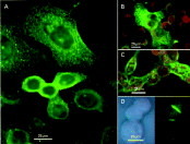
The serum pool from S. mansoni-infected mice reacted with AGS carcinoma cell culture giving a cytoplasmic and granular staining (Fig. 3A). Antibodies against TF reacted similarly with intracytoplasmic granules (Fig. 3B) and the periphery of cells (Fig. 3C), whereas anti-CD15 antibodies gave a different pattern of reactivity, being more intense in areas of cell-cell contact (Fig. 3D).

Fig. 3. Gastric carcinoma cell line AGS stained by immunofluorescence with serum from Schistosoma mansoni infected mice (A), anti-TF5 monoclonal antibodies against the Thomsen-Friedenreich (TF) epitope (B, C) and monoclonal anti-CD15 (Lewis x) (D).
Reactivity of monoclonal antibodies against carcinoma-associated antigens with S. mansoni
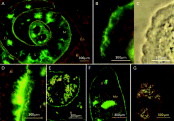
The results of immunofluorescence (IF) staining of S. mansoni adult worms and eggs in experimentally infected mice, using monoclonal antibodies against the described tumour-associated antigens, showed distinct staining of the adult worms with anti-TF antibodies (Fig. 4). The localization (surface syncytia, ductal epithelia and the intra- and perioval staining) suggests that the TF determinant is expressed on secretory/excretory products of the parasite. The lumpy staining of cells in the periphery of granulomas (Fig. 5) corresponds to the localization of Kupffer cells. In higher magnification the target antigen was seen in and outside the autofluorescent egg-shell. No staining of normal liver tissue could be seen. Among the tested antibodies against carbohydrate epitopes, only those antibodies recognizing TF reacted.

Fig. 4. Localization by immunofluorescence of the Thomsen-Friedenreich (TF) epitope in Schistosoma mansoni worm pairs. Excretory ducts (D) of male (M) and female (F) worms (A), surface syncytium (B, C). Staining of material in the gynecophoric channel between adjacent male and female worm (D). Surface spines seen in phase contrast (S). Vitelline cells (Vi) of female worm are stained (E) but partly obscured by autofluorescent vitelline granules demonstrated in an unstained preparation (G). Staining of excretory ducts (D), Mehlis gland (Me) by anti-TF antibodies (F). No staining of TF antigen in the gut (G) (A and E). B, blood; En, endothelium.

Fig. 5. Localization by immunofluorescence of the Thomsen-Friedenreich (TF) epitope in Schistosoma mansoni egg in the liver of infected mouse. Amorphous intraoval staining, staining of the egg-shell and -surface (insert) and staining of Kupffer cells in the periphery of the perioval granuloma is seen.
Anti-TF-antibodies in S. mansoni-infected mouse sera
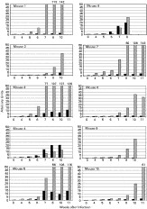
The reactivity in sera from experimentally S. mansoni-infected mice against AGP and KLH was demonstrated in ELISA (Fig. 6). Anti-KLH antibodies were used as a marker for infection. After 6 weeks of infection an increased serum anti-TF antibody titre was seen in 4 out of 10 animals. When GP was used as antigen, no reaction was seen.

Fig. 6. Serum anti-TF antibodies in mice experimentally infected with Schistosoma mansoni detected by ELISA using asialoglycophorin (AGP) as antigen. Anti-KLH antibodies (grey) were used as control for infection.
DISCUSSION
The results show that the TF epitope is shared by schistosomes and carcinomas and that this epitope is immunogenic in S. mansoni-infected mice. The TF antigen being present in bladder carcinomas (Baldus et al. 1992; Langkilde et al. 1992) was confirmed in our study. As seen previously (Thors and Linder, 1998) and here in this study, antibodies cross-reacting with KLH develop during schistosome infections. Even if the TF epitope is present in KLH, anti-KLH antibodies in schistosomiasis appear to be directed mainly towards antigenic epitopes distinct from TF, the major one being identified as Fuc(alpha1-->3)GalNAc- (Kantelhardt et al. 2002). It is not known, however, if this epitope is responsible for the previously demonstrated reactivity of anti-KLH antibodies with tumours (Harris and Markl, 1999). The relative importance of the anti-TF antibody specificity in explaining the observed carcinoma-reactivity of sera from mice with schistosomiasis remains to be studied.
TF antigen in primary and metastatic cancers as a pancarcinoma-associated antigen has attracted scientific interest for more than 65 years (Hanisch and Baldus, 1997). In mature mammalian tissues the TF epitope is masked by terminal sialic acid. It is an oncofetal antigen, which is expressed in fetal mucosa, but concealed by further glycosylation during normal development (Campbell et al. 1995; Ragupathi, 1996). Several studies have described its expression in relation to tumour grade, metastasis and likelihood of relapse or tumour aggression (MacLean and Longenecker, 1991). The results showing the presence of the Gal(beta1-3) GalNAc-O- epitope in schistosomes is consistent with the observed common feature, absence of sialic acid, in both helminth glycans and oncofetal antigens (Yu et al. 1997; Cummings and Nyame, 1999).
Schistosome glycans inducing an anti-KLH response appear to be excretory/secretory products produced by the different life-stages of the parasite (Thors and Linder, 2003). The TF epitope seems to be one of several such cross-reacting glycotopes present in both molluscs and in schistosomes, not surprisingly in both S. mansoni and S. haematobium (Kantelhardt et al. 2002). The widespread distribution of the TF-epitope in the tissues of the schistosome makes it potentially involved in several aspects of the host-parasite interactions. They are likely to play a role in granuloma formation around schistosome eggs in host tissues, as was seen in the egg-shell. At the surface of the intravascular parasite the TF epitope might be involved in protecting vital tegumental structures and, as an excreted product, it may occupy the host immune system in various ways, e.g. by adhering to host cells. By binding to asialoglycoprotein receptors, shown to preferentially recognize GalNAc- and galactose, present at the surface of both Kupffer cells and hepatocytes (Fadden, Holt and Drickamer, 2003), the TF-epitope may interfere with the functions of these host cells in various ways. An anti-TF antibody response might also have effects on parasite fecundity, a possible consequence of an immune response to glycans expressed in the gynecophoric channel and reproductive organs of female and male schistosomes as shown in Fig. 4, even if it appears more likely that this type of immunity requires release of parasite components such as GP50 (Linder et al. 1991) following chemotherapy as suggested (Polman et al. 2002).
Parasite glycans, on the other hand, appear to be similar to the TF-expressing antigens shed by tumour cells both in vitro and in vivo (Samuel et al. 1990). Carbohydrates seem to be involved in the progression, dissemination and invasion of cancer cells (Sell, 1990; Springer, 1997; Barchi, 2000). Thus parasite and tumour glycans may compete for carbohydrate receptors e.g. on host vascular endothelial cells and hepatocytes and an anti-schistosome immune response may interfere with the carcinogenetic process and with tumour cell binding to hepatocytes, a TF-mediated interaction leading to liver metastases (Shigeoka et al. 1999).
Our observations may be of some relevance to the well-known association between urinary schistosomiasis and carcinomas (WHO, 1994; Mostafa et al. 1999). It has been proposed that schistosomiasis could supply the proliferative stimulus necessary to accelerate cancer growth from latent tumour foci (Hicks, James and Webbe, 1980). In fact it has been shown that the parasite can induce epithelial proliferation (Cheever et al. 1988) and that anti-TF antibodies may have a proliferative effect on cells expressing the TF antigen (Yu et al. 1997). Considering the complexity of host-parasite interactions in schistosomiasis, we cannot exclude the possibility that schistosomiasis, under some circumstances, may have a beneficial effect based on the observed immunotherapeutic potential of the TF antigen (Cao et al. 1996; Dahlenborg et al. 1997; Zhang et al. 1998) and the therapeutic local effects of intravesical instillation with KLH in bladder carcinoma patients (Jurincic-Winkler et al. 1996; Harris and Markl, 1999).