Introduction
Tetralogy of Fallot is the most prevalent form of cyanotic congenital heart disease.Reference Apitz, Webb and Redington1 Significant advances in early management of tetralogy of Fallot have remarkably improved the survival of patients with tetralogy of Fallot,Reference Valente, Gauvreau and Assent2 with current 30-year survival rates reaching 89%.Reference Nollert, Fischlein, Bouterwek, Bohmer, Klinner and Reichart3 However, late ventricular dysfunction related to residual haemodynamic and electrophysiological abnormalities remains a problem, contributing to increasing morbidity and mortality rates beginning in the fourth decade of life.Reference Valente, Gauvreau and Assent2,Reference Babu-Narayam, Kilner and Li4–Reference Chiu, Wang and Chen6
The importance of left ventricular dysfunction as a risk factor for long-term adverse outcomes is increasingly acknowledged.Reference Davlouros, Kilner and Harming7–Reference Diller, Kempny and Liodakis10 Recently, Diller et al. showed that left ventricular longitudinal dysfunction, such as impairment in global longitudinal strain, was associated with an increased risk of sudden cardiac death and/or life-threatening ventricular arrhythmias in patients with repaired tetralogy of Fallot.Reference Diller, Kempny and Liodakis10 Therefore, the assessment of left ventricular function in patients with tetralogy of Fallot is crucial because it is the strongest determinant of a poor clinical outcome in these patients.
Layer-specific strain analysis is a recent development, which allows separate quantification of deformation in the endocardial, midmyocardial, and epicardial layers of the myocardium.Reference Yamada, Takahashi and Kobayashi11 This new sensitive indicator has been reported to be highly effective in detecting cardiac dysfunction in various cardiac diseases.Reference Yamada, Takahashi and Kobayashi11–Reference Becker, Ocklenburg and Altiok14
Exercise stress echocardiography enables the detection of subclinical cardiac dysfunction that is not obvious at rest. Mese et al. showed significant changes in global longitudinal strain during exercise in patients with repaired tetralogy of Fallot.Reference Mese, Guven, Yilmazer, Demirol, Coban and Karadeniz15 However, to the best of our knowledge, no previous study has assessed layer-specific deformation of the left ventricle during exercise in patients with repaired tetralogy of Fallot. Thus, our aim was to evaluate layer-specific strains and torsion parameters during exercise in order to investigate the characteristics of cardiac dysfunction in patients with repaired tetralogy of Fallot and to detect subclinical left ventricular dysfunction.
Materials and methods
Study population
We prospectively recruited 15 patients (age 13–39 years) with repaired tetralogy of Fallot and scheduled for elective outpatient echocardiography at the Juntendo University. The exclusion criteria were age <10 years, <140 cm in height, inability to cycle, significant intellectual disability, and poor acoustic windows.Reference Ait-Ali, Siciliano and Passino16 Patients with residual intracardiac shunts, known ventricular arrhythmias, and implanted pacemakers were also excluded. Left ventricular strain analysis at rest and during exercise was performed in all patients. A group of healthy subjects was recruited as controls for left ventricular myocardial mechanical analysis at rest and during exercise. Controls were healthy volunteers who had no history of cardiovascular disease, showed normal blood pressure and sinus rhythm on electrocardiography, and had normal findings on echocardiography. All participants or their guardians provided informed consent as established by the Research Ethics Board at Juntendo University.
Exercise echocardiography
Exercise echocardiography was conducted on a supine bicycle (Lode BV, the Netherlands). All participants pedalled the bicycle, initially with minimal resistance at rates that caused a slow and steady increase in heart rate without excessive chest movement or fatigue. The pedalling rate was kept constant at 70 rpm for all subjects. Resistance was gradually added until the heart rate reached a maximum of 100 bpm (i.e. submaximal exercise to maximise frame rates) within 5 minutes.Reference Tan, Wenzelburger, Sanderson and Leyva17 Heart rate, symptom status, brachial blood pressure, and heart rhythm were monitored continuously during the exercise. Exercise echocardiography images were recorded within 1 to 5 minutes after reaching 100 bpm. Therefore, the total time on the bicycle was 25 minutes. After 10 minutes of rest and before the exercise, the baseline heart rate and resting echocardiography images were recorded.
Echocardiography
Echocardiography was performed using a Vivid E9 ultrasound system (GE Healthcare, Milwaukee, WI, USA) with an M5S or 6S probe, as appropriate for patient size. Images were optimised for gain, compression, depth, and sector width, and acquired at frame rates of 70–125 frames/second. Apical four- and two-chamber views and parasternal short-axis views at the basal, papillary, and apical ventricular levels were acquired at rest and during exercise. In each plane, images from three consecutive cardiac cycles were acquired during a breath hold at end expiration, if possible.
Mitral inflow E-wave, A-wave, and E/A ratio were measured at rest and during exercise. The degree of pulmonary regurgitation was assessed by measuring the jet size using colour Doppler, jet density, and deceleration rate with continuous wave, and pulmonary systolic flow compared with systemic flow using pulse-wave Doppler.Reference Zoghbi, Enriquez-Sarano and Foster18 The pressure gradient at the right ventricular outflow tract was estimated by the peak continuous-wave Doppler velocity of the right ventricular outflow tract. Right ventricular end-diastolic area and right ventricular end-systolic area were measured in the apical four-chamber view by tracing the endocardial border of the right ventricle and the tricuspid annular plane.Reference Lang, Badano and Mor-Avi19 Right ventricular fractional area change was calculated as (right ventricular end-diastolic area minus right ventricular end-systolic area)/right ventricular end-diastolic area, which was correlated with right ventricular ejection fraction by cardiac magnetic resonance.Reference D’Anna, Caputi and Natali20 Left ventricular end-diastolic volume and left ventricular end-systolic volume were calculated from the apical four-chamber and two-chamber views using the modified Simpson rule. Left ventricular ejection fraction was calculated as (left ventricular end-diastolic volume minus left ventricular end-systolic volume)/left ventricular end-diastolic volume. Left ventricular diastolic function was quantified using the ratio between the E-wave velocity of the pulsed-wave Doppler mitral flow and early diastolic velocity of the septum and left ventricular free wall at the mitral annulus level (e’ wave) on tissue Doppler imaging. These measurements were taken at rest and during exercise.
Left ventricular deformation analysis
Strain was measured using two-dimensional speckle-tracking echocardiography. The left ventricular endocardium was manually traced, and the region of interest was manually adjusted to the thickness of the left ventricular wall. The software tracks myocardial motion through the cardiac cycle and calculates strain from the echogenic speckles in the B-mode image. From the basal, papillary, and apical short-axis views and the apical four-chamber view, one cardiac cycle was selected for subsequent analysis of the circumferential and longitudinal strains. The system used for this study allows calculation of mean strain values for the total wall thickness and for three separate myocardial layers (endocardium, midmyocardium, and epicardium), as described previously (Figure S1 and S2).Reference Leitman, Lysyansky and Sidenko21 Left ventricular torsion and untwisting rate were analysed, as described previously.Reference Takahashi, Naami, Thompson, Inage, Mackie and Smallhorn22 All strain and torsion parameters were measured at least three times, and the average of these values was used as strain and torsion values. Analysis was performed offline using a commercially available software package (EchoPAC 113 1.0; GE Vingmed Ultrasound AS, Horton, Norway). Strain analysis was performed by two observers (K.Y. and M.Y.) who were unaware of the clinical data.
Statistical Analysis
Normally distributed continuous variables are expressed as mean ± standard deviation; non-normally distributed variables, as median (interquartile range). Independent student t-test or Wilcoxon test was used as appropriate to compare the differences in continuous variables between patients with repaired tetralogy of Fallot and controls for baseline characteristics. Between groups at rest or during exercise and within each group at rest and during exercise differences were assessed using a one-factor analysis of variance with a post hoc comparison using the Bonferroni correction for data with normal distribution or the Steel-Dwass test for data with non-normal distribution in each group. Statistical analyses were performed using JMP software (version 14.2.0; SAS Institute Inc., Cary, NC, USA). A p value of < 0.05 was considered statistically significant.
Intra- and interobserver agreements for the left ventricular layer-specific strain and torsion were calculated using the Bland-Altman approach, including the calculation of mean bias (average difference between measurements), and the lower and upper limits of agreement (95% limits of agreement of mean bias) in 10 randomly selected patients and controls at more than 6 months apart. The coefficient of variation was also determined (i.e. the standard deviation of the difference in paired samples divided by the average of the paired samples). Important differences were not observed in the variability scores of all parameters at rest and during exercise (Table S1).
Results
Study population
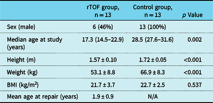
Two patients were excluded because of poor acoustic windows. Therefore, 13 patients with repaired tetralogy of Fallot and 13 healthy volunteers, used as controls, were enrolled from October 2014 to November 2017. Demographic data are summarised in Table 1. All patients had sinus rhythm and were classified as having New York Heart Association functional class 1 with no clinical symptoms. Seven patients (53.8%) had transannular patch. One patient (7.7%) had undergone the Rastelli procedure. Information regarding the surgical procedure was unavailable for one patient. The mean right ventricular outflow pressure gradient was 22.2 ± 11.8 mmHg. The degree of pulmonary regurgitation in six patients (46.2%) was moderate or severe.
Baseline characteristics

rTOF, repaired tetralogy of Fallot; BMI, body mass index; N/A, not applicable.
Data are expressed as mean ± standard deviation, or median (interquartile range).
Haemodynamic and left ventricular and right ventricular functional responses to exercise
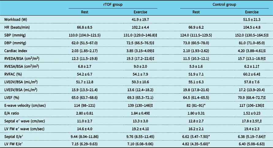
All the patients successfully completed exercise stress echocardiography without adverse events. The control group showed greater increase in left ventricular ejection fraction and right ventricular fractional area change during exercise, while the repaired tetralogy of Fallot group had a lower response. However, no significant differences were detected between patients and controls for left ventricular ejection fraction and right ventricular fractional area change both at rest and during exercise (Table 2).
Change in echocardiographic indices from rest to exercise in the repaired tetralogy of Fallot and control groups

* Patients with repaired tetralogy of Fallot versus controls at rest, p < 0.05.
† Patients with repaired tetralogy of Fallot versus controls during exercise, p < 0.05.
‡ Significant difference between rest and exercise, p < 0.05.
rTOF, repaired tetralogy of Fallot; HR, heart rate; SBP, systolic blood pressure; DBP, diastolic blood pressure; RVEDA, right ventricular end-diastolic area; BSA, body surface area; RVESA, right ventricular end-systolic area; RVFAC, right ventricular fractional area change; LVEDV, left ventricular end-diastolic volume; LVESV, left ventricular end-systolic volume; LVEF, left ventricular ejection fraction; FW, free wall.
Data are expressed as mean ± standard deviation, or median (interquartile range).
Left ventricular deformation during exercise
Papillary circumferential strain
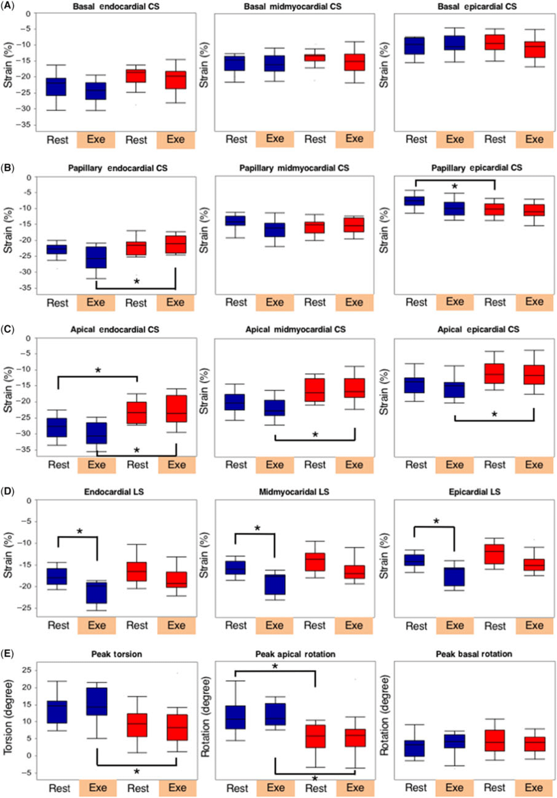
The peak endocardial papillary circumferential strain was significantly lower in the repaired tetralogy of Fallot group than in the control group during exercise though no significant difference was found at rest (−21.1 ± 2.6% vs. −25.8 ± 3.8%, p = 0.007).
Apical circumferential strain
The peak endocardial apical circumferential strain was significantly lower in the repaired tetralogy of Fallot group than in the control group both at rest and during exercise. The peak midmyocardial and epicardial apical circumferential strains were significantly lower in the repaired tetralogy of Fallot group than in the control group during exercise, though no significant difference was found at rest (midmyocardial, −11.1 ± 4.0% vs. −15.6 ± 3.2%, p = 0.001; epicardial, −11.1 ± 4.0% vs. −15.6 ± 3.2%, p = 0.021).
Basal circumferential strain and longitudinal strain
No significant difference was detected between patients and controls in all three layers at rest and during exercise.
Torsion and untwisting rate
Torsion (8.9 ± 6.0 vs. 14.9 ± 4.8 degree, p = 0.021) and untwisting rate (−98 ± 49 vs. −167 ± 45 degree/s, p = 0.012) were significantly lower in the repaired tetralogy of Fallot group than in the control group during exercise, though no significant difference was found at rest. Changes in left ventricular deformation by exercise are shown in Fig 1.
Box plots depicting the left ventricular layer-specific strain analysis of (A) basal circumferential strain (CS), (B) papillary CS, (C) apical CS, (D) longitudinal strain (LS), (E) torsion, apical rotation, and basal rotation in control subjects and patients with repaired tetralogy of Fallot at rest and during exercise. For each plot, the central line represents the median rate of change; box, the interquartile range. The whiskers extend from the ends of the box to the outermost data point that falls within the distances computed as follows: 1st quartile – 1.5*(interquartile range) and 3rd quartile + 1.5*(interquartile range). Blue, normal controls; red, patients with repaired tetralogy of Fallot; Rest, at rest; Exe, during exercise. * p < 0.05.

Discussion
To our knowledge, the present study is the first to use layer-specific strain analysis during exercise to detect subclinical left ventricular dysfunction in patients with repaired tetralogy of Fallot. Our results showed that peak endocardial papillary circumferential strain, midmyocardial and epicardial apical circumferential strains, torsion, and untwisting rate were significantly lower in the repaired tetralogy of Fallot group than in the control group during exercise, though no significant difference was found between patients and controls at rest.
Decrease in myocardial contractile reserve in patients with repaired tetralogy of Fallot
These findings suggest that no significant response to exercise was detected for left ventricular deformation in the repaired tetralogy of Fallot group. This indicates a decrease in myocardial contractile reserve in the repaired tetralogy of Fallot group. Three main mechanisms may explain the decrease in myocardial contractile reserve in the repaired tetralogy of Fallot group. First, the left ventricular contractile force does not increase in patients with repaired tetralogy of Fallot during exercise according to Frank–Starling law. In normal human subjects, increased left ventricular preload during exercise increases left ventricular contractile force according to Frank–Starling law. However, patients with repaired tetralogy of Fallot commonly experience progressive pulmonary regurgitation, pulmonary stenosis, right ventricular dilatation, and right ventricular dysfunction.Reference Davlouros, Kilner and Harming7 As a result, left ventricular preload does not increase during exercise, and left ventricular contractile force was decreased in patients with repaired tetralogy of Fallot compared with controls.Reference Friedberg, Fernandes and Roche23 In this study, left ventricular end-diastolic volume/body surface area increased in the control group during exercise; however, it did not increase in the repaired tetralogy of Fallot group, possibly due to the decrease in left ventricular preload with pulmonary regurgitation. These results are consistent with those of previous reports.Reference Davlouros, Kilner and Harming7,Reference Friedberg, Fernandes and Roche23 Conversely, these findings suggest that impairment of strain was caused not only by the decrease in left ventricular preload but also by cardiac dysfunction since left ventricular preload did not significantly decrease during exercise. Second, in left ventricular force–frequency relationships, at any given heart rate, the left ventricular isovolumic contraction or force generated by patients with repaired tetralogy of Fallot was lower than that in the controls.Reference Roche, Grosse-Wortmann, Friedberg, Redington, Stephens and Kantor24 Compared with the controls, patients with repaired tetralogy of Fallot exhibited a markedly depressed force–frequency response, with a flattened isovolumic contraction response to increasing heart rate. Healthy human myocardia respond to rising heart rate by increasing its inotropic state,Reference Endoh25 a response augmented by exerciseReference Miura, Miyazaki, Guth, Kambayashi and Ross26 and known as a positive force–frequency relationship. The premature plateau of force development in patients with repaired tetralogy of Fallot suggests a marked limitation of contractile reserve, notably evident, while left ventricular ejection fraction remains normal and is not reflected by any symptoms or neurohormonal activation. Third, patchy areas of fibrosis within the ventricles of patients with repaired tetralogy of Fallot are associated with both decreased exercise capacity and adverse outcomes. Roche et al. suggested that these regions, and possibly subclinical ischaemia, become significant during exercise stress and contribute to worsening ventricular mechanics.Reference Roche, Grosse-Wortmann and Redington27
Characteristics of cardiac dysfunction in patients with repaired tetralogy of Fallot
We defined the characteristics of cardiac dysfunction in patients with repaired tetralogy of Fallot by layer-specific analysis at rest and during exercise. First, the present study suggests that impairment in the left ventricular performance may start at the apical level. Van der Hulst et al. reported that only deterioration of right ventricular apical strain was related to impairment in left ventricular mechanics, indicating an adverse ventricular–ventricular interaction at the apical level.Reference van der Hulst, Delgado and Holman28 Sheehan et al. concluded that apical right ventricular dilatation may lead to distortion of the apical left ventricular geometry and altered fibre orientation at the apex of the heart and regional longitudinal strain, and rotation at the left ventricular apex may decrease at an earlier stage than in the left ventricular basal segments.Reference Sheehan, Ge and Vick29 Second, the present study suggests that when focusing on the apical level at rest and the papillary level during exercise, impairment of left ventricular peak strain may begin with endocardial circumferential strain before epicardial circumferential strain in patients with repaired tetralogy of Fallot as we have previously reported.Reference Yamada, Takahashi and Kobayashi11 Several studies have shown vulnerability in the endocardial layers in other diseases.Reference Yazaki, Takahashi and Shigemitsu13,Reference Becker, Ocklenburg and Altiok14,Reference Enomoto, Ishizu and Seo30 Hamada et al. reported that endocardial circumferential strain is a powerful predictor of cardiac events in patients with chronic ischaemic cardiomyopathy.Reference Hamada, Schroeder and Hoffmann12 Although vulnerability of the endocardial layers in patients with repaired tetralogy of Fallot has not been clarified physiologically or pathologically, a previous study by our group demonstrated that potential myocardial damage occurs in the endocardial layers before the midmyocardial and epicardial layers in patients with repaired tetralogy of Fallot using layer-specific strain analysis.Reference Yamada, Takahashi and Kobayashi11 Finally, the present study suggests that contraction impairment affects circumferential strain more than longitudinal strain when myocardial damage is not very severe. Orwat et al. reported that in patients with repaired tetralogy of Fallot (mean age: 16 years), the strongest prognostic marker was the left ventricular circumferential strain.Reference Orwat, Diller and Kempny31 Our previous study indicated that impairment of circumferential strain before longitudinal strain was associated with the fibre orientation in the left ventricle.Reference Yamada, Takahashi and Kobayashi11
Clinical implications
Patients with repaired tetralogy of Fallot and residual pulmonary regurgitation exhibit a slowly progressive postoperative pathophysiology that may ultimately result in biventricular contractile dysfunction with clinical relevance to outcomes in adult life.Reference Roche, Grosse-Wortmann, Friedberg, Redington, Stephens and Kantor24,Reference Valente, Gauvreau and Assenza32 Although this has been understood for some time, specific indices for monitoring the subtle evolution of these processes are lacking. In patients with repaired tetralogy of Fallot with residual haemodynamic sequelae, detecting early cardiac dysfunction is of paramount importance in the decision-making process. Early identification of individuals at greatest risk, and timely intervention, will most likely improve late outcomes. Exercise stress echocardiography is emerging as an important tool for cardiac assessment and has been shown to be of prognostic value in patients with left bundle branch block,Reference Bouzas-Mosquera, Peteiro and Alvarez-Garcia33 coronary artery disease,Reference Bouzas-Mosquera, Peteiro and Alvarez-Garcia34 and atrial fibrillation.Reference Bouzas-Mosquera, Peteiro and Broullon35 Furthermore, our previous study reported that layer-specific strains and torsion parameters were highly effective in detecting cardiac dysfunction in patients with repaired tetralogy of Fallot.Reference Yamada, Takahashi and Kobayashi11 Namely, the present study showed that in patients with repaired tetralogy of Fallot, layer-specific strains, and torsion parameters during exercise could detect subclinical left ventricular dysfunction earlier despite normal values in these parameters at rest. Nonetheless, a longitudinal study is important to establish whether these early parameters of impairment foreshadow clinically relevant dysfunction. In a clinical situation, the change over time of layer-specific strains and torsion during exercise stress echocardiography in the regular follow-up of a patient with repaired tetralogy of Fallot may allow early detection of cardiac dysfunction in that patient. Additionally, this may help in the clinical decision-making process to start early therapeutic intervention, such as β-blocker treatment, to prevent severe left ventricular dysfunction over the long-term.
Study limitations
We experienced some noteworthy limitations to the study. First, it included a small number of patients. Future studies with a larger sample size are necessary to provide a more robust conclusion. Second, age and sex were significantly different between the two groups. Previous studies have shown that the effect of age on several parameters during peak exercise, including left ventricular global longitudinal strain, appeared to be marginal, and that no significant difference was noted between women and men for these parameters.Reference Larsen, Clemmensen, Wiggers and Poulsen36,Reference Arruda-Olson, Juracan, Mahoney, McCully, Roger and Pellikka37 These findings may suggest that differences between patients with repaired tetralogy of Fallot and controls based on exercise are much larger than the differences based on age and sex. However, further large-scale studies are needed to confirm whether early detection of potential myocardial damage allows early therapeutic intervention to improve late ventricular dysfunction.
Conclusions
The analysis of layer-specific strains and torsion parameters during exercise could detect subclinical left ventricular dysfunction in patients with repaired tetralogy of Fallot, which might reflect potential myocardial damage, at a stage where these parameters have normal values at rest. This is a new insight into the mechanisms of cardiac dysfunction in patients with repaired tetralogy of Fallot.
Supplementary material
To view supplementary material for this article, please visit https://doi.org/10.1017/S1047951120000888
Acknowledgements
None.
Financial support
This research received no specific grant from any funding agency or from commercial or not-for-profit sectors.
Conflicts of interest
None.
Ethical standards
All participants or their guardians provided informed consent, as established by the Research Ethics Board at Juntendo University.