Congenital tracheal stenosis represents significant morbidity and mortality in children both with and without CHD.Reference Anton-Pacheco, Cano and Comas1, Reference Chiu and Kim2 Long-segment congenital tracheal stenosis constricts the trachea for greater than 50% of its length, and is typically associated with complete tracheal rings and often coexistent cardiac lesions.Reference Butler, Speggiorin and Rijnberg3 Although initially simple resection and cartilage or pericardial patch augmentation were described, the outcomes of these patients were often suboptimal.Reference Grillo, Wright, Vlahakes and Mac Gillivray4, Reference Backer, Mavroudis, Gerber and Holinger5 Increasingly surgical techniques have evolved to favour slide tracheoplasty, thereby improving survival among children with this serious congenital lesion.Reference Loeff, Filler and Vinograd6–Reference Chung, Yang and Jun10 Although 3D printing of complex congenital cardiac defects has increased recently, there are limited data on the use of 3D printing in the pre-operative planning for these complex patients.Reference Arcieri, Giordano, Bellanti, Chappino and Murzi11 Management of such children requires a multi-disciplinary team approach including cardiology, otolaryngology, anaesthesiology, intensive care, cardiothoracic surgery, dietetics, and speech and language teams.
Methods and materials
We retrospectively reviewed all children with congenital tracheal stenosis who underwent surgical repair of congenital tracheal stenosis at Our Lady’s Children’s Hospital, Dublin, Ireland, between January 2002 and February 2019. We excluded four children who underwent repair of tracheal stenosis for reasons other than tracheal ring stenosis. Medical records, operative notes, and echocardiograms were retrospectively reviewed. The outcomes for this patient cohort was collected in terms of survival, duration of ventilation, duration of hospital stay, and need for further airway intervention.
Statistics: Data were presented as median and range. A Kaplan–Meier curve was generated to analyse survival after tracheal repair. The ethics review board approved the study and generation of 3D printed models of congenital cardiothoracic patients.
Pre-operative evaluation and 3D printing of congenital tracheal stenosis
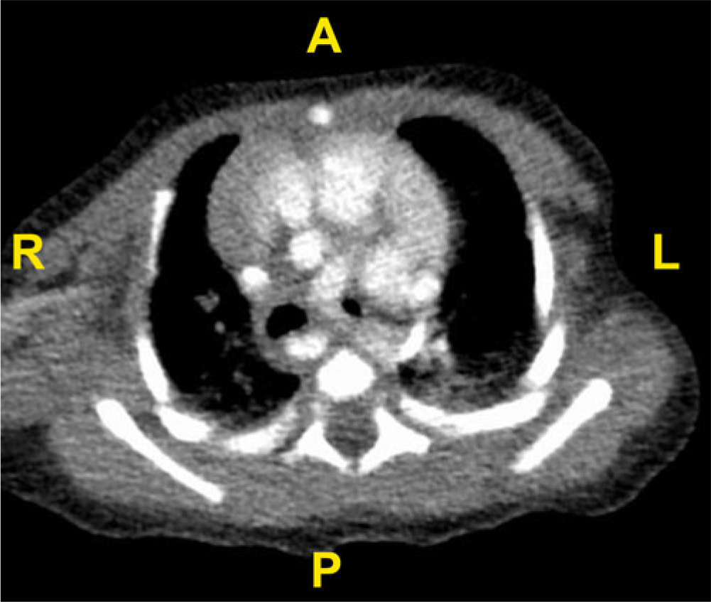
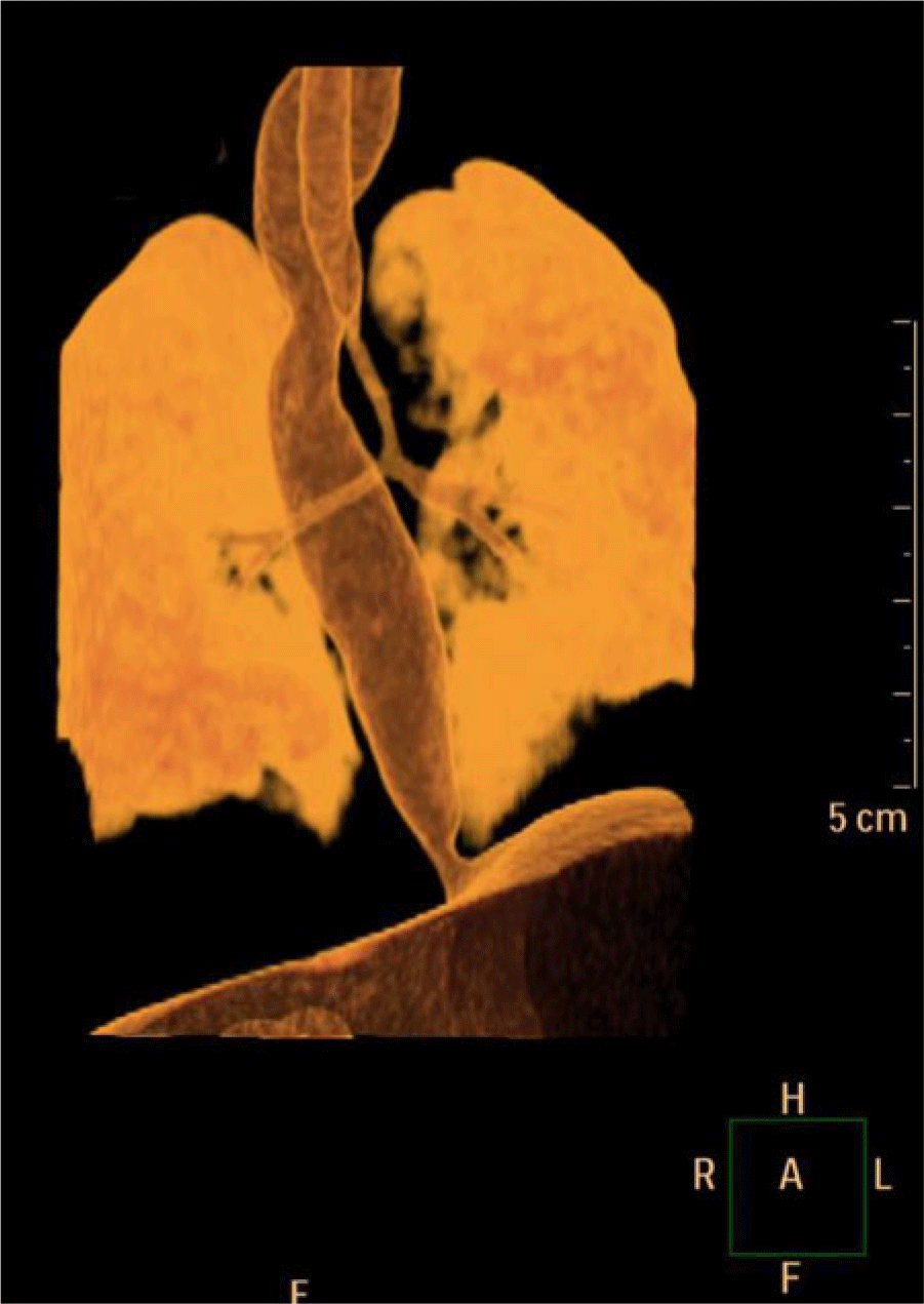
Twenty-one patients (81%) underwent CT of the trachea showing varying degrees of tracheal stenosis from discrete to long-segment stenosis (Fig 1). In one patient the tracheal stenosis comprised a long segment extending down into both bronchi (Fig 2). Microlaryngobronchoscopy was performed in all patients prior to surgery.
CT of severe congenital tracheal stenosis (axial image).

CT coronal image of long-segment tracheal stenosis.

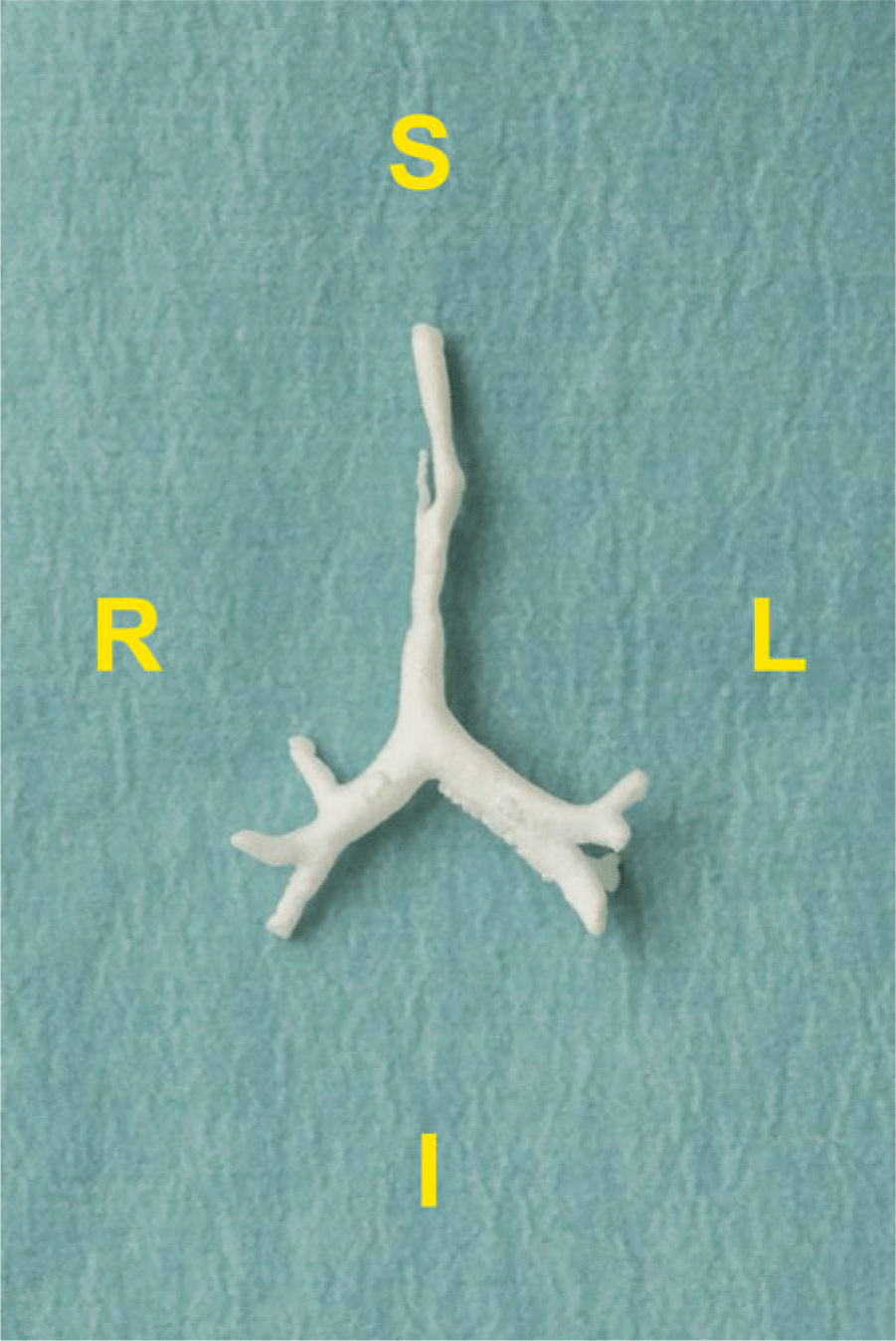
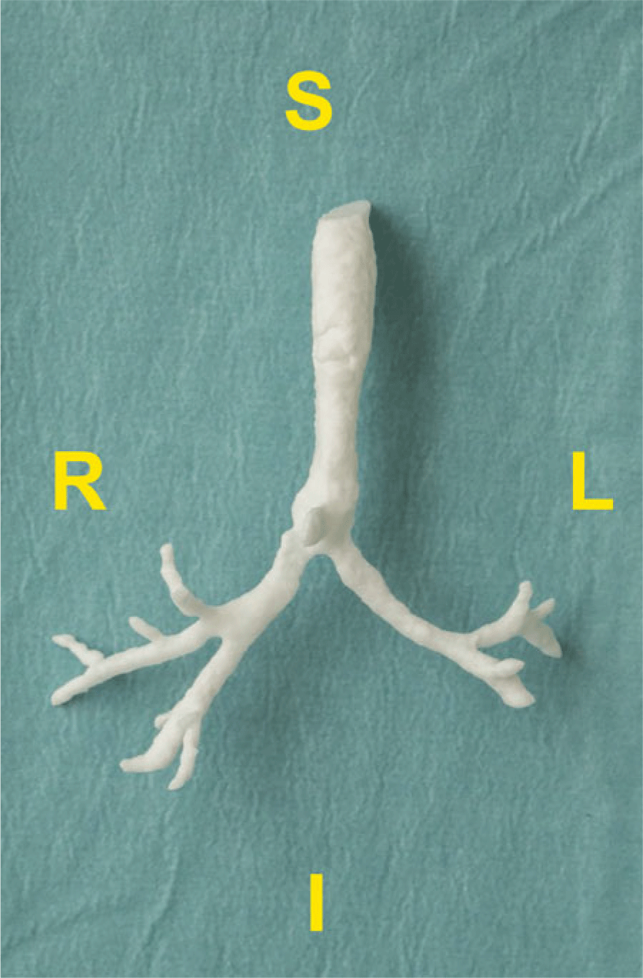
In two patients digital imaging and communication in medicine data derived from CT was used to generate a surface tessellation language file. This was used to print a 3D solid model of the trachea and the bronchi (Figs 3 and 4).
3D print of the trachea demonstrating long-segment stenosis.

3D print of the trachea demonstrating severe lower tracheal stenosis extending into both bronchi.

Surgical technique
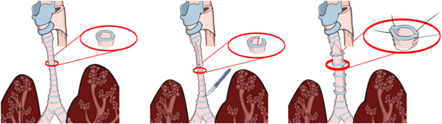
A median sternotomy was performed and cardiopulmonary bypass was instituted with either a standard cardiopulmonary bypass circuit requiring full heparinisation or an extracorporeal life support circuit with no reservoir allowing low-level anticoagulation depending on whether an intra-cardiac repair was required. Congenital cardiac defects were corrected by the same procedure. The trachea was exposed along its entire length and the area of stenosis identified by inspection. The thyroid isthmus was divided and the thymus sub-totally removed to aid exposure. The trachea was mobilised along the relevant length and all the tracheal rings exposed. Additional release may be required by division of the suprahyoid muscles (laryngeal release) and by division of the inferior pulmonary ligaments. For patients undergoing slide tracheoplasty, the trachea was divided at the mid-stenotic point and longitudinal incisions were placed on opposite sides of the anterior and posterior sections. A sliding oblique anastomosis was performed using continuous polydioxanone sutures (Fig 5). A mediastinal drain was placed in addition to pleural drains. The child was then returned to the ICU and mechanical ventilation continued with the child paralysed and sedated.
Schematic illustration of slide tracheoplasty repair.

The role of the paediatric otolaryngology surgeon
The paediatric otolaryngologist carried out a rigid and flexible bronchoscopy prior to a slide tracheoplasty. The objective was to confirm the CT/MRI diagnosis or to make the diagnosis. The length of the stenotic segment, the diameter of the complete rings, and the involvement of the carina and bronchi were also determined. The information was then discussed with the cardiothoracic surgeon to plan the surgery. A transcervical infrahyoid laryngeal release was carried out at the start of the procedure prior to going on bypass or extracorporeal membrane oxygenation. This allowed a 1–2 cm descent of the larynx and reduced the tension on the anastomosis.
Once the cardiac surgeon had mobilized the trachea, the otolaryngologist passed a 2.2 mm flexible bronchoscope via the endotracheal tube to the mid-point of the tracheal stenosis. This helped the surgeon identify the mid-point of the complete ring stenosis externally via illumination from the bronchoscope. This allowed division exactly at the mid-portion of the stenosis. Once the slide was performed, a repeat flexible bronchoscopy showed the cardiac surgeon the anastomosis from the inside. This provided good feedback on how good the anastomosis was and avoided missing a figure-of-8 deformity, which can be rectified easily at this stage.
In the paediatric ICU, prior to extubation a repeat flexible bronchoscopy was performed. This ensured that the airway was good and that no granulations or granulomas had formed that might prevent extubation. Once successfully extubated, the child underwent a repeat bronchoscopy as a day case usually 4–6 weeks later. This assessment again was to rule out granulation, granuloma, or stricture formation, which could be easily dealt with at that time. The child then underwent a series of six monthly evaluations under general anaesthesia for 2 years. After 2 years the child was usually followed up at the clinic yearly for the next 3 years.
Results
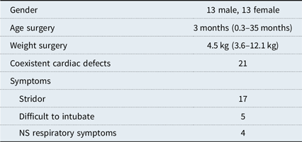
A total of 26 children underwent surgical treatment for congenital tracheal stenosis (Table 1). There were 13 boys and 13 girls. The median age was 3 months (range 0.3–35 months) and the median weight was 4.7 kg (range 2.5–13 kg) at the time of surgical intervention. Stridor was the most common presenting symptom in 17 patients (65% patients). Twenty-one patients (83%) had concurrent cardiac anomalies, with pulmonary arterial sling being the most common, present in nine patients (28%).
Demographics of 26 congenital tracheal stenosis patients.

NS = non-stridor.
Extracorporeal life support was utilised in seven patients preoperatively (27%). Laryngeal release was required in 16 patients. In 7 patients an end-to-end anastomosis was performed, in 18 patients slide tracheoplasty, and 1 patient had a double slide tracheoplasty. The median cardiopulmonary bypass time was 106 minutes (range 25–255 minutes). The median cross-clamp time was 30 minutes (range 5–67 minutes). A semi-continuous polydiaxanone 6-0 suture was used for the anastomosis in 22 patients and 4 had repair with interrupted 6-0 polydiaxone suture.
Congenital cardiac defects
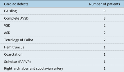
In 21 patients (81%) there were coexistent cardiac defects (Table 2). The most common defect was pulmonary arterial sling in nine patients (34%), followed by atrioventricular septal defect, ventricular septal defect, and atrial septal defect. Three-dimensional reconstructed CT images aided in cardiac diagnoses, including right aortic arch with aberrant subclavian artery causing a vascular ring.
Congenital cardiac defects in association with congenital tracheal stenosis in 21 patients.

ASD = atrial septal defect; AVSD = atrioventricular septal defect; PA = pulmonary artery; PAPVR = partial anomalous pulmonary venous return; VSD = ventricular septal defect.
Post-operative course
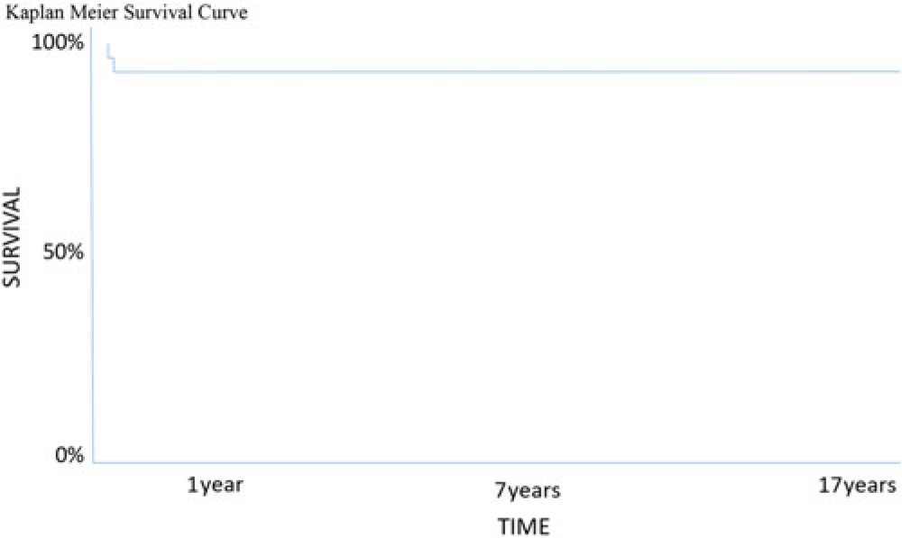
The median post-operative duration of ventilation was 5 days (range 0.5–16 days). Median ICU length of stay was 12.5 days (range 2–60 days). There were three hospital mortalities with 88% survival. One patient only required re-intervention with balloon dilation. One patient required placement of an external bioabsorbable stent immediately after the slide procedure. Twenty-two patients (83%) remained symptom-free on median follow-up of 7.6 years (range 0.2–17 years).
Survival
There were three deaths during the study period (Fig 6). One 4-month-old child who underwent pulmonary arterial sling and slide tracheoplasty failed to wean from bypass and was placed on extracorporeal life support. She developed a cerebrovascular accident, renal failure requiring peritoneal dialysis, and sepsis and succumbed 3 days in the post-operative period. A 2-month-old boy with tetralogy of Fallot status post a right-sided Blalock–Taussig shunt underwent an end-to-end tracheal anastomosis but developed sepsis and acute kidney injury and succumbed 1 week after surgery. A third child ventilated from birth underwent slide repair and developed bleeding complications after remaining on extracorporeal life support. A Kaplan–Meier survival curve is presented in Fig 6.
Kaplan–Meier graph of survival after congenital tracheal stenosis repair.

Complications of surgical repair
Vocal cord paresis occurred in two children (8%). In two patients (8%) there were significant intra-procedural problems. One patient developed a figure-of-8 hour-glass deformity requiring external stent placement using a Rapiasorb strut plate (Synthes, West Chester) modelled around a 10 Hegar dilator. A second patient developed tracheal obstruction and required balloon dilation. One patient manifested a right-sided cerebrovascular accident after extubation. Two patients accidentally self-extubated on day 1 and developed a respiratory arrest but recovered after resuscitation. Two patients developed chylothoraces but settled with pleural drainage. One child developed myocardial infarction and a stroke.
Post pulmonary arterial sling repair stenting
Of the nine patients after pulmonary arterial sling repair, three required left pulmonary arterial stenting for restenosis (33%). One patient with hemitruncus required right pulmonary arterial balloon angioplasty after re-implantation.
Discussion
This study demonstrates favourable outcomes for 22 of 26 patients with congenital tracheal stenosis using either a tracheal resection or, more recently, a slide tracheoplasty procedure over two decades at a single national surgical centre. In recent years we have increasingly adopted the slide tracheoplasty approach with good clinical outcomes. A running or interrupted suture technique may be used with excellent outcomes. As reported in several previous studies, significant cardiac defects can be corrected by the same surgical procedure, the most common association being pulmonary arterial sling.Reference Hong, Zhou, Liu, Liu, Wang and Feng12–Reference Matsuhisa, Oshima, Kadowaki and Yokoi18 Some centres have even undertaken repair of congenital tracheal stenosis in the setting of a single lung.Reference Yong, Harrison, Berkowitz, Ranganathan and Konstantinov19, Reference Yokoi, Oshima and Nishijima20
We advocate a single national centre for operating on these children given the complexity of the airway and often coexistent cardiac defects. The surgical procedure, critical as it is, is one part of the child’s care. These children require extensive multi-disciplinary and inter-disciplinary collaboration across multiple care givers, including intensive care, anaesthesiology, otolaryngology, dietician, speech and language, and nursing. Despite excellent surgical alternatives there is often a prolonged period of recuperation in a small subset of these children. These children typically have a component of bronchomalacia which requires time to recover sufficiently to allow weaning from mechanical support.
The use of extra-corporeal life support pre-slide or in the post-operative period may improve survival for these patients.Reference Martin, Holinski, Noga, El-Hakim and Aklabi21 We employed pre-operative extra-corporeal life support in seven children with a late presentation of tracheal stenosis who developed profound respiratory compromise.Reference Kelleher, Nolke and McMahon16
Lastly, 3D printed models may supplement the data derived from CT and microlaryngobronchoscopy in the pre-operative planning for this complex cohort of patients. The use of multiple imaging modalities may supplement each other in providing the surgical team with the best strategy for surgical repair. Arcieri et al11 reported a case of 3D printed assistance in pre-operative planning of congenital tracheal stenosis in association with a pulmonary sling. Our experience extends this experience with two further cases. One potential advantage of the 3D model is further demonstration of extension of tracheal stenosis into one or both bronchi, which may require an extended surgical repair. For severe long-segment congenital tracheal stenosis with complex cardiac defects, we would advocate the use of multiple imaging modalities including 3D printing to provide maximal pre-operative datasets in this highly complex set of patients. With the increasing availability of 3D printers and excellent collaboration with industry, this technology should become more available for our patients.
Limitations
There are several limitations of this study. It is retrospective in nature and only reflects the experience of this one unit, despite it representing one national centre’s outcome managing all children with congenital tracheal stenosis over two decades. Although some groups have reported on the benefit of fusion imaging combining CT, 3D printing, and microlaryngobronchoscopy, we did not undertake that strategy in the imaging of our patient cohort.
Conclusion
In the current era slide tracheoplasty represents a highly effective strategy for long-segment tracheal stenosis. The use of multiple imaging modalities (CT, microlaryngobronchoscopy, echocardiography, MR and 3D printing) facilitates optimal imaging of the severity and extent of congenital tracheal stenosis in addition to defining coexisting cardiac defects.
Acknowledgements
We are grateful to Mr. Andrew Pendred for providing graphic illustration of the slide and double slide procedure. We are also grateful to Materialise, Leuven, for assistance in the generation of the 3D printed models.
Financial Support
This research received no specific grant from any funding agency, commercial or not-for-profit sectors.
Conflicts of Interest
None.
Ethical Standards
All procedures were in accordance with the Declaration of Helsinki. Ethical approval for 3D printing of congenital defects was received from the Institutional Review Board at Our Lady’s Children’s Hospital, Crumlin, Dublin, Ireland.