The incidence of the left ventricular diverticulum is reported to be 0.05% of all congenital heart malformations. It is currently believed that it occurs as a result of impaired development of the endocardial tube during the first 8 weeks of embryologic development. Reference Jnah, Newberry and England1 Although it may present as an isolated disorder, this entity is often associated with other cardiac abnormalities or with Cantrell Pentalogy in around 70% of cases. Reference Yang, Zhu, Chen and Guo2,Reference Morales-Quispe, Aguilar and Ganiku-Furujen3 Cantrell’s Pentalogy is a rare syndrome with an estimated 5.5 per 1 million live births. It was defined by Cantrell et al in 1958 as a condition with different degrees of five defects: supraumbilical abdominal wall defect, lower sternal cleft, defect of the diaphragm in the central tendon, pericardium defect, and various intracardiac anomalies. They considered the intracardiac defects as an integral of this association and further described the frequency as 100% for ventricular septal defect, 53% for atrial septal defect, 33% for pulmonary stenosis (either valvular or infundibular), 20% for left ventricular diverticulum, and described Tetralogy of Fallot as the only established cardiac syndrome to be accompanying this syndrome with a frequency of 20%. Reference Cantrell, Haller and Ravitch4 In 1972, Toyama et al proposed a classification of this syndrome into three subtypes. The first subtype is the complete syndrome with all five defects. The second subtype is the probable syndrome with four defects, including intracardiac and ventral wall anomalies. The third subtype is the incomplete syndrome with various combinations of defects sternal abnormality. Reference Toyama5
We describe a 6-year-old male patient with a pulsatile mass extending from the lower chest to the upper abdomen.
Case report
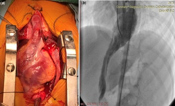
A 6-year-old male patient was admitted to our clinic. The history obtained from his family revealed that he was referred to a paediatric cardiologist in his hometown with the complaint of a pulsating mass in his abdomen before his admission to our department. The patient had no other complaints except pulsatile mass, and other physical examinations were found to be normal. ECG, 24 hour Holter monitor, and abdominal USG were performed for further investigation. ECG and 24-Holter monitoring were found to be normal. Abdominal USG revealed a 6 mm wide and 20 mm long pulsatile lesion and a 12 mm wide umbilical herniation. The combination of umbilical herniation and left ventricular diverticulum led to a probable diagnosis of Cantrell’s Pentalogy. Figure 1 shows the operative view (a) and the pre-operative angiographic view (b) of the left ventricular diverticulum.
Pre-operative angiographic view and operative view of the left ventricular diverticulum.

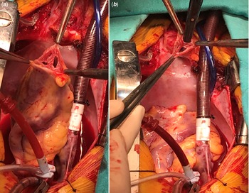
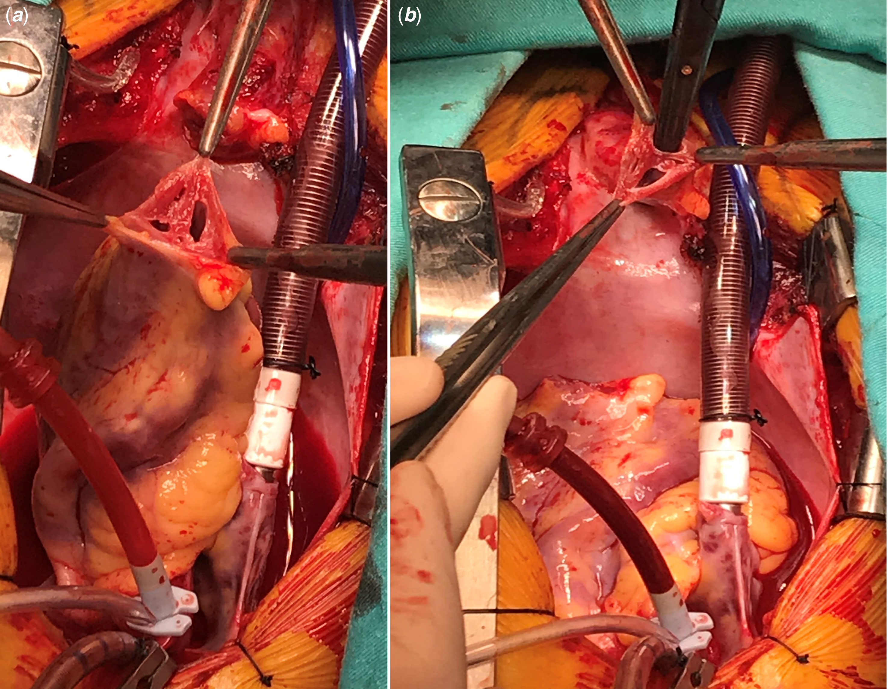
The patient underwent corrective surgery without delay at 6 years of age. A midsternal incision and median sternotomy were performed. The lower third of the sternum was absent, and the anterior pericardium was observed as a thin membrane. The pericardium was opened, and a piece was reserved to be used as a pledget. The apical left ventricle diverticulum with a length of 8 cm was observed, and the incision was extended to the umbilicus. The diverticulum was separated from the surrounding fascia. Aortic and two-stage venous cannulations were performed to initiate the cardiopulmonary bypass. The diastolic arrest was provided with a cross-clamp to ascending aorta using antegrade tepid blood cardioplegia. The diverticulum was excised, and the three layers (endocardium, myocardium, and epicardium) were observed. Figure 2 shows the operative view of the three layers of the diverticulum on both sides. The ventricular side of the defect was closed with 4-0 prolene sutures and pericardial pledgets. After gradual reduction, the cardiopulmonary bypass was ended, and the heart was decannulated. Wires for the temporary ventricular pacemaker and the drainage tubes were placed. After primary closure of the umbilical defect and the anterior abdominal wall, sternotomy and the abdominal incision were closed.
Operative view of the three layers of the diverticulum on both sides.
Discussion
Cantrell’s Pentalogy is a rare syndrome, and those with complex cardiac defects requiring surgical intervention in neonatal and infantile age may end up with a high mortality rate and poor prognosis. The long-term prognosis mainly depends on the severity of the cardiac and accompanying defects and requires multi-disciplinary management due to the involvement of multisystem anomalies. Reference Aydın, Suzan, Cevik, Odemis and Erek6 In contrast with most Cantrell’s patients, our patient had only ventricular diverticulum, and referring for operation at 6 years of age might increase procedural success. The patient’s parents postponed the surgery until the age of 6. This decision might increase the risk of ventricular arrhythmia and embolic events; fortunately, the child has never experienced such complications. The syndrome is characterised by a partial sternal cleft, anterior abdominal wall defects, anterior diaphragmatic defect, and intracardiac defects. Reference Vazquez-Jimenez, Muehler and Daebritz7 The steps for closure are usually chest wall closure, closure of the sternal defect, closure of the omphalocele, placement of the heart in the thorax, and repair of the intracardiac defect. Reference Pius, Abubakar Ibrahim, Bello and Bashir Tahir8 Nearly one-third of the cases with this syndrome have left ventricular diverticulum, which is a life-threatening abnormality that may cause systemic embolisation, fatal ventricular arrhythmias, and sudden death due to ventricle rupture. Early detection and surgical intervention for this pathology might be lifesaving. Reference Scardigno, Chieppa, Deluca and Carbonara9
Vazquez-Jimenez et al published a literature review of 153 Cantrell’s Pentalogy cases in 1998. According to the analysis, a ventricular septal defect was the most common cardiac malformation (72%). Atrial septal defect was present in 44 (34.6%) patients, left ventricular diverticulum in 41 (32.3%), pulmonary stenosis or atresia in 40 (31.5%), Tetralogy of Fallot was present in 22 patients (17.3%), dextrocardia in 19 (15.0%), and various malformations accompanied it in lesser numbers. Reference Vazquez-Jimenez, Muehler and Daebritz7
The analysis showed three cases of Cantrell’s Pentalogy, of which the intracardiac defect is limited with left ventricular diverticulum (6%), without any accompanying intracardiac defects. First of these three cases was a 2-year-old female patient with a lower sternal defect, an umbilical hernia, and a ventral diaphragmatic defect; the second case was a 2-year-old male patient with a short sternum, an omphalocele, and a diaphragmatic defect; and the third case was a 7-month-old male patient with a huge ventral hernia, a diaphragmatic defect, and a pericardial defect coherent with the incomplete syndrome. In addition, a case of complete syndrome of which the intracardiac defect is limited to the left and right ventricle along with a bifid sternum, an omphalocele, a ventral hernia, and absent pericardium has been reported. Reference Vazquez-Jimenez, Muehler and Daebritz7
Turbendian et al reported another incomplete case of Cantrell’s Pentalogy with an intracardiac defect limited to the left ventricular diverticulum along with a Patent Ductus Arteriosus, an omphalocele, a pericardial, and a diaphragmatic defect. Reference Turbendian, Carroll and Chen10
According to the Toyama classification, our case fulfils all requirements of the complete syndrome with supraumbilical abdominal wall defect, lower sternal cleft, defect of the diaphragm in the central tendon, pericardium defect, and left ventricular diverticulum. According to the previous research, the left ventricular diverticulum is a defect related to Cantrell’s syndrome almost always accompanies other intracardiac defects. Reference Halbertsma, van Oort and van der Staak11 However, the intracardiac defect is limited to the left ventricle diverticulum in our patient.
In summary, our case is interesting because of the presence of the left ventricular diverticulum alone. Also, isolated ventricular diverticulum often accompanies incomplete Cantrell’s cases, and our case is one of the rarest complete Cantrell’s syndromes with no other intracardiac defects.
Acknowledgements
None.
Financial support
This research received no specific grant from any funding agency, commercial, or not-for-profit sectors.
Conflicts of interest
None.
Ethical standards
Institutional Review Board approval, consent statement, and clinical trial registration are not applicable for this study.