Introduction
Approximately 1 in every 350 individuals living in the United States will develop cancer before the age of 20.1 Since the 1960s, a variety of treatment modalities have been used against cancer, including combinations of surgery, chemotherapy and radiation. Currently, over 70% of children diagnosed with cancer will survive for years, if not decades, after treatment.Reference Horner, Ries and Krapcho2 However, cancer therapy carries a high morbidity rate, and may affect organs not targeted during treatment.Reference Armstrong, Stovall and Robison3
During radiation therapy, the highest radiation doses are directed to the tumour or target tissues, but radiation may scatter to nearby healthy tissue.Reference Taylor, Kron and Franich4 Children are more sensitive to radiation injury, perhaps because of higher cell replication rates and their longer life expectancy.Reference Ron, Lubin and Shore5 The thyroid gland is particularly susceptible to the effects of radiation and is often located near the target field of radiation therapy.Reference Chow, Friedman and Stovall6 Hypothyroidism is the most common consequence of thyroid radiation injury, and early detection is paramount in the prevention of changes in growth and development.Reference Bonato, Severino and Elnecave7, Reference Sklar, Whitton and Mertens8 Although several studies have focused on this topic, the exact timing of these changes is still unknown, as is their potential correlation with radiation doses.
This study consisted of a prospective assessment of children and adolescents who underwent radiation therapy directed to a variety of organs and structures other than the thyroid to evaluate the occurrence of any functional and structural changes in this gland within 3 months after completion of radiation treatment.
Materials and methods
The study sample comprised 31 children and adolescents, with a median age of 7·0 years (interquartile range (IQR): 3·6–16; range: 1·3–20·0), treated between January 2009 and December 2011. Patients were consecutively enroled into a contemporary cohort for early diagnosis of acute functional or volumetric thyroid changes (the latter diagnosed by ultrasonography) after external radiation therapy not directed to the thyroid gland. The exclusion criteria were: glucocorticoid therapy, history of thyroid dysfunction, history of radiation therapy, a planned radiation therapy regimen in which the thyroid gland fell within the main beam, or a planned radiation therapy regimen involving total body irradiation. The irradiated sites encompassed several different body segments, for the treatment of a wide range of cancers. Radiation was administered with one of two linear accelerators (Siemens Mevatron MDE, S/N 3054, nominal photon energy 6 MV, or Varian 23EX, S/N 3595, nominal photon energies 6 and 15 MV; Siemens, Concord, USA) at the Hospital de Clínicas de Porto Alegre (HCPA) Radiation Therapy Center.
Thyroid-stimulating hormone (TSH), thyroxine (T4), free thyroxine (fT4), triiodothyronine (T3), anti-thyroperoxidase antibodies (anti-TPO) and thyroglobulin (Tg) levels were measured before radiation therapy, on the last day of radiation therapy and at 1 and 3 months after the end of radiation therapy. Measurements were obtained by electrochemiluminescence immunoassay (ECLIA) or chemiluminescence immunoassay (CLIA). Both methods exhibited good correlation and agreement on analysis at the HCPA Clinical Pathology Laboratory. All ECLIA assays were performed on Roche equipment (Roche Diagnostics GmbH, Mannheim, Germany) and reference ranges were as follows: TSH, 0·27–4·2 μUI/mL; T4, 5·1–14·1 μg/dL; fT4, 0·93–1·7 ng/dL; T3, 80–200 ng/dL; anti-TPO antibodies, <34 UI/mL; and Tg, 1·4–78 ng/mL. CLIA assays were performed on Siemens equipment (Siemens Healthcare Diagnostics, Erlangen, Germany) and reference ranges were as follows: TSH, 0·35–5·50 μUI/mL; T4, 3·2–12·6 μg/dL; fT4, 0·70–1·90 ng/dL; T3, 60–181 ng/dL; anti-TPO antibodies, <35 UI/mL; and Tg, <55 ng/mL.
Thyroid volume was measured by ultrasound before and shortly after the completion of radiation therapy by a staff radiologist at the HCPA Radiology Service, using an ALOKA ProSound 4000 ultrasonography system (Hitachi-Aloka, Japan) with a 15 Hz linear transducer.
Six- and 24-hour 131I uptake (10 μCi) was measured at baseline and after the last radiation therapy session. The tests were performed at HCPA Nuclear Medicine Service using a GE Millennium gamma camera (GE Healthcare, Milwaukee, WI, USA).
Patients’ chemotherapy regimens were recorded, but not analysed, as there is controversy regarding the potential isolated or add-on effect of chemotherapy on thyroid dysfunction.Reference van Santen, Thonissen and de Kraker9–Reference Schmiegelow, Feldt-Rasmussen and Rasmussen12
In patients who underwent computerised planning of 3D conformal radiation therapy, a radiologist contoured the thyroid gland in their planning CT scans. The estimated scattered dose to the thyroid during radiation therapy was then calculated with the Eclipse 10·0 TPS (VARIAN Medical Systems, Palo Alto, CA, USA). This software package uses pencil beam convolution and anisotropic analytical algorithms for dose calculation.
The linear accelerator was calibrated with a Farmer-type ionisation chamber (Freiburg, Germany; PTW TN30004, S/N 244) and a PTW Unidos E electrometer (Freiburg, Germany; S/N T10010-00055), in accordance with the IAEA TRS-398 protocol.
Scattered doses to the thyroid gland and to the skin overlying the thyroid isthmus, where thermoluminescent dosimeters (TLDs) were placed, were estimated in the treatment planning system. Field and total radiation doses were calculated. Dose–volume histograms were used to determine the mean dose to the gland.
During treatment, TLDs were placed onto the skin overlying the thyroid isthmus to measure the radiation dose from each treatment field that reached the thyroid gland. As the thyroid is located only a few millimetres from the surface (skin), one may estimate that the entrance dose (by definition, the absorbed dose at depth of maximum ionisation) is the dose absorbed by the gland. Therefore, doses measured by TLDs during treatment provide reliable estimates of the radiation dose deposited in the region of the gland. At least two measurements were obtained for each treatment field in some sessions.
TLDs were provided and analysed by the Radiation Therapy Quality Program of the José de Alencar Gomes da Silva National Cancer Institute (INCA), affiliated with and operated by the Brazilian Ministry of Health, in Rio de Janeiro. The dosimeters were Harshaw TLD-100 (Thermo RMP, Waltham, USA) chips (lithium fluoride doped with magnesium and titanium [LiF:MgTi], dimensions: 3 × 3 × 0·9 mm3). One pair of TLDs was used for each treatment field, and the mean of two measured values calculated for analysis. The pair of TLDs was placed into a chrome-nickel steel hemispherical build-up cap (radius 15 mm) to accomplish electronic equilibrium and to ensure maximal readings.
TLD readouts were performed in a PCL3 (Fimel) reader at the Radiation Therapy Quality Program, INCA, Rio de Janeiro. Annealing was performed with an EDG1800 oven (EDG Equipamentos, Brazil) and a Fanem 315SE drying oven (Fanem, Guarulhos, Brazil).
The procedures carried out on the patients of this study were reviewed and approved by the Research Ethics Committee of the Hospital de Clínicas de Porto Alegre (protocol number 04-376), where the study was conducted, and were performed in accordance with standards set by the committee and in compliance with the 1975 Helsinki Declaration and its 2000 revision. Assent from patients and informed consent from their legal guardians was obtained before the study.
Statistical analysis
Based on Nishiyama et al.,Reference Nishiyama, Kozuka and Higashihara13 who estimated an effect size (E/S) of 1·23 standard deviation units (which is considered a large effect), we chose to use a smaller effect. Thus, for a 40% smaller effect size (E/S = 0·74), a significance level of α = 0·05 and a statistical power of 90% to detect TSH changes before and after radiotherapy, the required sample size was estimated at 21 patients. Considering a maximum loss to follow-up of 20%, the sample size was expanded to 27 patients.
Quantitative laboratory test values (TSH, T4, fT4, T3, Tg and anti-TPO antibodies) were skewed and thus log-transformed before analysis. A mixed-effects model for repeated measures was used, with comparison across all four time points of assessment. A similar model was used to assess the effects of sex, age (as a dichotomous variable, with the cut-off set at 15 years), the interaction between sex and age, and the total radiation dose scattered to the thyroid. Values were expressed as the mean antilog of log-transformed values (i.e., the geometric mean).
Thyroid volumes were expressed as medians, interquartile ranges (P 25-P 75) and ranges (minimum–maximum). The Wilcoxon signed-rank test was used for between-group comparisons of this variable.
Six-hour and 24-hour 131I uptake values, before and after radiation therapy, were expressed as means and standard deviations and subjected to mixed-effects repeated measures analysis.
The significance level was set at α = 0·05 for all analyses. However, for multivariable analyses, values of 0·10> p > 0·05 were considered borderline significant.Reference Katz14 Data were processed and analysed with SPSS version 18·0 and R version 2·14·1 softwares.
Results
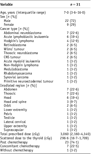
The overall sample profile is described in Table 1.
Sample profile

Note: Data are expressed as n (%) or median (interquartile range).
Abbreviation: CNS, central nervous system.
There were no statistically significant differences across the four time points of assessment with regard to levels of TSH (p = 0·575), T4 (p = 0·950), fT4 (p = 0·510), T3 (p = 0·843), Tg (p = 0·620) (Figure 1) or anti-TPO antibodies (p = 0·546).
Line plots with geometric means and 95% confidence intervals representing the variation observed along four measurements for selected variables. Abbreviations: T3, triiodothyronine; fT4, free thyroxine; T4, thyroxine; TSH, Thyroid-stimulating hormone.
Regarding T4, there were borderline differences in the rate of change between younger subjects (0·44 μg/dL at measurement 1–10·15 μg/dL at measurement 4; a 7·5% increase) and older subjects (8·28 μg/dL at measurement 1–7·67 at measurement 4; a 7·3% decrease) (p = 0·082). Although there was no statistically significant effect of sex on the rate of change of T4 (p = 0·998), or an interaction effect between age and sex over time, data indicate that younger girls would have the highest levels and boys the lowest across all four time points (p = 0·05) (Figure 2). When the radiation dose scattered to the thyroid was added to the T4 model, which already included age and sex, its effect did not show a significant impact on the rate of change across all four measurements (p = 0·322).
Line profiles of estimated marginal means obtained in the linear mixed model for thyroxine (T4).
Although subjects under the age of 15 years showed significantly higher geometric mean T3 levels (161·5 ng/dL at measurement 1 and 148·3 ng/dL at measurement 4) as compared with older subjects (100·69 ng/dL at measurement 1 and 138·04 ng/dL at measurement 4) (p = 0·035), the impact of age on the rate of change was not statistically significant (p = 0·306). In addition, in a model adjusting for the combined effects of age (<15 or ≥15 years), sex, and radiation dose scattered to the thyroid, none of the factors was significantly associated with T3.
Geometric mean thyroglobulin levels were higher in under-15s (21·03 ng/mL at measurement 1 and 29·02 ng/mL at measurement 4) than in older subjects (12·3 ng/mL at measurement 1 and 12·6 ng/mL at measurement 4) (p = 0·026). Furthermore, the rate of change was higher in younger subjects (38% increase for under-15s versus 2·3% increase for over-15s; p = 0·085) and higher among girls (51% increase for girls versus 3% increase for boys; p = 0·065) (Figure 3). The addition of radiation dose scattered to the thyroid to the Tg model did not show any significant effect.
Line profiles of estimated marginal means obtained in the linear mixed model for thyroglobulin.
Stratification of patients into age groups (<15 and ≥15 years) failed to show any differences in the rates of change of TSH (p = 0·812), fT4 (p = 0·488) or anti-TPO antibodies (p = 0·250). In addition, sex and radiation dose scattered to the thyroid did not show any significant effect when added to these models.
Median thyroid volumes were 3·50 cm3 (IQR: 1·60–4·41; range: 1·14–12·78) before radiation therapy and 3·87 cm3 (IQR: 2·06–4·56; range: 1·25–15·83) shortly thereafter. There was no significant difference between these values (p = 0·692).
Mean 131I uptake at 6 and 24 hours was 25·4 ± 10·2% and 25·1 ± 8·9%, respectively, before radiation therapy and 27·2 ± 12·0% and 25·1 ± 8·6%, respectively, after radiation therapy. There were no statistically significant differences among these four values (p = 0·92).
Discussion
Most studies of the effects of radiation on the thyroid are retrospective, which hinders assessment of the dose–injury relationship. In children, the distance between the thyroid gland and other organs is shorter because of body size, and cell replication is greater. These factors might increase the susceptibility of the gland even if scattered radiation doses were lower.
In this study, there were no differences in TSH, T4, fT4, total T3, Tg or anti-TPO values over the 3-month assessment period. The radiation dose scattered to the thyroid may have been too low for detection of any changes within this period, which does not rule out the possibility of such changes surfacing during long-term follow-up. Furthermore, the sample size may have been too small to demonstrate changes. Nishiyama et al.Reference Nishiyama, Kozuka and Higashihara13 and Bakhshandeh et al.Reference Bakhshandeh, Hashemi and Mahdavi15 reported decreased TSH and increased T4 levels, the former occurring shortly after radiation therapy and the latter during treatment. In these studies, radiation was directed to the neck or head and neck, respectively, and the dose scattered to the thyroid was higher. The highest dose scattered to the thyroid in the present study was 296·6 cGy, at the end of treatment, whereas the first changes in the Bakhshandeh et al.Reference Bakhshandeh, Hashemi and Mahdavi15 study were only detected at 1,200 cGy.
In the Childhood Cancer Survivor Study,Reference Sklar, Whitton and Mertens8 the incidence of hypothyroidism was higher in children receiving radiation therapy after the age of 15 than in younger patients. In our study, using the same stratification strategy, detectable differences were observed in T4, T3 and Tg levels, which were higher in patients under the age of 15. T4 and Tg levels were also higher in female patients. These differences may correspond to physiological behaviour before treatment.Reference Djemli, Van Vliet and Belgoudi16, Reference Elmlinger, Kuhnel and Lambrecht17 Acutely increased T4 and Tg levels after radiation therapy in younger subjects may indicate greater cell sensitivity. Further studies involving more patients are warranted to answer these questions.
In our sample, there were no differences in thyroid volume before and shortly after radiation therapy. This may have been due to the rather short study interval. In a prior, retrospective study assessing late effects of non-directed radiation therapy on the thyroid (mean follow-up time: 7·3±3·62 years), we detected hypothyroidism and smaller thyroid volume, and we inferred these two findings were associated.Reference Bonato, Severino and Elnecave7
In this study, 131I uptake was performed to detect cell injury, which could not be observed by this method. However, the optimal timing for scans was unknown, and there was no evidence as to whether radiation scattered to the thyroid would produce any significant changes. This technique was previously used in patients who received radiation therapy to the neck and presented with clinical signs and symptoms of thyrotoxicosis within 2 weeks of treatment, and were thus diagnosed with thyrotoxic thyroiditis.Reference Blitzer, Paolozzi and Gottlieb18, Reference Bryer-Ash, Lodhi and Robbins19
This was the first prospective study assessing early effects of scattered radiation on the thyroid in children and adolescents undergoing radiation therapy targeted to different sites. Perhaps a more sensitive method would be required to detect radiation injury induced by the low doses to which patients were exposed in this study – a method such as colour Doppler ultrasonography, which is capable of demonstrating vascular changes and changes in echogenicity.Reference Bakhshandeh, Hashemi and Mahdavi15 It is important that such early changes are known so that prophylactic measures can be instigated in an attempt to prevent the development of hypothyroidism. We plan to continue follow-up of the patients included in this study so as to observe the long-term incidence of functional and structural changes and their correlation with the dose of radiation scattered to the thyroid gland.
Conclusion
Despite the increasingly targeted nature of radiation therapy and ever increasing care concerning protection of tissues and organs outside the target field, incidental radiation exposure still occurs. This may produce delayed sequelae that must be known ahead of time, so they can be prevented and treated. In this study, the scattered doses measured had no acute effect on thyroid function or volume in children and adolescents. Larger studies using more sensitive methods are required to confirm these findings.
Acknowledgements
The authors thank the Nuclear Medicine Service of HCPA, Dr Ilza Vasques de Moraes for 131I uptake, Dr Alvaro Porto Alegre Furtado for the thyroid ultrasound examinations and Dr Günther Alex Schneider for the precious assistance in radiotherapy.