Introduction
Grid therapy, which is also known as spatially fractionated Grid radiation therapy (SFGRT), is a method of radiation therapy for treatment of bulky malignant tumours (size > 6 cm). This technique is more effective for the radio-resistant tumour cells than the radio-sensitive ones.Reference Nedaie, Darestani and Banaee1,Reference Zwicker, Meigooni and Mohiuddin2 In this method, a high dose of radiation is delivered in a single fraction by dividing the treatment field into a set of small open areas. The remainder of the field is blocked by a Grid block or by multi-leaf collimators.Reference Ayyangar, Lakshmanan and Chandra3 It has been proven that a single fraction of a high-photon dose (10–20 Gy) with Grid therapy has advantages on normal tissue sparing while it is killing the tumour cells.Reference Sohrabi and Hakimi4–Reference Wang, Charlton and Esquivel6 The Grid blocks are generally made of cerrobend or lead. Several investigatorsReference Sohrabi, Hakimi and Mahdavi7,Reference Meigooni, Dou and Meigooni8 have studied the dosimetric properties of 6-MV photon beam energy in Grid therapy (e.g., dose profiles and the output factors) by using different dosimeters such as radiographic films, TLDs an ionization chamber in water or solid water equivalent phantom material.
The merit of using a high-photon beam energy (i.e., >10 MV) is its larger depth dose value at greater depths and larger skin sparing properties for delivering high doses of radiation to bulky and deeply seated tumours.Reference Klett and Leuschner9,Reference Mitev and Suntharalingam10 However, when using a high-energy photon beam with a Grid block, in addition to obtaining photon dosimetric properties, photo-neutron (PN) contamination should be considered due to a high-Z material of the Grid block.Reference Neuner, Mohiuddin and Vander Walde11 Avoiding secondary cancers associated with neutron production using the high-Z accessory in radiotherapy techniques like in Grid therapy is critical. Therefore, it is recommended to determine the PN dosimetric properties before the clinical use of high-energy photon beans during Grid therapy.Reference Neuner, Mohiuddin and Vander Walde11
Different PN dosimeters have been introduced to measure equivalent dose produced by thermal and fast neutrons. A combination of thermoluminescence dosimetry (TLD) types 600 and 700 have been used to determine equivalent dose from thermal neutron from photon irradiation in megavoltage linear accelerators.Reference Sohrabi and Hakimi12,Reference Gholami, Nedaie and Meigooni13 These TLDs were manufactured by Fimel Company (Fimel, Velizy, France).
Polycarbonate track dosimeters (PCTD) without a cadmium cover are suitable for fast neutron dose measurements.Reference Marks14,Reference Reiff, Huq and Mohiuddin15 This polycarbonate dosimeter detects fast neutrons in a PN spectrum by counting fast-neutron-induced tracks when the dosimeters processed by electrochemically etching (ECE), compared with other methods using PCTD is a simple, sensitive and inexpensive method to measure fast neutrons in photon fields.Reference Reiff, Huq and Mohiuddin15
The aim of this study is experimental to determine the dose contributions from a photon and PN in a Grid therapy technique with 18-MV photon beam from a Varian Clinac 2100c linear accelerator (Varian Medical Systems, Palo Alto, CA, USA). For photon dosimetry, pinpoint ionization chamber (PTW, Freiburg, Germany) was used in a water phantom. The combination of TLD types 600 and 700, and Polycarbonate Film dosimeters were used to measure PN contamination on the surface and in the maximum depth dose (d max) of the beam in Solid Water™ slabs.
Materials and Methods
This section includes various parts including Grid block charecteristics, 18 MV photon dose measurements method and Photon-neutron contamination measurements (TLD and polycarbonatedosimetry measurement).
Grid block
A Grid block was previously constructed and used at our department for Varian Clinac 2100C linear accelerator and its dosimetric characteristics [e.g., dose profile, percent depth dose (PDD), output factor, etc…] has been measured for 6-MV photon beam energy.Reference Sohrabi and Morgan16 This Grid consists of 149 divergent apertures with a hexagonal pattern in a 7.5-cm thick block of lead and can provide a maximum field size of 20 × 20 cm at the isocentre. The projection of each hole in the Grid field has a diameter of 13 mm with a centre-to-centre distance of 17 mm.
18-MV photon dose measurements
The PTW 310014 pinpoint ionization chamber was used in a water phantom to obtain dosimetric characteristics of 18-MV photon beam energy produced by the linear accelerator. PDD, in-plane and cross-plane dose profiles (at d max, 5- and 10-cm depth) and output factor were measured, using a water phantom for field size of 20 × 20 cm2.
Photon–neutron contamination measurements
TLD dosimetry measurement
The combination of TLD-600 (Harshaw, 6LiF: Mg, Ti) and TLD-700 (Harshaw, 7LiF: Mg, Ti) were used to measure thermal neutrons equivalent dose in open and Grid fields. TLD-600 has a high-thermal neutron capture cross-section while TLD-700 is much more sensitive to the photon beam. Both dosimeters have the same dimensions of 3 × 3 × 0.9 mm3. For the dosimeter’s annealing, calibration, and obtaining the glow-curves, we have followed the procedure that was described by Nedaie et al.Reference Zhang, Johnson and Zwicker17 The TLD reader (Fimel, Velizy, France) was used to obtain the responses of the irradiated TLDs. An element correction factor (ECC) for both types of TLD dosimeters were measured by irradiating all dosimeters with the same dose (100 cGy), using the 18-MV photon beam.
The ECC is known as a correction factor, showing the differences in the responses of each TLD element respect to the average value from the same TLD setReference Zhang, Wang and Mayr18 and can be given by:
 $$1/{\rm{EC}}{{\rm{C}}_j} = {{{\rm{TL}}{{\rm{E}}_j}} \over {\langle{\rm{TLE}\rangle}}}$$
where, ECCj is related to the TLD number j, TLE is the average reading of all the dosimeters and TLEj is the response of the dosimeter j.
For the next step, the calibration curves of TLDs should be obtained for neutron and photon doses, which was performed by 241Am-Be sources and 18-MV X-ray beam, respectively. The area under the glow curves were considered as TL intensities to definite doses in the calibration curve zone. Assuming a linear TL intensity (I) function, we can calculate that according to the following equation:
 $$I = \alpha D$$
where, α is the calibration factor. For photon calibration curve, doses of 2, 4, 8, 10, 20, 40, 60, 80 and 100 cGy were used for irradiation of the TLDs using the 18-MV photon beam produced by the linear accelerator at 2-cm depth in a solid water™ phantom. In this irradiation, source to surface distance (SSD) of 98 cm with a field size of 10 × 10 cm2 was used. The 241Am-Be source (activity of 10 Ci) was utilized to obtain neutron calibration curve. In this case, the TLDs were placed on the surface of a solid phantom with dimension of 30 × 30 × 15 cm3 with SSD = 50 cm. TLD pairs were irritated by neutron beam in different time intervals: 0.565, 1.123, 2.26, 9.711, 14.37 and 19.432 h, corresponding to 0.655, 1.31, 2.65, 10.511, 15.646 and 20.593 mSv. Then the related dose values were measured using a Berthold LB 6411 neutron probe.Reference Sohrabi and Morgan19
A determination of photon and thermal neutron dose values in a mixed irradiation field contains neutron and photon absorbed doses at each point requires one pair of TLD dosimeters (one TLD-700 and one TLD-600). After obtaining ECC of the TLD dosimeters and the calibration factors for neutron and photon beam, the D photon photon dose and Dn neutron dose in a mixed radiation field can be obtained by the following equations:
 $$\hskip -17pt{D_{{\rm{photon}}}} = \left\langle{\left( {R_{700}^{{\rm{photon}}}} \right)_j} \times {\rm{EC}}{{\rm{C}}_j}\right\rangle \times \alpha _{700}^{{\rm{photon}}}$$
 $${D_{{\rm{photon}}}} = \left\langle{\left( {R_{600}^{n + {\rm{photon}}}} \right)_j} \times {\rm{EC}}{{\rm{C}}_j}\right\rangle \times \alpha _{600}^n - {{\alpha _{700}^{{\rm{photon}}}\alpha _{600}^n} \over {\alpha _{600}^{{\rm{photon}}}}}\left\langle{\left( {R_{700}^{{\rm{photon}}}} \right)_j} \times {\rm{EC}}{{\rm{C}}_j}\right\rangle$$
where, the response of the TLD-600 is
 $R_{600}^{n + {\rm{photon}}}$
and the calibration factors for neutrons and photons are
 $\alpha _{600}^n$
and
 $\alpha _{600}^{{\rm{photon}}}$
, respectively.
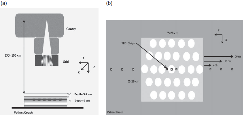
The schematic diagram of TLD set-up for measuring thermal neutron and photon doses by TLDs in a Grid field is shown in Figure 1. TLDs were placed at the centre of the hole along the central axis of the beam and in the blocked areas of the Grid field at depth of 3.5 and 5 cm. In addition, measurements were performed out of field at the distance of 5, 10, 20 and 40 cm from the beam edge on the surface of the slab phantom. It has been considered that the maximum field size in the Grid field was (20 × 20 cm2). A dose rate of 2000 MU was delivered to irradiate TLDs with and without the Grid. Finally, the results from with and without Grid block were compared at several points.
(a) Schematic view of the linear accelerator with the Grid block and TLD chips positions, using solid water™ slabs. (b) Axial view of TLD chips positions.

Polycarbonate dosimeters for fast neutron
PCTD dosimeters were used for fast neutron dose measurements in and out of the Grid field. The responses (tracks/neutron cm2) of this dosimeter are independent of the neutron spectrum energy.Reference Triolo, Marrale and Brai20
Therefore, the track density from fast neutron can be simply converted to PN dose equivalent by a conversion factor. However, in order to further check this conversion factor, the dosimeters were calibrated in the field of a 252Cf standard neutron source in ambient dose equivalent.
The PCTD dosimeters used were 250-mm thick and were cut from a larger polycarbonate sheet to 2 × 2 cm2 size. Similar to TLDs experiments, PCTD dosimeters were placed at the centre of the hole along the central axis of the beam and in the blocked areas of the Grid field at a depth of 3.5 and 5 cm. In addition, measurements were performed out of the Grid field at the distance of 5, 10, 20 and 40 cm from the beam edge on the surface of the slab phantom. For both TLD and PCTD dosimetry, each measurement repeated three times, in order to reduce dosimetry uncertainty. A dose rate of 2000 MU was delivered to irradiate PCTD dosimeters with and without the Grid. Finally, the results from with and without Grid block were compared at several points.
For processing, the optimized ECE [2 kHz – 32 kV·cm−1, 15 g KOH + 40 g C2H5OH + 45 g H2O at 26 °C for 3 h] was utilizedReference Sohrabi and Katouzi21–Reference Sohrabi and Hakimi23. Then, the dosimeters were washed in distilled water and dried in ambient air.
The PN dose equivalent of each position on a dosimeter was obtained by determining track density (tracks cm2) of each position through counting the tracks of PN-induced recoils for PCTDs have a dose equivalent neutron energy response matching well with that of International Commission on Radiological Protection (ICRP) ambient dose equivalent response from 1 to 20 MeV, which makes the dosimeters independence of the neutron spectrum.Reference Sohrabi and Katouzi21,Reference Mukherjee24–Reference Gholami, Nedaie and Longo26 Nevertheless, the PCTDs were further calibrated to fission neutrons from a 252Cf neutron source.
The mean value and related standard deviations (SDs) of the recoils of fast PN induce tracks from 10 microscopic field measurements were determined, using relevant conversion factors obtained from the calibration process the fast PN dose equivalent was obtained based on the mean track density ± SD (tracks cm−2).
According to ICRP dose equivalent, the conversion factor of 9.67 ± 0.13 tracks cm2/mSv was considered to convert the track density counts to PN dose equivalent.
Results
18-MV photon beam dosimetry
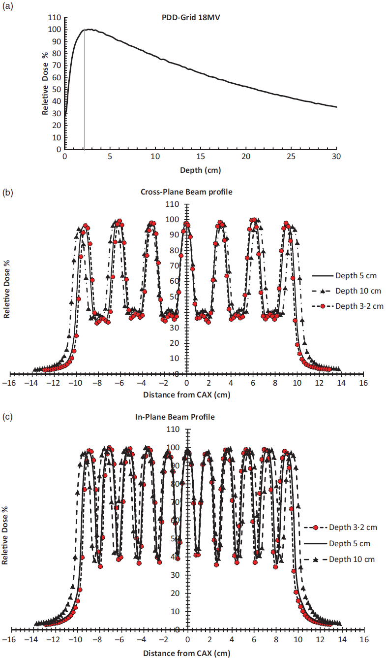
The PDD and beam profile results of Grid block in two in-plane and cross-plane directions are shown in Figure 2. The valley dose in grid therapy refers to the minimum dose from blocked areas as a result of scatter radiation. The valley-to-peak ratio quantifies the degree of spatial fractionation (14). The valley-to-peak ration obtained from the beam profiles was ~35%.
(a) PDD (b) in-plane and (c) cross-plane beam profiles of the Grid block for 18 MV photon beam.
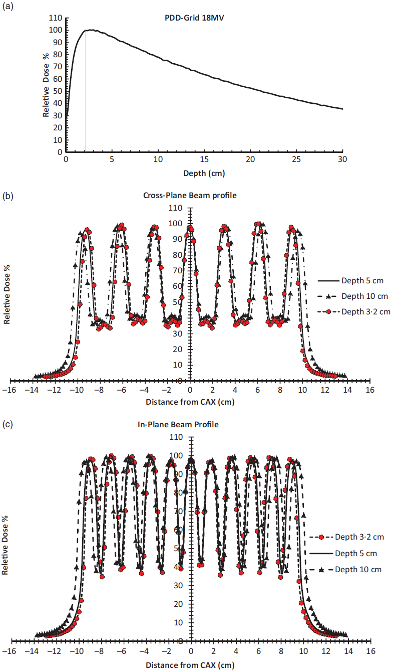
The Grid output factor for 18-MV X-ray beam calculated as a ratio of pinpoint chamber reading along central axis with and without Grid for the same value of MU was equal to 0.72 for the 20 × 20 cm2 field size.
The TLDs calibration curves
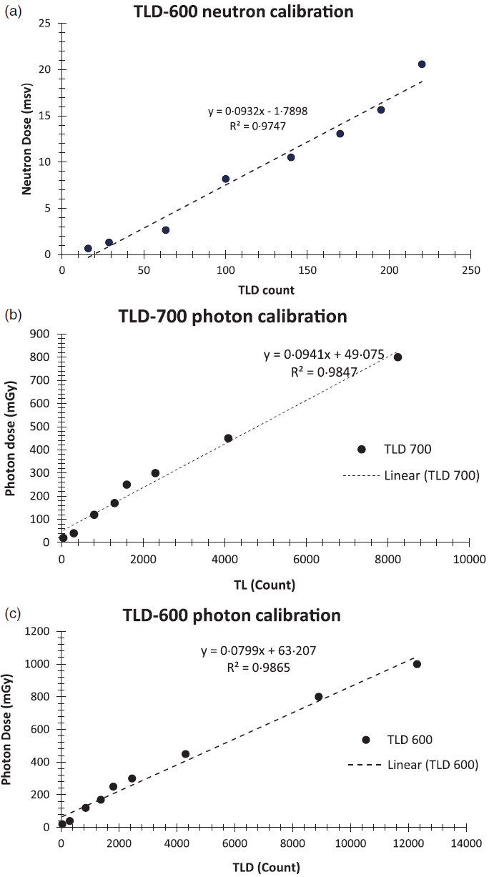
The calibration curves of TLDs for neutron and gamma doses performed by 18-MV X-ray beam are shown in Figure 3.
(a) TLD-600 neutron calibration curve calculated by using the 241Am-Be source. Photon calibration curve for (b) TLD-700 and (c) TLD-600.

Thermal and fast photon–neutron with and without the Grid block
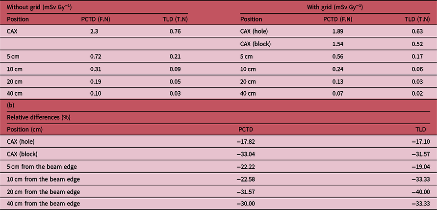
Table 1a shows the results obtained from the TLDs and PCTD dosimeters for thermal and fast neutron, respectively. The decrease in both fast and thermal PN equivalent dose can be seen in the Grid field compared to an open field (without Grid). Equation 4 was used to calculate the relative difference of fast/thermal neutron equivalent dose in the Grid field (Table 1b).
 $${\rm{Relative\ differences}} = {{{R_{{\rm{with\ Grid}}}} - {R_{{\rm{without\ Grid}}}}} \over {{R_{{\rm{without\ Grid}}}}}} \times 100$$
(a) Fast-neutron (F.N) and thermal-neutron (T.N) obtained from PCTD and TLDs 600/700 dosimeters with/without the Grid block on the surface of solid water phantom. (b) Relative differences (%) of the PN results on the surface of solid water phantom due to the presence of a Grid block

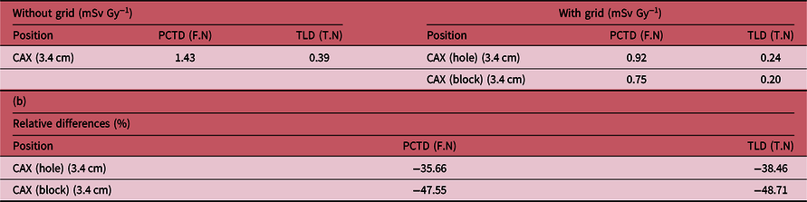
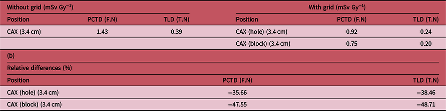
Table 2 represents the results of fast and thermal dosimetry at d max depth (3.4 cm) with corresponding relative differences (%).
(a) Fast-neutron (F.N) and thermal-neutron (T.N) obtained from PCTD and TLDs 600/700 dosimeters with/without the Grid block at a d max depth of solid water phantom. (b) Relative differences (%) of the PN results at d max due to the presence of a Grid block
Discussion
Using the high-energy photon beam in radiotherapy has better dosimetric properties for delivering a high doses of radiation to bulky and deeply tumours in a Grid therapy technique. In the present study, dosimetric characteristics of a Grid block were obtained for 18-MV X-ray beam. Moreover, the TLDs and PCTD dosimeters were used to detect PN contamination on the surface and at a d max depth of water equivalent phantom.
For our Grid block, the valley-to-peak ratio was 35% for 18-MV X-ray beam, while it has been previously obtained 20% for 6-MV X-ray beam, using the same type of linear accelerator and similar designed Grid block.Reference Meigooni, Dou and Meigooni8 However, similar valley-to-peak ratio was reported for the Grid block designed by JE Reiff et al.Reference Meigooni, Dou and Meigooni8 Actually, the longer forward electron range in higher energy photon beams results in a longer lateral electron range that increases the penumbral width and valley-to-peak ratio significantly. In addition, the Grid output factor was 0.72 for 18-MV photon beam, while was reported 0.87 for 6-MV photon beam, using the same type of linear accelerator and similar designed Grid block.Reference Sohrabi and Morgan16
Results from PCTD and TLDs measurements have demonstrated the reduction for both fast and thermal neutrons due to the presence of a Grid block in comparison with the open field. This reduction was obtained 17.82 and 17.10% for PCTD and TLD dosimeters at the centre of the whole area of the Grid on the phantom surface, respectively. In addition, the amount of neutron dose reduction on block region of the Grid field on the phantom surface was 33.04 and 31.57% for PCTD and TLD dosimeters, respectively. In general, using the Grid block decreases the amount of both fast and thermal PN contamination. Similar results have been previously reported by Wang et al.Reference Neuner, Mohiuddin and Vander Walde11 In their study, the obtained PN dose equivalent in the presence of a Grid block was 44.9% lower than the obtained value without the Grid block, using in the same field size and monitor units.Reference Neuner, Mohiuddin and Vander Walde11 This reduction is reasonable because the large fraction of the primary beam is blocked by the Grid and the scattered photons are decreased. The block shields a fraction of the photons as well as reduces neutron in this treatment technique.Reference Neuner, Mohiuddin and Vander Walde11
In a Monte Carlo simulation study by Chegeni et al.,Reference Hakimi, Sohrabi and Rabie Mahdavi27 a decrease of neutron dose on several distances from a Grid field edge relative to an open field was reported as well, for 18-MV photon beam.
Conclusion
To conclude based on the findings of this study, the production of both fast and thermal neutron dose equivalent was reduced in the Grid therapy compared to the conventional radiotherapy. Therefore, the use of Grid block with higher energy of photon beam for deep and bulky tumours seems safe. However, it should be considered that continuously using of a Grid block in such high energy remove of photon beam for a long period of the treatment might activate the Grid block and, therefore, produce neutrons for patients. In addition in this particular study, in vivo dosimetry for patients has not been considered, further investigations are recommended in this subject.
If a guideline is introduced for SFGRT in 18-MV photon beam, our article will help the patients with the amount of contamination in 18-MV photon beam energy for treatment.
Acknowledgements
This research is supported by Tehran University of Medical Sciences and Health Services with grant number of 97-01-30-37972.