The first heterosporous lycopsids appeared in the Middle Devonian (Givetian), and from the Frasnian to the latest Famennian became a diverse group. Most were tree-like with monosporangiate strobili (Fairon-Demaret Reference Fairon-Demaret1977, Reference Fairon-Demaret1991; Cai & Chen Reference Cai and Chen1996; Wang Reference Wang2001; Berry et al. Reference Berry, Wang and Chongyang2003; Wang et al. Reference Wang, Xu, Basinger and Wang2012; Meng et al. Reference Meng, Wang, Xue and Zhu2013, Reference Meng, Wang and Yao2015, Reference Meng, Liu, Wang and Yao2016; Wang et al. Reference Wang, Meng, Xue, Basinger, Guo and Liu2014, Reference Wang, Qin, Meng, Liu and Ferguson2016). Finds of bisporangiate strobili and cone structures of early heterosporous lycopsids are very rare (Chitaley & McGregor Reference Chitaley and McGregor1988; Senkevitsch et al. Reference Senkevitsch, Jurina and Arkhangelskaya1993; Schweitzer & Li Reference Schweitzer and Li1996; Hao et al. Reference Hao, Xue, Wang and Liu2007). The present restudy of the Frasnian bisporangiate strobili of Kossoviella gives us an opportunity to increase our knowledge about the early evolution of heterosporous lycopsids.
1. Material and methods
The material studied was collected from several localities in North Timan, Russia (Orlova et al. Reference Orlova, Jurina and Snigirevsky2016). Most specimens come from the Ust'bezmoshitsa Formation (upper Frasnian) exposed in the Suvojnyj area of the eastern coast of Cheshskaya Bay, between Suvojnyj Cape and Krestovyi Cape, and the Ludovatyj area of the same region between the East Ludovatyj Nos Cape and the mouth of the Velikaya River. A few specimens of Kossoviella were also recovered from the Grubyj Ruchej and Rassokha formations (middle Frasnian and lower part of the upper Frasnian) of the Vyuchejskij borehole. Most of the specimens from the aforementioned formations were collected by Kossovoj in 1957–1964; some were collected by Snigirevsky in 1993 from the Ust'bezmoshitsa Formation.
The plant remains are preserved as adpressions of stems, vegetative axes with leaves and numerous strobili with sporangia full of mega- and microspores, in grey siltstones and mudstones. Some sporangia were extracted from the strobili using steel needles. They were then macerated in two different ways using (1) standard HF/HCl acid digestion, and (2) standard Schultze solution. Spore masses with tetrads, separate micro- and megaspores and sporangial walls were recovered by these means. In total, more than 7000 spores were extracted from eight strobili or strobilus fragments. We examined the majority of these spores under a light microscope (LM) in order to trace possible trends in dimensions and morphology. In particular, we studied and measured 87 megaspores from different parts of three strobili, and 2419 microspores and 4805 microspore tetrads from different parts of five strobili. Megaspores were also macroscopically observed in strobili. The general morphology and sculpture of more than 30 megaspores extracted from strobili were examined under a scanning electron microscope (SEM). Two of these spores were studied in semithin sections under an LM and SEM, and in ultrathin sections under a transmission electron microscope (TEM), and two more were examined only under a TEM. The sculpture of microspores was studied in more than 20 monads and tetrads under an SEM, six of which were later examined under a TEM; one microspore was studied in semithin sections in an SEM and ultrathin sections in a TEM; and one more was studied only under a TEM.
The material was photographed using a Leica MZ16 stereomicroscope and an Olympus CX 31 microscope. Fragments of strobili, sporangia with in situ spores and sporophylls were investigated using a TESCAN SEM in the Laboratory of Electron Microscopy, Borissiak Palaeontological Institute, Russian Academy of Sciences (PIN RAS), Moscow, without coating in low-vacuum mode; extracted, coated spores were observed in the same SEM in high-vacuum mode. The inner structure of sporoderms was studied in semithin sections in transmitted light (Axioplan 2 Zeiss microscope, PIN), under the TESCAN SEM and in ultrathin sections under Jeol 100B and Jeol JEM–1011 TEMs in the Laboratory of Electron Microscopy, Lomonosov Moscow State University (MSU). The methods used are after Zavialova & Karasev (Reference Zavialova and Karasev2017). The terms used to describe the strobilus are after Berry et al. (Reference Berry, Wang and Chongyang2003).
The palaeobotanical collections studied are housed in the Palaeontological Museum of the Department of Sedimentary Geology, Institute of Earth Sciences, St. Petersburg University (SPBU; Petrosjan and Snigirevsky collections); the Central Scientific Research Geological Survey Museum, named after Academician F. N. Chernyshev (TSNIGR Museum) in the A. P. Karpinsky Russian Geological Research Institute, St. Petersburg (VSEGEI); and the Department of Palaeontology, Geology Faculty, MSU.
2. Results
2.1. General morphology
2.1.1. Branches and sterile leaves
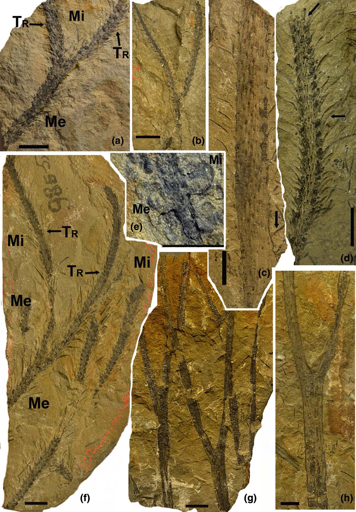
Stems are at least 150mm long and 5–20mm wide. They dichotomise at least twice into branchlets 2–6mm wide Fig. 1g). The angles between the dichotomising axes are 20–55°. Some axes bear poorly preserved, long, fusiform leaf bases (Fig. 1c). They are 6–7mm high and 1mm wide on wide axes, which are at least 7–10mm wide and 3.5–4mm high, and 0.5–0.7mm wide on narrow axes, which are 2mm wide. A ligule is not seen. Leaf bases are arranged in a tight spiral or, occasionally, in pseudo-whorls. Leaves are attached to the leaf bases. As a result of differences in preservation, some stems are smooth, without leaf bases and leaves, or bear parallel longitudinal ridges (Fig. 1h).
Morphology of sterile axes and strobili of Kossoviella timanica Petrosjan, emend. (a) Incomplete bisporangiate strobilus; arrow indicates transition (Tr) between megasporangiate (Me) and microsporangiate (Mi) parts, 21-418-2. (b) Poorly preserved strobilus: only axes with triangular bases of sporophylls are observed on their apices, 21-420. (c) Fragment of wide sterile axis with poorly preserved long, fusiform leaf bases, long sterile leaves with a single leaf vein (arrow), 21-428. (d) Incomplete megasporangiate part of strobilus; arrow indicates a sporophyll with crenulated margins, 21-422. (e) Transition zone of a strobilus; note megasporangium (Me) to the left of the axis and microsporangium (Mi) to the right, 21-430. (f) Holotype of Kossoviella timanica suggested by Petrosjan, dichotomously branched bisporangiate strobilus (left) and two incomplete strobilus fragments; megasporangia are situated proximally (Me), microsporangia distally (Mi); arrow indicates transition (Tr) between megasporangiate (Me) and microsporangiate (Mi) parts, 1-10654. (g) Dichotomously branched axes, 21-445. (h) Dichotomously branched sterile axis without leaf bases, 21-440. Scale bars = 1cm.

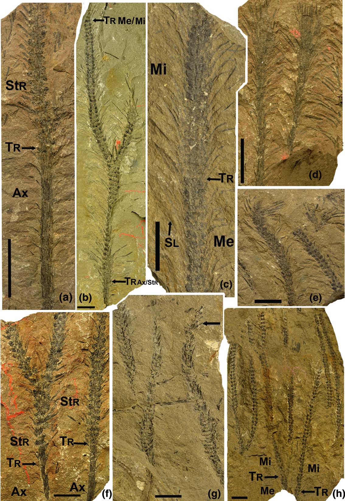
Vegetative axes usually bear persistent leaves up to 5–20mm long and 1mm wide. The leaves depart at an angle of 40–90° from the axes and often curve downwards (Fig. 2d). Leaves with smooth margins are simple, linear, rarely lanceolate and often prostrate. Leaves are rare on wide axes, 3–7mm between each other vertically, but are closely arranged on distal axes. Rarely, a single leaf vein is visible (Fig. 1c). We have found only one specimen with a whorl of narrow lanceolate leaves up to 12mm long at the apex of a sterile axis (Fig. 2d). Narrow axes are usually terminated with long strobili (Figs 2a, b, f).
Morphology of sterile axes and strobili of Kossoviella timanica Petrosjan, emend. (a) Transition between sterile axis (Ax) and basal megasporangiate strobilus part (Str), indicated by arrow, 21-437. (b) The largest incomplete bisporangiate strobilus preserved with a transition (arrow at the bottom, TrAx/Str) to the sterile axis; megasporangia are situated proximally, microsporangia distally; transition between megasporangiate and microsporangiate parts (TrMe/Mi) is indicated by arrow, 21-439. (c) Incomplete bisporangiate strobilus; megasporangia are situated proximally (Me), microsporangia distally (Mi); transition (arrow Tr) between megasporangiate (Me) and microsporangiate (Mi) parts; sporophyll lamina (Sl) with a single vein is indicated by arrow, 21-441. (d) Apices of sterile, narrowly lanceolate leaves, 10-10654 (from type collection of Petrosjan). (e) Possible loose strobilus apex, 21-436. (f) Two incomplete megasporangiate parts of strobili: dichotomously branched strobilus on the left and unbranched strobilus on the right; transitions from vegetative parts of axis (Ax) to the strobili (Str) are indicated by Tr and arrows, 7-10654. (g) Two strobili with well-preserved crenulated sporophylls (arrow), 21-426. (h) Several incomplete bisporangiate strobili and fragments of parts of incomplete microsporangiate strobilus; transitions (Tr) between megasporangiate (Me) and microsporangiate (Mi) parts are indicated by arrows, 4-10654 (from type collection of Petrosjan). Scale bars = 1cm.

2.1.2. Strobili
Many strobili are preserved attached to vegetative axes (Figs 2a, b, f); other strobili are broken at both ends (Figs 1a, d, f, 2c, h). We found only one strobilus apex in our material (Fig. 2e). Strobili are narrowly cylindrical, at least 50–160mm long and 2–8mm wide. As a rule, they are bisporangiate (Figs 1a, e, f, 2b, c, h). The microsporangiate part of strobili is usually narrower (2–4mm wide) than the megasporangiate part (4–8mm wide). The maximum length of the axis with an incomplete dichotomous strobilus is 176mm, of which 16mm belong to the vegetative axis and the remaining 160mm correspond to the strobilus part (Fig. 2b).
Strobilus axes are narrow, decreasing in width acropetally, from 1.5–3 to 0.5–2mm wide. Occasionally, the strobili are dichotomously branched (Figs 1a, b, f, 2b, f–h). The angles between the dichotomising strobilus parts are 20–50° (35° on average). In many specimens, a megasporangiate part is situated both below and slightly above the dichotomy of the strobilus (Fig. 1a).
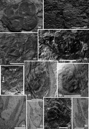
Most often, sporophylls are very densely arranged on axes (Fig. 2c). They are lanceolate, 8–18mm long, with a spoon-like basal part. Entire-margined sporophylls are occasionally present in the transition zone between the vegetative part and strobilus base, from the first to the third basal levels (Fig. 2f). Their margins then become crenulated (Fig. 1d). Sporophylls are usually attached at an angle of 30–60° (40° on average) to the strobilus axis. In the basal part of some strobili, the angle is nearly 90°; in the apical part, sporophylls are attached to the strobilus axis at more acute angles (20–30°). Occasionally, incomplete strobili show in their lower part sporophylls that are attached perpendicularly without sporangia on the axes (Fig. 2g). Sporophylls are bent upwards, reaching the middle part of the sporophylls of the next level. The sporophyll lamina has a single vein (Fig. 2c). Rarely, the sporophyll cuticle is preserved (Fig. 3b). Epidermal cells are elongate, narrow and arranged in regular rows. Stomatal apertures are parallel to the normal epidermal cells and the leaf length. The length of the stomatal aperture reaches 15–20μm. Guard cells are bean-shaped, 35–40×10μm in size. Two strobili in a single specimen (Fig. 1b) are poorly preserved, and only axes with triangular bases of sporophylls are seen on their apices.
Morphology of sporophylls and megasporangia of Kossoviella timanica Petrosjan, emend. SEM micrographs: (a) two overlaid megaspore tetrads, 21-441; (b) sporophyll cuticle, stomatal apertures are parallel with ordinary epidermal cells and the length of the leaf, 21-414-2; (c) two tetrads in megasporangium, 21-427; (d) sessile megasporangium, 21-418-2; (e) strobilus fragment, note microsporangium (Mi) to the left of the axis (Ax) and megasporangium (Me) to the right of the axis, 21-436; (f) sessile megasporangium with destroyed sporangial wall, 422-44; (g) megasporangium with partly preserved sporangial wall (in the upper part), 7-10654; (h) sporophylls with crenulated margins, 21-422; (i) crenulated sporophyll in lateral view, 6-10654; (j) megasporangium with destroyed sporangial wall, 7-10654; (k) lanceolate sporophyll with a spoon-like basal part and crenulated margins, 6-10654. Scale bars = 500μm (a, f, j); 100μm (b); 1mm (c–e, g, i, k); 2mm (h).

Sporophylls are arranged in distinctly spiral phyllotaxis on the axis of the upper part of the strobilus, but in many cases they resemble pseudo-whorls. The axes bear four to eight sporophylls (or sporophyll scars) in a spiral (or horizontal row). There are four or six sporophylls per gyre in the apical part of the strobili (Fig. 1f), and six or eight in the basal and central parts (Fig. 2b). In some cases, sporophyll scars are not preserved, and only two to four longitudinal ribs are observed on the surface of the axis.
Each sporophyll bears a single adaxial, sessile sporangium (Figs 3d, g, 4h–j). Usually, megasporangia are situated proximally, and microsporangia occur distally (Figs 1a, f, 2b, c, h). Some incomplete strobili contain only megasporangia (Figs 1d, 2f) or microsporangia (Fig. 2g). Occasionally, a microsporangium is located on one side of the axis and a megasporangium is on the other side in the transition zone between megasporangiate and microsporangiate parts (Figs 1e, 3e). A sessile megasporangium is attached to the adaxial surface of a spoon-like part of the sporophyll. The sporangial walls of megasporangia are usually destroyed (Fig. 3j). Megasporangia are ovoid, elliptical, heart-shaped and globose in outline (Fig. 3d, f, g, j). They are 1.5–3mm long and 1.3–2mm high. Megasporangium sizes and shapes depend on the size of the megaspores. Large megasporangia are elliptical; smaller ones vary from ovoid to globose. Each megasporangium contains two tetrads (Figs 3a, c, g), although the megasporangia of two basal rows contain only one tetrad in each sporangium.
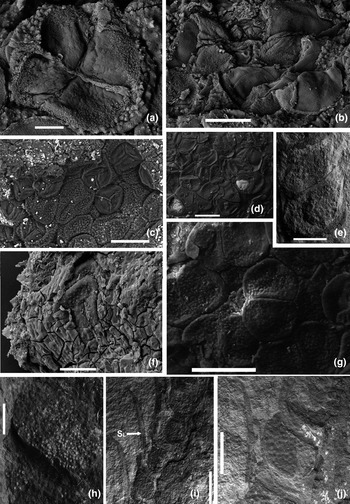
Microsporangia are globose, ovoid, occasionally with an acute distal part (Figs 4e, i, j). Their tubercular surface (Fig. 4j) is formed by microspore impressions on the inner part of sporangium. Sporangial walls are thin and one-layered (Fig. 4f). Microsporangia are 1–2.4mm long and 0.9–1.5mm high. They become smaller from the transitional part to the apex. Numerous tetrads of rounded-triangular microspores are present in the microsporangia (Figs 4b–d, g).
2.2. Description of in-situ spores
2.2.1. Microspores
Microspores are numerous, mostly in tetrads (Figs 4a–d, g, 5a). Their diameter varies from 66 to 119μm. After maceration, there were a lot of microspore tetrads and a few microspores in each specimen. Microsporangia from transitional parts of the strobilus (from mega- to microsporangia) usually contain microspores measuring 75–106μm in diameter (mean 92μm); 80% of the microspores are in tetrads. Upwards, the microspore diameter in the unbranched strobili gradually increases slightly and the number of spores preserved in tetrads decreases. In apical parts of such strobili, the microspore diameter varies from 81 to 109μm (averaging 95–97μm). Tetrads of spores are two or three times less numerous than monads. However, if a strobilus dichotomises, microspores from the sporangia of one of its branchlets transform in a manner similar to that of microspores in unbranched strobili, whereas microspores from the other branchlet become smaller (from 91 to 81–85μm on average) towards the apex and are mostly preserved in tetrads.
Microspores and microsporangia of Kossoviella timanica Petrosjan, emend. SEM micrographs: (a) fragmentary microspore tetrad, note arms of the proximal scar and sculptural elements of the equatorial region, 4-10654; (b) microspores in the microsporangium, 4-10654; (c) numerous microspores in the microsporangium, proximal and distal spore faces are visible, 4-10654; (d) numerous tetrads and monads of microspores in sporangium, trilete scars are observed proximally, 21-418-2; (e) microsporangia with an acute distal part, 21-418-2; (f) microsporangium with partly destroyed sporangial wall; distal echinate microspore surface is visible in the centre, 4-10654; (g) numerous tetrads of microspores show distal echinate surface, 21-418-2; (h) sessile microsporangium with numerous microspores, 21-416; (i) several sessile microsporangia with an acute distal part, arrow points to sporophyll lamina (Sl) with a single vein, 21-418-2; (j) sessile microsporangium with tubercular surface, formed by microspore impressions from the inner part of the sporangium, 21-418-5. Scale bars = 20μm (a); 50μm (b, f); 100μm (c, d, g); 1mm (e, j); 500μm (h); 2mm (i).

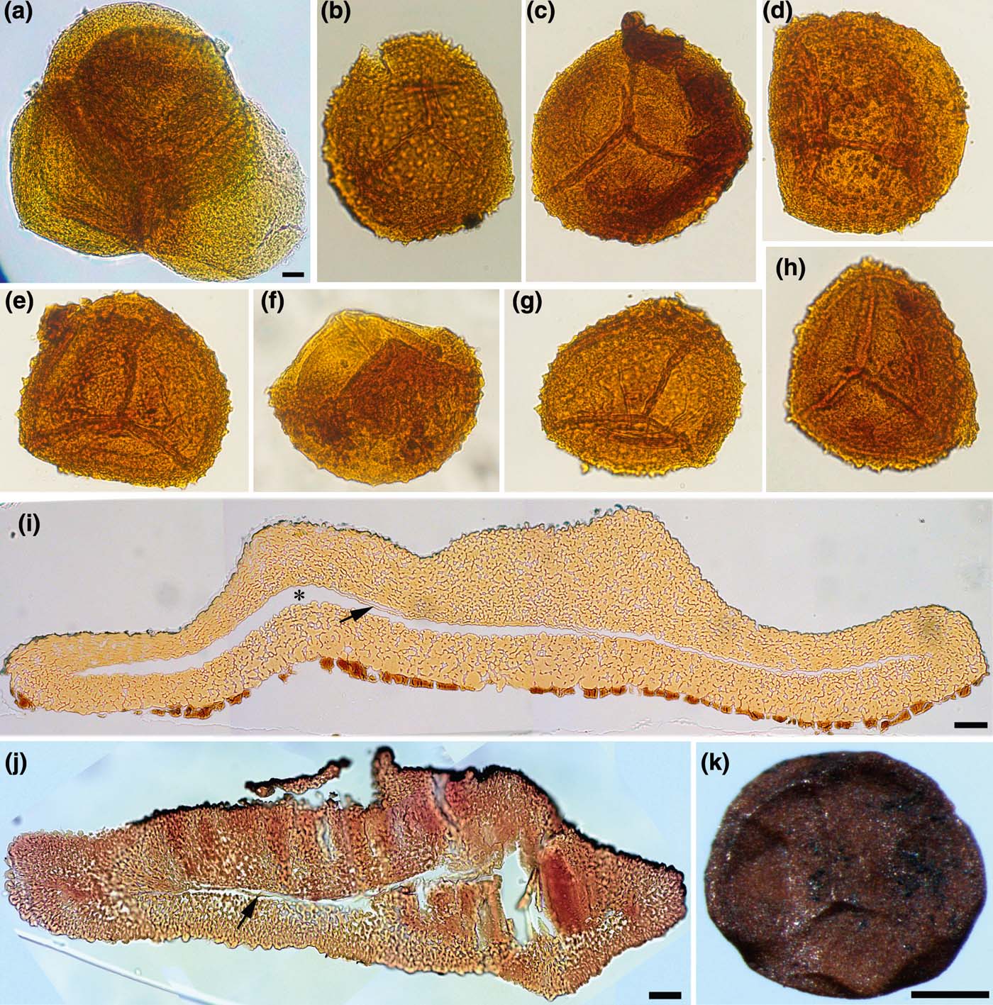
General morphology of microspores and inner structure of megaspores, LM photographs. (a) Tetrad of microspores, 21-435-1-2. (b–h) Microspores: (b, d, g) distal sculpture is clearly visible; (b) 4-10654-6-5; (c) elevated scar is distinct, 4-10654-6-2; (d) 4-10654-1-12; (e) 4-10654-1-9; (f) microspore in equatorial view, 4-10654-6-1; (g) 4-10654-1-10; (h) 4-10654-6-3. (i) Semithin section of full-sized megaspore; fused basal lamina is discernible (arrow); sporoderm cavity is clearly visible (asterisk); darker remnants of sporangial tissue are visible on the distal surface, 418-2-5-3. (j) Semithin section of abortive megaspore; basal lamina (arrow) is visible in places, 1-10654-08 U. (k) Megaspore, 21-418-2-6. Scale bars = 10μm (a–h), the bar is shown in Fig. 5a; 20μm (i, j); 200 μm (k).

Most of the observed microspores are attributable to the dispersed miospore genus Cristatisporites. In two fragments of apical sporangial walls we found larger microspores of another type that slightly resemble the miospore Membranibaculisporis. These spores are probably not associated with the studied plant.
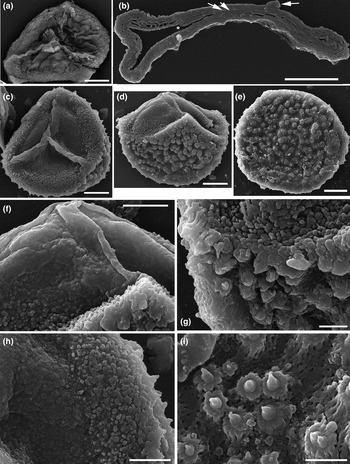
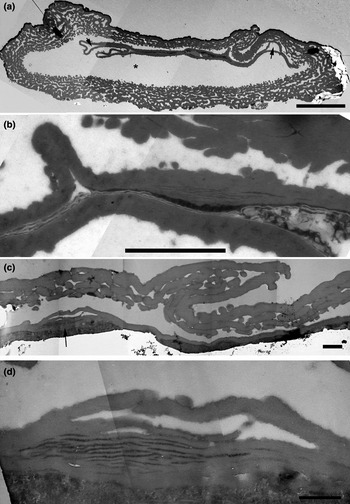
The microspores are rounded-triangular and trilete. The scar is distinct, closed and relatively elevated (Figs 5b–h, 6a, c). Their surface differs proximally, equatorially and distally. The surface of the arms is smooth (Fig. 6f). We have observed a rugulate proximal surface in microspores that we detached from tetrads (Fig. 6a). More often, the proximal surface is more weakly sculptured and slightly folded (Figs 6c, d, f). There are numerous small verrucae about 0.5–1μm in diameter close to the equator (Figs 6c, d, f–h). Distally, unusual sculptural elements are present: these have a regular polylobate base about 3μm in diameter and a pointed central tip about 3μm high (Figs 6e, g, i). They give the microspores an echinate appearance in transmitted light (Figs 5b, d, g). The sporoderm is two-layered: the thicker outer layer is composed of a net of interconnected branching elements that are rounded in cross section, and a thinner homogeneous inner layer (Fig. 7a). The spores are cavate. The cavity can occupy the entire sporoderm, but is much wider distally and equatorially than proximally (Fig. 7a), or is developed only distally and equatorially (Fig. 6b). The sporoderm is separated in such a way that some elements of the outer layer remain attached to the inner layer (Fig. 7b). The outer layer of the sporoderm is 4.7–6.5μm thick distally (Figs 6b, 7a). Proximally, its thickness decreases from about 5.1 to 1.4μm in the polar region (Fig. 7a, c). The inner layer is of constant thickness, about 0.42–0.47μm, except where it is multilamellate (Fig. 7c, arrow) near the arms of the proximal scar. These multilamellate zones are about 3.8μm long and 1.1μm thick (Figs 7b–d). The homogeneous layer becomes thicker and is split into about a dozen intervening lamellae, each about 0.03–0.04μm thick (Fig. 7d). These lamellae are quite loosely arranged, and more electron-dense matter is present between them; the outermost and innermost lamellae are thicker than most of the others (Fig. 7d). The inner layer may be folded into the outer layer in areas that are relatively close to the proximal pole and directly in the area of the pole (Fig. 7b), but this is not always the case (Fig. 7c).
Microspore morphology, surface sculpture and inner structure, SEM micrographs. (a) Microspore mechanically detached from a tetrad; note distinctly rugulate proximal surface, 21-435-1-2. (b) Semithin section, proximal surface to the top; arrow points to an arm of the proximal scar, asterisk indicates a voluminous sporoderm cavity; gametophyte cavity disappeared between pressed proximal and distal portions of the inner layer (two arrows), 21-418-2-1-3. (c) Proximal view, small verrucae are visible in equatorial region, 4-10654-1-9. (d) Equatorial view, 4-10654-6-1. (e) Distal view, 4-10654-6-2. (f) Proximal and equatorial surfaces, enlargement of (d), 4-10654-6-1. (g) Equatorial and distal surface patterns, enlargement of (d), 4-10654-6-1. (h) Enlargement of (c), 4-10654-1-9. (i) Distal sculptural elements abundantly occurring on foveolate surface, enlargement of (e), 4-10654-6-2. Scale bars=20μm (a–e); 10μm (f, h); 5μm (g, i).

Microspore ultrastructure, TEM micrographs. (a) Composite image of microspore section; proximal face is to the top of the figure; sporoderm cavity is well developed (asterisk); gametophyte cavity is discernible (arrows), 4-10654-1-9. (b) Enlargement of (a) showing a multilamellate zone; the folded electron-translucent lamella that is visible in the gametophyte cavity probably does not belong to the sporoderm, 4-10654-1-9. (c) Proximal sporoderm of a spore in the vicinity of an arm of the proximal scar; arrow points to a multilamellate zone, 21-418-2-1-2. (d) Enlargement of (c) (in the area of the arrow) showing a multilamellate zone, 21-418-2-1-2. Scale bars = 10μm (a); 2μm (b); 1μm (c); 0.5μm (d).

2.2.2. Megaspores
Megaspores were macroscopically observed in strobili (Figs 3a, c, f, g, j). They are trilete, cavate and more or less rounded in the proximal view. Those in proximal megasporangia (usually the first three rows) are comparatively small, measuring 450–700μm in diameter. Then they become larger, up to 800–1180μm in diameter. Large megaspores (up to 984μm in diameter) were also observed in the transition to the microsporangiate part of the strobili. Apparently abortive spores are between 350 and 500μm in equatorial diameter, most of which are apparently elongated along the polar axis (Fig. 8b). They co-occur in tetrads with fully developed spores (Fig. 8a).
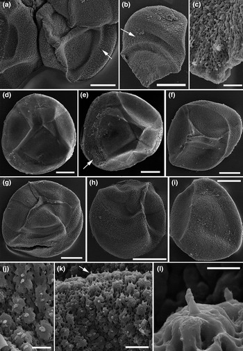
Megaspore general morphology and surface sculpture, SEM micrographs. (a) Abortive spore (to the right) co-occurs with full-sized spores; arrow indicates the position of polylobate sculptural elements, 1-10654-08 U. (b) Abortive spores in lateral position; arrow indicates the position of polylobate sculptural elements, 1-10654-01 D. (c) Enlargement of (b) showing the surface of the proximal pole, 1-10654-01 D. (d) Spore with a weakly elevated proximal scar, 21-418-2-5-1. (e) Spore with a depressed proximal scar, arrow points to scaly remnants of sporangial tissue, 21-418-2-5-3. (f–h) Spores with elevated scars: (f) 1-10654-08-M; (g) 1-10654-01 S; (h) 1-10654-08 T. (i) Spore in equatorial-distal orientation, 1-10654-08 P. (j) Polylobate sculptural elements, 1-10654-01 H. (k) Polylobate sculptural elements: top and lateral views, 1-10654-01 I. (l) Enlargement of (k) showing a polylobate sculptural element in lateral view, 1-10654-01 I. Scale bars = 200μm (a, b, d–i); 20μm (c, k); 10μm (j); 5μm (l).

The megaspores show a relatively high level of morphological variation (Fig. 8a, b, d–i). Although all of the proximal scars we have observed are narrow and closed, they vary in the degree of elevation. We have observed weakly elevated scars (Figs 5k, 8d), and even one in which the arms are represented by narrow grooves (Fig. 8e). The spore with a depressed scar and that with an insignificantly elevated scar once belonged to a tetrad, which we mechanically disintegrated: we saw that the depressed scar of the former was a counterpart of the scar of the latter (Figs 8d, e). Most megaspores bear scars that are significantly elevated over the proximal pole but approach the level of the rest of the sporoderm towards the equator (Figs 8f–h). Spores with weakly elevated and depressed scars came from an uppermost megasporangium situated below the microsporangiate zone (sample 21-418-2-5). Those with a distinctly elevated scar were observed in megasporangia in the lowermost preserved megasporangiate portion of the strobilus (1/10654-1 and 1/10654-02), and also in a sporangium from an uppermost megasporangium situated below the microsporangiate zone (1/10654-08). Abortive spores always demonstrate distinctly elevated scars (Figs 8a, b). We observed an abortive spore in which the elevation of the trilete arms close to the equator is similar to that over the pole (Fig. 8a).
The contact area and curvaturae are usually distinct but irregular (and can even transect arms of the proximal scar), probably because members of tetrads were pressed irregularly over each other (Fig. 8a).
The surface sculpture is not consistent over the distal, proximal and equatorial regions. Differences between spores were also detected. The distal sculpture in all spores is reticulate-foveolate, with isodiametric foveolae (Figs 8i, 9g), but the reticulum on spores from an uppermost megasporangium situated below the microsporangiate zone (21-418-2-2) is slightly coarser than in megaspores from other samples. The proximal surface varies from verrucate to rugulate-foveolate, both among spores and, occasionally, over the surface of a single spore. With respect to the latter, densely distributed verrucae cover the surface near the pole between the trilete arms, whereas rugulate-foveolate sculpture is present proximally, but away from the pole. This rugulate-foveolate pattern can be more or less stretched towards the equator (Fig. 8g). Since the same elements constitute the bulk of the sporoderm as well as its surface, this stretching is easily observed in sections (e.g., Fig. 9h). Some spores have a rugulate-foveolate or reticulate-foveolate proximal surface and lack verrucae (Figs 8d, e, 9e, f): these are from an uppermost megasporangium situated below the microsporangiate zone (21-418-2-2). Many spores are sculptured with verrucae over the proximal pole and rugulae-foveolae closer to the equator (Figs 8g, h, 9b–d). Abortive spores are always verrucate over the proximal hemisphere (Figs 8a, b, 9a).
Megaspore surface sculpture and inner sporoderm structure, SEM micrographs. (a–f) Proximal surface sculpture: (a) verrucate sculpture of the proximal surface of the abortive spore, 1 10654-08 U; (b) rugulate-foveolate sculpture with verrucae also present, 1-10654-08 S; (c) rugulate-foveolate sculpture, again with verrucae in association, 1-10654-08 R; (d) rugulate-foveolate sculpture with just a few verrucae present, 1-10654-08 N; (e, f) rugulate-foveolate sculpture, 21-481-2-5-1. (g) Distal reticulate-foveolate sculpture, 1-10654-08 P. (h) Outer layer of the sporoderm in the proximal-equatorial area; constituting elements are laterally stretched; proximal and distal portions of the basal lamina are fused (arrows) and the gametophyte cavity that existed between them is invisible; black background to the bottom of the figure is a cavity between the distal portion of the basal lamina and the distal outer layer, 21-418-2-6. (i) Proximal sporoderm in the vicinity of an arm of the proximal scar; outer layer and basal lamina (arrow) are visible, 21-418-2-6. (j) Sporoderm section in the equatorial region showing, from the top to the bottom of the figure, proximal outer layer, basal lamina (arrow), obliterating gametophyte cavity (asterisk), distal portion of basal lamina and distal outer layer, 21-418-2-5-3. (k) Section showing, from the top to the bottom of the figure, the lowermost portion of the outer layer, proximal and distal portion of the basal lamina (arrows) with a slit of gametophyte cavity between them and a cavity between the distal basal lamina and the outer layer, 21-418-2-5-3. (l) Proximal sporoderm; many cylindrical structural elements are cut transversely, 21-418-2-6. (m) Enlargement of (l), 21-418-2-6. Scale bars = 50μm (a–g); 10μm (h, l); 20μm (i, j); 5μm (k); 2μm (m).

The surface of the trilete arms does not differ from the proximal surface away from the arms in spores in which the arms are depressed (Fig. 8e), and scarcely differs in spores with weakly elevated arms: it becomes solid and smooth only over the pole (Fig. 8d). It is quite similar in spores with elevated trilete arms: the surface at the pole and the ridges of the arms are solid and smooth, and the surface of the slopes resembles the proximal surface away from the scar, but the surface elements can be elongated perpendicular to the arm (Fig. 8c).
Polylobate elevated sculptural elements were observed in the equatorial area of some megaspores: these are about 10–12μm in size and terminated by a short appendage about 1μm in diameter (Figs 8j–l). Such elements may be scarce or quite numerous and form an equatorial band (Figs 8a, b). Appendages are clearly evident in some specimens, but less distinct in others. They were not detected in megaspores from an uppermost megasporangium situated below the microsporangiate zone (Figs 8d, e), but they are present on most megaspores, and always distinct and quite numerous on abortive spores (Figs 8a, b).
Irregular scales, which are visible in places, represent remnants of under-macerated sporangial tissue (Figs 5i, 8e). When they occur in sections, they strongly differ from the sporoderm by their higher electron density. The sporoderm of megaspores consists of a thick outer layer and a thin basal lamina (Figs 5i, 9i, j, 10a, b, d). The sporoderm is thicker proximally than distally (Figs 5i, j, 9j, 10g). The outer layer is a complex spongy network of large solid, irregular, cylindrical elements, which branch occasionally and often fuse with each other (Figs 9j–m, 10a–g). Rounded elements that are occasionally visible in ultrathin sections at the boundary of the outer layer and basal lamina (Figs 9k, 10b, e) are cross sections of the cylindrical elements as revealed by our examination of semithin sections under the SEM (Figs 9k, m), and by tracing outlines of these elements in a ribbon of adjacent ultrathin sections. A cavity is present throughout the distal face between the outer sporoderm layer and basal lamina (Figs 5i, 10g).
Megaspore ultrastructure, TEM micrographs. (a) Composite image of a sporoderm section; proximal face is to the top of the figure, arrow points to fused proximal and distal portions of the basal lamina; outermost elements of the distal sporoderm are larger than innermost elements, 21-418-2-5-3. (b) Inner area of outer sporoderm layer; arrow points to slit-like remnants of the gametophyte cavity between proximal and distal portions of the basal lamina, 21-418-2-5-3. (c) Composite image of a longitudinal section of an abortive spore preserved in lateral orientation; only the outer sporoderm layer is present because the section did not reach the basal lamina, 1-10654-01 D. (d) The area indicated by arrow in (a); enlargement of the adjacent section of the same ribbon of sections, 21-418-2-5-3. (e) Deep area of sporoderm in equatorial region; basal lamina forms numerous folds; arrow is placed within the gametophyte cavity, 21-418-2-5-3. (f) Enlargement of (g), 1-10654-08 M. (g) Composite section of a megaspore; proximal face is to the top of the figure; the spore was mechanically damaged and the distal face is partly missing, 1-10654-08 M. Asterisk indicates sporoderm cavity in all figures. Scale bars = 6μm (a); 1μm (b, d, e); 50μm (c, g); 10μm (f).

The sporoderm varies in thickness within a single spore and from section to section. In general, the sporoderm is thicker proximally than distally (Figs 5i, j, 10g). The basal lamina is poorly preserved and often broken, or its proximal and distal portions are fused, hiding the gametophyte cavity (Fig. 10b, d). The proximal sporoderm becomes thicker in the vicinity of the trilete arms, but no other ultrastructural differences were detected in these areas. The basal lamina is often folded, but the folds are not restricted to a particular area of the spore (Figs 10d, e).
The outer sporoderm layer of spores that we think are more mature (21-418-2-2) appears denser (Fig. 10a) than that in other full-sized spores we have studied (Fig. 10g) as well as in abortive spores (Fig. 10c), which means that the ratio of sporopollenin structural elements to gaps between them seems higher. Sections of structural elements show less angular outlines (compare Fig. 10a and f). In addition, distally outermost elements appear thicker than innermost elements in the former group (Fig. 10a). No such size gradient was detected in other full-sized megaspores examined (Fig. 10g) or in abortive megaspores (Fig. 10c).
3. Systematic palaeobotany
Class Lycopsida
Order Incertae sedis
Family Kossoviellaceae Petrosjan, Reference Petrosjan, Kossovoj, Srebrodolskaya, Kolobova and Modzalevskaya1984 emend.
Type genus Kossoviella Petrosjan, Reference Petrosjan, Kossovoj, Srebrodolskaya, Kolobova and Modzalevskaya1984
Emended family diagnosis. Eligulate heterosporous lycopsids with dichotomously branched axes. Leaves simple, with smooth margins and a single vein. Leaf bases fusiform in tight spiral or in pseudo-whorls. Strobili bisporangiate: megasporangia proximal, microsporangia distal. Sporadic megasporangia and microsporangia occur at the same level on opposite sides of the axis in transition zone. Sporophylls long, crenulated, densely arranged on the axis. Each megasporangium contains four to eight trilete megaspores. Microsporangia with numerous tetrads of rounded-triangular, trilete microspores.
Genus Kossoviella Petrosjan, Reference Petrosjan, Kossovoj, Srebrodolskaya, Kolobova and Modzalevskaya1984 emend.
Type species Kossoviella timanica Petrosjan, Reference Petrosjan, Kossovoj, Srebrodolskaya, Kolobova and Modzalevskaya1984
Emended generic diagnosis. Heterosporous lycopsids with erect, dichotomously divided axes covered with leaves without a ligule. Leaves with smooth margins, linear, rarely lanceolate, with a single vein. Leaves in tight spiral or in pseudo-whorls. Leaf bases fusiform. Strobili bisporangiate: megasporangia in proximal-middle part and microsporangia in middle-distal part of strobilus. Sporadic megasporangia and microsporangia occur at the same level on opposite sides of the axis in the transition zone. Sporophylls lanceolate, with spoon-like base and crenulated margins. Each sporophyll bears a single adaxial, sessile sporangium. Megasporangia large, with usually destroyed sporangial walls and with four to eight trilete megaspores. Megaspore sporoderm two-layered and cavate. Microsporangia globose, ovoid, with numerous tetrads of rounded-triangular, trilete microspores. Microspore sporoderm two-layered, cavate, with multilamellate zones near arms of the proximal scar.
Kossoviella timanica Petrosjan, Reference Petrosjan, Kossovoj, Srebrodolskaya, Kolobova and Modzalevskaya1984 emend.
Orlova & Zavialova
Holotype. 1/10654 TSNIGR Museum, VSEGEI, St. Petersburg (Petrosjan & Kossovoj Reference Petrosjan, Kossovoj, Srebrodolskaya, Kolobova and Modzalevskaya1984, pl. 1, fig. 1; Fig. 1f of the present study).
Type locality. Western slope of North Timan, Russia, south-eastern sea coast of Cheshskaya Bay, 500m to the north of the mouth of the Suvojnaya River.
Localities. Suvojnyj part of the east coast of Cheshskaya Bay between Suvojnyj Cape and Krestovyi Cape; Ludovatyj area of the east coast of Cheshskaya Bay between the East Ludovatyj Nos Cape and the mouth of the Velikaya River; and Vyuchejskij borehole.
Stratigraphy. Grubyj Ruchej, Rassokha and Ust'bezmoshitsa formations.
Age. Middle–Late Frasnian, Late Devonian.
Repository. TSNIGR Museum, VSEGEI, St. Petersburg; Palaeontological Museum of the Department of Sedimentary Geology, Institute of Earth Sciences, SPBU (Petrosjan and Snigirevsky collections); Department of Palaeontology, Geology Faculty, MSU.
Emended diagnosis. Heterosporous lycopsids with erect axes up to 20mm wide, dichotomising at least twice into branchlets up to 2–6mm wide. Leaf bases small, long, fusiform, arranged in tight spiral or in pseudo-whorls. Linear (rarely lanceolate) sterile leaves with smooth margins. Narrow axes terminated by long, narrowly cylindrical strobili at least 50–160mm long and 2–8mm wide. Strobili bisporangiate. Microsporangiate strobilus part narrower than megasporangiate part. Strobilus axes narrow, decreasing in width acropetally. Occasionally, strobili dichotomously branched. Angles between dichotomising strobilus parts averaging 35°. Sporophylls lanceolate, with spoon-like base, 8–18mm long, arranged very densely on axes. Sporophylls with crenulated margins, attached at an angle averaging 40° to strobilus axis. Sporophyll lamina with single vein. Sporophylls in distinctly spiral phyllotaxis on axis, often in pseudo-whorls. Each sporophyll with a single adaxial, sessile sporangium. Megasporangia mostly situated in proximal and middle parts, and microsporangia in middle and distal parts. Megasporangia and microsporangia occur sporadically at the same level on opposite sides of the axis. Megasporangia ovoid, elliptical, 1.5–3mm long and 1.3–2mm high, with very thin or destroyed sporangial walls, with one or two tetrads. Megaspores 450–1180μm in diameter, trilete, most often with an elevated scar, more or less rounded in the proximal view, cavate. Proximal surface most often rugulate-foveolate; verrucae may be present or be prominent. Distal surface reticulate-foveolate. Sporoderm with thick outer layer and basal lamina. Outer layer composed of a network of occasionally branching cylindrical elements; basal lamina separated from outer layer by an extended cavum. Microsporangia globose, ovoid, occasionally with acute distal part, 1–2.4mm long and 0.9–1.5mm high. Sporangial walls thin and one-layered. Microspores numerous, 66–119μm in diameter, rounded-triangular in outline, trilete, of Cristatisporites-type. Proximal scar narrow, closed, elevated. Spore surface rugulate to smooth proximally, verrucate subequatorially, and echinate distally. Sporoderm two-layered, cavate, with multilamellate zones near arms of proximal scar.
4. Discussion
4.1. Previous studies of Kossoviella
The family Kossoviellaceae and genus Kossoviella with the type species K. timanica were established by Petrosjan in Petrosjan & Kossovoj (Reference Petrosjan, Kossovoj, Srebrodolskaya, Kolobova and Modzalevskaya1984). The genus was named in honour of the outstanding Russian geologist Kossovoj, who collected the rich palaeobotanical material from North Timan. Petrosjan attributed the Kossoviellaceae to lycopsids and believed that Kossoviella was similar to plants of the Selaginellales. Petrosjan (Petrosjan & Kossovoj Reference Petrosjan, Kossovoj, Srebrodolskaya, Kolobova and Modzalevskaya1984) pointed out that plants of the family Kossoviellaceae differed from other members of the Selaginellales by the presence of fertile zones of mega- and microsporangia, irregularly alternating on axes. She thought that plants of the Kossoviellaceae were herbaceous, heterosporous, with dichotomous axes bearing simple leaves with serrated leaf margins and a ligula. She remarked that sterile and fertile leaves did not differ from each other. Our study has corrected some of Petrosjan's conclusions (Table 1). Firstly, we did not find a ligule on the axes and strobili studied. We suppose that Petrosjan mistook a vascular scar on the leaf bases for a ligula. Secondly, we found that sporophylls differ morphologically from sterile leaves. Sterile leaves are narrower, shorter and with smooth leaf margins, whereas sporophylls are lanceolate, long and with crenulated leaf margins (Table 1). Thirdly, we believe that the reproductive structures of Kossoviella were arranged in strobili, which terminated lateral axes. Megasporangia are mainly situated in the proximal and middle parts of the strobilus, with microsporangia occurring above them. Only rarely is the arrangement of megasporangia and microsporangia irregular. Lastly, we think that Kossoviella was not a herbaceous plant, because some preserved axes are relatively wide (20mm) and maintain their thickness through their visible length (up to 150mm). Incomplete strobili are up to 160mm long, so intact strobili were probably significantly longer. We suppose that Kossoviella was a small arborescent plant that reached a height of more than 1–1.5m. Unfortunately, anatomical structures are not preserved in the material studied to substantiate this suggestion.
Main features of Kossoviella timanica Petrosjan, emend.: a comparison between our results and the description made by Petrosjan (Petrosjan & Kossovoj Reference Petrosjan, Kossovoj, Srebrodolskaya, Kolobova and Modzalevskaya1984).

–, no information.
We corrected the description of Kossoviella and supplemented it with more detail. Sizes of mega- and microsporangia and mega- and microspores differ slightly from the sizes provided by Petrosjan (see Table 1). We also found differences in distal and proximal surface sculpture (Table 1). Spores of Kossoviella were previously studied under the SEM and TEM by Telnova (Telnova & Meyer-Melikyan Reference Telnova and Meyer-Melikyan2002; Telnova Reference Telnova2005, Reference Telnova2007). Telnova reported a scaly-reticulate proximal sculpture; however, some of the illustrations show a megaspore surface that is partly covered by scaly remnants of under-macerated sporangial tissue. She described a peculiar thin outer layer of scales and an underlying thick alveolate layer in TEM sections, but did not mention either a basal lamina or a sporoderm cavity. Our observations contradict Telnova's description of the megaspore ultrastructure. The only explanation we can think of is that she examined a single micrograph of a region of the sporoderm and confused the top and bottom of the image. In addition, she reported the presence of granules in the sporoderm. In our opinion, she observed cross sections of cylindrical elements of the outer layer, which indeed appear similar to granules if one examines a single micrograph rather than micrographs of a series of adjacent sections. We report for the first time a sporoderm cavity in both megaspores and microspores, and multilamellate zones in the inner sporoderm layer of microspores. In general, multilamellate zones are known in spores of Selaginellales, Isoetales and Pleuromeiales, and are considered to be characteristic of these groups of heterosporous lycopsids (Lugardon et al. Reference Lugardon, Grauvogel-Stamm and Dobruskina1999; Grauvogel-Stamm & Lugardon Reference Grauvogel-Stamm and Lugardon2001). They have also been reported in spores of the homosporous lycopsids Leclercqia andrewsii and L. complexa from the Lower Devonian of Canada (Wellman et al. Reference Wellman, Gensel and Taylor2009). Although both megaspores and microspores of Kossoviella are characterised by a two-layered cavate sporoderm, they show significant differences in the ultrastructure. This particularly concerns the basal lamina of megaspores and the multilamellate zone of microspores. We conclude that we are dealing with a relatively advanced stage of heterospory.
4.2. Comparison with the Devonian heterosporous lycopsids
The first findings of heterosporous lycopsids come from the Middle Devonian (Givetian). Only two heterosporous genera are represented by bisporangiate strobili or fertile structures in the Givetian. Petrification of the bisporangiate strobilus Mixostrobus givetensis was described from the Givetian of Kazakhstan (Senkevitsch et al. Reference Senkevitsch, Jurina and Arkhangelskaya1993). It is small (38mm long, 26mm wide) and elongated ovate. Similar to Kossoviella, sporophylls are arranged in a spiral or in pseudo-whorls. Unlike Kossoviella, sporophylls with smooth margins have a composite structure (pedicel and lamina), and micro- and megasporangia are irregularly distributed. Differences also exist between the spores of these genera. Megaspores of Mixostrobus are numerous, of Lagenicula-type, with a gula, and smaller (480–650μm) than those of Kossoviella (450–1180μm), which lack a gula and occur in much lower numbers (up to eight) in a sporangium. Microspores of Mixostrobus are generally smaller and less variable in size than those of Kossoviella (70–90μm vs. 66–119μm, respectively); they are reticulate, unlike the microspores of Kossoviella, which are rugulate proximally, verrucate equatorially and echinate distally.
A bisporangiate lycopsid, Yuguangia ordinata, was described from the Upper Givetian of China (Hao et al. Reference Hao, Xue, Wang and Liu2007). Like Kossoviella, it has dichotomously branching axes, sporophylls with spiny margins and strobili with proximal megasporangia and distal microsporangia. Yuguangia differs from Kossoviella by the presence of a ligula, sterile leaves with spiny margins, sporophylls with a composite structure (pedicel, lamina and heel) and in the size and structure of their spores.
The Late Devonian can be considered as the heyday of heterosporous lycopsids. More than ten heterosporous lycopsid genera (Fairon-Demaret Reference Fairon-Demaret1977, Reference Fairon-Demaret1991; Chitaley & Reference Chitaley and McGregorMcGregor 1988; Cai & Chen Reference Cai and Chen1996; Wang Reference Wang2001; Berry et al. Reference Berry, Wang and Chongyang2003; Wang et al. Reference Wang, Li, Geng and Chitaley2003; Wang et al. Reference Wang, Xu, Basinger and Wang2012; Meng et al. Reference Meng, Wang, Xue and Zhu2013, Reference Meng, Wang and Yao2015, Reference Meng, Liu, Wang and Yao2016; Wang et al. Reference Wang, Meng, Xue, Basinger, Guo and Liu2014, Reference Wang, Qin, Meng, Liu and Ferguson2016) appeared at this time, but only three of these (including Kossoviella) were bisporangiate. Chamaedendron multisporangiatum from the Lower Frasnian of China (Schweitzer & Li Reference Schweitzer and Li1996) is similar to Kossoviella in sporophyll morphology (crenulated margins and spoon-like base). However, the fertile bisporangiate zones or cone-like structures of Chamaedendron, which bear mega- and microsporangia, are much shorter than Kossoviella strobili (up to 8mm and at least 160mm, respectively).
A bisporangiate strobilus of Bisporangiostrobus harrisii was described from the Famennian of the US (Chitaley & McGregor Reference Chitaley and McGregor1988). Kossoviella resembles Bisporangiostrobus in lacking a ligula, and in having a similar arrangement of sporangia on the axis (in a close, low spiral and pseudo-whorls; proximal megasporangia and distal microsporangia). Unlike Kossoviella, sporophylls of Bisporangiostrobus have smooth margins and are of composite structure; microspores are smaller (46–69μm in diameter) and of Geminospora-type; and megaspores are slightly smaller (474–758μm in diameter) and of Duosporites-type.
The first find of a heterosporous, monosporangiate lycopsid, namely Longostachys latisporophyllum (Cai & Chen Reference Cai and Chen1996), is also from the Middle Devonian (Givetian). In common with Kossoviella, the strobili of Longostachys have spoon-like sporophylls with spiny margins and sessile globose or elliptical sporangia of similar sizes. Longostachys differs from Kossoviella in that the strobili are larger (up to 225mm) with wider axes (2.9mm), the sporophylls are longer (up to 30mm) and the spores are different (megaspores of Laevigatisporites-type).
Most Devonian monosporangiate lycopsids are known from the Famennian. Minostrobus chaohuensis has been repeatedly reported from the Famennian of South China (Wang Reference Wang2001; Wang et al. Reference Wang, Xu, Basinger and Wang2012; Meng et al. Reference Meng, Wang, Xue and Zhu2013, Reference Meng, Wang and Yao2015). The monosporangiate strobili of Minostrobus have sporangia that are similar in shape and size, and sporophylls that are similarly arranged on the axis. Minostrobus differs from Kossoviella in that the strobili are smaller, the sporophylls have smooth margins and a ligula, the megaspores are of Lagenicula-type and the microspores of Lycospora-type.
The monosporangiate lycopsid Changxingia longifolia (Wang et al. Reference Wang, Meng, Xue, Basinger, Guo and Liu2014, Reference Wang, Qin, Meng, Liu and Ferguson2016) from the Famennian of China resembles Kossoviella timanica in having twice dichotomous axes and in the manner of attachment of sterile leaves with smooth margins. By comparison with Kossoviella, Changxingia has smaller strobili with a composite structure of sporophylls with smooth margins, megaspores of Lagenicula-type and foveolate microspores 18–26μm in diameter. Other monosporangiate lycopsids from the Famennian are very different from Kossoviella.
4.3. Remarks on sporoderm ontogenesis
Since we have numerous well-preserved spores of various sizes, many of which are preserved as tetrads and some as monads, in sporangia on relatively long strobili, we had hoped to obtain some information about sporoderm ontogenesis. We succeeded for the megaspores. We suspect that those from specimen 21-418-2, an uppermost sporangium situated below the microsporangiate zone, are at a slightly later stage of maturation than other megaspores we examined. Abortive megaspores served as a source of information about earlier stages of development.
The proximal surface of supposedly more mature spores is rugulate-foveolate, that of the supposedly least mature spores is densely verrucate and the intermediate surface pattern is verrucate-rugulate-foveolate. The distal surface of supposedly more mature spores shows a slightly coarser reticulum than that of other spores. We know that the sporoderm of the megaspores lacks a solid tectum; therefore, the inner structure of the sporoderm is, in part, visible on intact spores, and it is a reticulum of branching units. If enlargement and lateral stretching of structural units and spaces between them took place during the final stages of ontogenesis of megaspores, then densely spaced verrucae could have gradually transformed into rugulae on the proximal surface of megaspores, and gaps between structural elements, which earlier were densely packed, become visible as foveolae. The proximal scar is more elevated in supposedly least developed spores, and is less elevated in apparently more mature spores. The lips of the scar could also have become less elevated, owing to stretching of the sporoderm. The accumulation of amorphous sporopollenin on most accessible areas of the sporoderm (outer distal areas) during the latest stages of ontogenesis could explain why in supposedly more mature megaspores the distal surface pattern is coarser, and larger structural elements are observed in sections closer to the distal surface. Accumulating sporopollenin made the outlines of structural elements of supposedly more mature sporoderm more rounded.
We checked the literature for any modern analogues and found indirect support for our idea in Rowley & Morbelli (Reference Rowley and Morbelli1995), at least where the transformation of verrucate into rugulate-foveolate sculpture is concerned. Rowley & Morbelli (Reference Rowley and Morbelli1995) reported that structural units of the sporoderm of Selaginella megaspores enlarge considerably during the ontogenesis, as well as spaces between these units, and lateral stretching occurs during this process. We have not found any modern analogue showing that arms of the proximal scar can become less elevated when mature. However, all underdeveloped megaspores that we observed in our material bear higher scars in comparison to those of full-sized megaspores, which suggests that our explanation might be correct.
The sporoderm varies significantly in thickness within the same megaspore and from one section to the next, so we failed to detect whether there is a difference in thickness among spores from different levels of strobili. Sections of one abortive spore did not reveal a basal lamina (1-10654-01 D), but we found a basal lamina in a different abortive spore (1-10654-08 U; Fig. 5j). This means that both layers of the sporoderm, as well as the cavity between them, developed early in the ontogenesis.
5. Conclusions
We have reinvestigated a unique heterosporous, bisporangiate lycopsid from the Frasnian of North Russia. For the first time, we noted that this plant was eligulate, with smooth-margined sterile leaves and sporophylls with crenulated margins; strobili were long, cylindrical, terminal and occasionally dichotomously branched; megasporangia and microsporangia were situated proximally and distally, respectively, and some incomplete strobili contained only megasporangia or microsporangia. Megasporangia were ovoid, elliptical, with very thin or destroyed sporangial walls, with one or two tetrads of trilete, cavate megaspores (450–1180μm in diameter). The megaspores were rugulate-foveolate proximally and reticulate-foveolate distally. Microsporangia were globose, ovoid, with a tubercular surface and thin sporangial walls. Numerous trilete microspores of Cristatisporites-type, mostly in tetrads, occurred in the microsporangia. The microspore surface was rugulate to smooth proximally and echinate distally. We tried to trace the dynamics of spore development. Both megaspores and microspores were found to be cavate, with a two-layered sporoderm. The outer sporoderm layer was formed by intertwined and occasionally branching cylindrical elements. The inner layer of the megaspore sporoderm is a basal lamina. The inner homogeneous layer of the microspore sporoderm is split into multilamellate zones near the arms of the proximal triradiate scar.
6. Acknowledgements
We thank Dr. Roman Rakitov and Dr. Elena Zhegallo (PIN RAS) for assistance with the SEM, Aleksandra Sokolova (PIN RAS) for discussion of the cuticle morphology, Dmitry Mamontov (MSU) for help with spore maceration and Aleksandr Yudin (MSU) for technical assistance. Georgii Davidovich (head of the laboratory of electron microscopy, MSU) is thanked for providing access to the TEMs and for the helpful assistance of his staff. We are grateful to Prof. David Batten (Aberystwyth University) for linguistic corrections and valuable suggestions. Comments of the reviewers helped us to improve the manuscript. This work is supported by the Russian Foundation for Basic Research, project no. 15-04-09067.