Introduction
Microtubules (MTs) are a major component of the cytoskeleton in cells, and play a crucial role in cell division, morphogenesis, motility, protein and organelle transport, signalling and multiple neuronal activities. To complete these functions, MTs remain highly dynamic via a well-balanced assembly and disassembly process, which is regulated by numerous proteins (Nogales and Wang, Reference Nogales and Wang2006; Walczak and Heald, Reference Walczak and Heald2008; Gardner et al., Reference Gardner, Zanic and Howard2013; Brouhard, Reference Brouhard2015), including MT-severing proteins (MTSPs) (Errico et al., Reference Errico, Ballabio and Rugarli2002; Sharma et al., Reference Sharma, Bryant, Wloga, Donaldson, Davis, Jerka-Dziadosz and Gaertig2007; Nakamura et al., Reference Nakamura, Ehrhardt and Hashimoto2010; Mukherjee et al., Reference Mukherjee, Diaz Valencia, Stewman, Metz, Monnier, Rath, Asenjo, Charafeddine, Sosa, Ross, Ma and Sharp2012; Smith et al., Reference Smith, Milne, Nelson, Eddie, Brown, Atanassova, O’Bryan, O’Donnell, Rhodes, Wells, Napper, Nolan, Lalanne, Cheeseman and Peters2012). MTSPs are categorized into the ‘meiotic’ subfamily of ATPases Associated with diverse cellular Activity (AAA) superfamily (Vale, Reference Vale2000; Monroe and Hill, Reference Monroe and Hill2016). Furthermore, all MTSPs have an AAA domain that can bind and hydrolyze ATP to sever an MT along its length (Vale, Reference Vale2000; Roll-Mecak and Vale, Reference Roll-Mecak and Vale2008; Johjima et al., Reference Johjima, Noi, Nishikori, Ogi, Esaki and Ogura2015; Monroe and Hill, Reference Monroe and Hill2016). Therefore, MT severing is an efficient mechanism to reorganize MTs compared with end-limited MT depolymerization by kinesins (Wordeman, Reference Wordeman2005; Howard and Hyman, Reference Howard and Hyman2007; Roll-Mecak and McNally, Reference Roll-Mecak and McNally2010; Friel and Howard, Reference Friel and Howard2011; Tanenbaum et al., Reference Tanenbaum, Macurek, van der Vaart, Galli, Akhmanova and Medema2011; Sharp and Ross, Reference Sharp and Ross2012).
Mitosis is the fundamental process for eukaryote cell proliferation. Moreover, abnormal chromosome segregation can cause aneuploidy, which is one of the primary causes of tumorigenesis (Meraldi et al., Reference Meraldi, Honda and Nigg2004; Lee, Reference Lee2006). Mitosis, particularly during anaphase in which two sets of sister chromatids are moving away from each other, requires rapid kinetochore-MT disassembly from both ends of the spindle (Rogers et al., Reference Rogers, Rogers, Schwimmer, Ems-McClung, Walczak, Vale, Scholey and Sharp2004; Zhang et al., Reference Zhang, Rogers, Buster and Sharp2007; Rath and Sharp, Reference Rath and Sharp2011). Previous studies have shown that MTSPs play important in mitosis via their MT-severing activity. Furthermore, katanin 60, the first identified MTSP (McNally and Vale, Reference McNally and Vale1993), is targeted to the centrosome (Hartman et al., Reference Hartman, Mahr, McNally, Okawa, Iwamatsu, Thomas, Cheesman, Heuser, Vale and McNally1998) and promotes γ-tubulin redistribution in human cells (Buster et al., Reference Buster, McNally and McNally2002). Katanin activity is also an important determinant of spindle length, as reported in a comparison study with Xenopus (X.) tropicalis and X. laevis egg extracts (Rose et al., Reference Rose, Wilbur, McNally and François2011). The second identified MTSP member, spastin, is mutated in the most frequent form of autosomal dominant spastic paraplegia (Hazan et al., Reference Hazan, Fonknechten, Mavel, Paternotte, Samson, Artiguenave, Davoine, Cruaud, Dürr, Wincker, Brottier, Cattolico, Barbe, Burgunder, Brice, Fontaine, Heilig and Weissenbach1999), and its centrosomal location indicates its centrosome-based mitotic function (Errico et al., Reference Errico, Ballabio and Rugarli2002, Reference Errico, Claudiani, D’Addio and Rugarli2004; Zhang et al., Reference Zhang, Rogers, Buster and Sharp2007). Moreover, Fidgetin (FIGN) (Zhang et al., Reference Zhang, Rogers, Buster and Sharp2007; Mukherjee et al., Reference Mukherjee, Diaz Valencia, Stewman, Metz, Monnier, Rath, Asenjo, Charafeddine, Sosa, Ross, Ma and Sharp2012), fign-like 1 (FIGNL1) (Luke-Glaser et al., Reference Luke-Glaser, Pintard, Tyers and Peter2007), katanin-like 1 (Sonbuchner et al., Reference Sonbuchner, Rath and Sharp2010) and katanin-like 2 (Ververis et al., Reference Ververis, Christodoulou, Christoforou, Kamilari, Lederer and Santama2016) are MTSPs that are considered important for mitosis, and their mitotic functions appear to be divergent as they show different localization patterns within cells, and a loss of function diversely affects MT dynamics and organization within the spindles.
Meiosis, although similar to mitosis, is a unique process. Meiosis is composed of two rounds of cell division in which homologous chromosomes are separated during the first round (meiosis I), and sister chromatids are separated during the second round (meiosis II). However, the final daughter cells, gametes or polar bodies cannot reproduce as they are haploid. Furthermore, oocytes do not have typical centrosomes, but some centrosomal components are found at the spindle poles. Therefore, a single protein could have different localization, function and regulation mechanisms for meiosis vs. mitosis. Moreover, normal meiosis, especially female meiosis, is crucial for genome stability, and dysfunctional female meiosis is a major cause of human genetic disease (Feng et al., Reference Feng, Sang, Kuang, Sun, Yan, Zhang, Shi, Tian, Luchniak, Fukuda, Li, Yu, Chen, Xu, Guo, Qu, Wang, Sun and Liu2006; Kogo et al., Reference Kogo, Kowa-Sugiyama, Yamada, Bolor, Tsutsumi, Ohye, Inagaki, Taniguchi, Toda and Kurahashi2010; Filges et al., Reference Filges, Manokhina, Peñaherrera, McFadden, Louie, Nosova, Friedman and Robinson2015). During female meiosis in Caenorhabditis elegans (C. elegans), the katanin 60 homologue MEI-1 is required for the translocation of the meiosis I spindle to the oocyte cortex and parallel/antiparallel MT organization in meiotic spindles (Yang et al., Reference Yang, McNally and McNally2003). However, the role of MTSPs in mammalian female meiosis, which is commonly used as a model of human meiosis, is not fully understood. FIGNL1 is a homologue of FIGN, both of which are members of the MTSP family. Furthermore, Fign has MT-severing activity and has been identified as a protein encoded by mutant genes in fidget mice (Cox et al., Reference Cox, Mahaffey, Nystuen, Letts and Frankel2000). Previous studies have also shown that FIGNL1 is mainly concentrated near the centrosome in mouse HEK293T cells (39). In addition, overexpression of Fignl1 can damage the integrity of the cytoplasmic MT network and inhibit MT-related ciliogenesis (Zhao et al., Reference Zhao, Jin, Wang, Sun, Hong, Cao and Wang2016). However, removal of Fignl1 by short hairpin RNA treatment can enhance ciliogenesis in HEK293T cells (Zhao et al., Reference Zhao, Jin, Wang, Sun, Hong, Cao and Wang2016). Therefore, these preliminarily results reveal the MT-severing activity of FIGNL1.
FIGNL1 has also been reported to play an important role in the transmission of information in nerve cells by severing MTs and regulating MT terminal kinetics and growth orientation (Coralie et al., Reference Coralie, Amélie, Laïla, Christian, Daniel, Stéphanie, Monica, Christophe, Philippe, Céline, Leticia, Susanne, Sylvie, Corinne, Fatiha, Jean-Christophe, Annie and Jamilé2018). In addition, as the ligand of RAD51 recombinase, FIGNL1 plays a role in repairing DNA double-stranded breaks in homologous recombination (HR), which is essential for maintaining genomic stability and preventing developmental disorders and cancer (Ma et al., Reference Ma, Li, Yao, Lin, Gu, Xu, Deng, Ma and Zhang2017; Matsuzaki et al., Reference Matsuzaki, Kondo, Ishikawa and Shinohara2019). Previous studies have also shown that Fignl1 expression in the ovaries of ageing rats decreases (Govindaraj and Rao, Reference Govindaraj and Rao2015), and Fignl1 has been screened as a possible gene associated with testicular weight loss and moderate malformed spermatozoa (L’Hôte et al., Reference L’Hôte, Vatin, Auer, Castille, Passet, Montagutelli, Serres and Vaiman2011). However, to the best of our knowledge, the study of Fignl1 in meiosis of mammalian and human oocytes has not been previously reported.
Materials and methods
General chemicals, reagents cells and animals
Chemicals and reagents were obtained from Sigma-Aldrich; Merck KGaA unless otherwise stated. The NIH3T3 cell line was purchased from the American Type Culture Collection. In total, 120 3-week-old female specific pathogen-free ICR mice (weighing 18–20 g) used in this study were obtained from Vital River Experimental Animal Technical Co., Ltd. Animals were housed at a temperature of 20–26˚C and a humidity of 40–70% under a 12 h light/dark cycle. The mice were fed in feeding boxes, and the frequency of food replacement was two times a week, and the frequency of water bottle replacement was three times a week. All animal experiments were approved by the Animal Care and Use Committee of Nanjing Medical University (Nanjing, China) and performed in accordance with institution guidelines.
Antibodies
Mouse monoclonal anti-β-actin (cat. no. A5316-100) antibody was obtained from Sigma-Aldrich; Merck KGaA, Germany. Mouse monoclonal anti-FIGNL1 (A-4) (cat. no. sc-398264) and mouse monoclonal anti-β-tubulin (cat. no. sc-5274) antibodies were purchased from Santa Cruz Biotechnology, USA. Human anti-centromere CREST antibody (cat. no. 15-234) was purchased from Antibodies Incorporated, China. Mouse monoclonal anti-EGFP (F56-6A1.2.3) (cat. no. ab184601) was purchased from Abcam, UK. Cy2-conjugated donkey anti-mouse IgG (code no. 715-225-150), tetramethylrhodamine (TRITC)-conjugated donkey anti-goat IgG (code no. 705-025-147), and Alexa Fluor 647-conjugated donkey anti-human IgG (code no. 709-605-149) were purchased from Jackson ImmunoResearch Laboratories, USA. Horseradish peroxidase (HRP)-conjugated rabbit anti-goat IgG (cat. no. 31402) and HRP-conjugated goat anti-mouse IgG (cat. no. 31430) were purchased from Invitrogen; Thermo Fisher Scientific, USA.
Oocyte collection and culture
Immature oocytes arrested in prophase I [germinal vesicle (GV) oocytes] were obtained from the ovaries of 3-week-old female ICR mice. The mice were first euthanized with CO2 and then sacrificed by cervical dislocation, and the ovaries were isolated and placed in operation medium (HEPES) with 2.5 nM milrinone and 10% fetal bovine serum (FBS) (Gibco; Thermo Fisher Scientific, USA). Oocytes were released from the ovary by puncturing the follicles with a hypodermic needle. Cumulus cells were washed off the cumulus–oocyte complexes, and 50 isolated denuded oocytes were placed in 100-µl droplets of culture medium under mineral oil in plastic dishes (BD Biosciences, USA). The culture medium was MEM+ [MEM with 0.01 mM EDTA, 0.23 mM Na-pyruvate, 0.2 mM pen/strep, 3 mg/ml bovine serum albumin (BSA) and 20% FBS]. The oocytes were cultured at 37.0°C, 5% O2, and 5% CO2 in a humidified atmosphere. Prior to in vitro maturation (IVM), all culture medium included 2.5 μM milrinone to prevent the resumption of meiosis.
siRNA production and microinjection
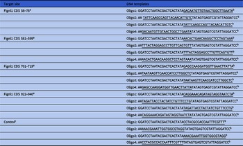
Sequences of all DNA templates used for siRNA production are listed in Table 1. The sequence of the control templates was a mock sequence that did not specifically bind to any mRNA from the mouse genome. DNA templates against four different DNA coding (sequence coding for the amino acids in a protein; CDS) regions of Fignl1 siRNA were designed online through BLOCK-iT™ RNAi Designer (http://rnaidesigner.invitrogen.com/rnaiexpress/) with some modifications. The sequence specificity was verified through a BLAST (http://blast.ncbi.nlm.nih.gov/Blast.cgi) homology search.
DNA oligos for siRNA production

a The numbers are the starting and ending position of the target sites in Fignl1 CDS (NM_001163359.1 in NCBI).
b Two pairs of DNA oligos are required for each double-stranded siRNA. Oligo2 is complementary with Oligo1 except for an ‘AA’ overhang at 5′; Oligo3 is complementary with Oligo4 except for an ‘AA’ overhang at 5′. In each oligo, gene-specific sequences are underlined, other sequences are for recognition and binding by T7 RNA polymerase.
c Control siRNA does not target any mRNA sequence in mouse.
siRNAs were produced using the T7 RiboMAX™ Express RNAi System (Promega Corporation, USA), according to the manufacturer’s instructions. Briefly, for each double-stranded siRNA against one of the four Fignl1 CDS regions, two pairs of synthesized complementary single-stranded DNA oligonucleotides were first annealed to form two double-stranded DNA templates. Subsequently, two complementary single-stranded siRNAs were separately synthesized in accordance with these two templates and then annealed to form a final double-stranded siRNA. Next, the siRNA was purified by conventional phenol/chloroform/isopropanol precipitation and then aliquoted and stored at −80°C after a quality check on an agarose gel. A ready-to-use siRNA mixture was prepared by mixing the siRNAs against four target regions together at an equal molar ratio to a final concentration of 5 µM.
Microinjection of siRNA into the cytoplasm of fully grown immature oocytes was used to knock down Fignl1. After injections, oocytes were arrested at GV stage with 2.5 μM milrinone for 20 h, and then were cultured in milrinone-free M2 medium for maturation.
Immunofluorescence
The oocytes were briefly washed in PBS with 0.05% polyvinylpyrrolidone (PVP), permeated in 0.5% Triton X-100/PHEM (60 mM PIPES, 25 mM HEPES pH 6.9, 10 mM EGTA, 8 mM MgSO4) for 5 min and washed three times rapidly in PBS/PVP. Next, the oocytes were fixed in 3.7% paraformaldehyde (PFA)/PHEM for 20 min at room temperature, washed three times (10 min each) in PBS/PVP and blocked with blocking buffer (1% BSA/PHEM with 100 mM glycine) at room temperature for 1 h. Then, the oocytes were in sequence incubated at 4°C overnight with a primary antibody diluted in blocking buffer, washed three times (10 min each) in PBS with 0.05% Tween-20 (PBST), incubated at room temperature for 45 min with a secondary antibody diluted in blocking buffer (1:750 in all cases), and washed three times (10 min each) in PBST. Finally, the DNA was stained with 10 µg/ml Hoechst 33258 (Sigma-Aldrich; Merck KGaA) at room temperature for 10 min, and the oocytes were mounted onto a slide with mounting medium (0.5% propyl gallate, 0.1 M Tris–HCl, pH 7.4, 88% glycerol) and covered with a cover glass (thickness, 0.13–0.17 µm). To maintain the dimension of the oocytes, two strips of double-stick tape (thickness, 90 µm) were placed between the slide and cover glass. The primary antibodies were diluted as follows: anti-FIGNL1, 1:200; anti-tubulin, 1:500; anti-human kinetochore, 1:500. The oocytes were examined with an Andor Revolution spinning disc confocal workstation (Oxford Instruments).
Western blotting
In total, 100 oocytes were lysed in Laemmli sample buffer (Bio-Rad Laboratories, USA) containing a protease inhibitor and boiled for 5 min before being subjected to 10% SDS-PAGE. The separated proteins were transferred to a PVDF membrane and then blocked in TBST (TBS containing 0.05% Tween-20) with 5% nonfat milk at room temperature for 1 h. The separated proteins were transferred to a PVDF membrane and then blocked in TBST (TBS containing 0.05% Tween-20) with 5% nonfat milk at room temperature for 1 h and incubated overnight at 4°C with mouse monoclonal anti-β-actin (cat. no. A5316-100, diluted with a blocking buffer to 1:1000) or mouse monoclonal anti-FIGNL1 (A-4, cat. no. sc-398264, diluted with a blocking buffer to 1:500). After being washed in TBST, the membranes were incubated with HRP-conjugated rabbit anti-goat IgG or HRP-conjugated goat anti-mouse IgG (diluted with a blocking buffer to 1:1000) for 1 h at room temperature and then processed using an ECL Plus Western Blotting Detection System (Vazyme, China). In the experiment, we used ImageJ 1.8.0 as data analysis software.
In vitro fertilization
Healthy wild-type ICR male mice at 8 weeks old that could mate with normal female mice to stably obtain offspring, were selected. Sperm were collected from the tail of epididymis and capacitated for 1–1.5 h in human tubal fluid (HTF; Millipore, USA) at 37°C. Then the sperm was diluted to the final concentration of 0.7–1.3 × 10⁶ sperm ml in the capacitation droplet. The collected matured oocytes were incubated with spermatozoa for 4 h and then washed to remove the excess spermatozoa. The oocytes were cultured overnight in separate dishes that contained a drop of modified potassium simplex optimized medium (KSOM; Sigma-Aldrich, St. Louis, MO, USA). At 24 h after fertilization, we determined the percentage of 2-cell embryos to evaluate the fertilization rate. The embryos were subsequently cultured at 37°C and 5.5% CO2 until they reached the blastocyst stage.
Data analysis and statistics
All experiments were repeated at least three times. Measurements on confocal images were performed using ImageJ software (National Institutes of Health). Results were expressed as mean ± standard error of the mean (SEM). Multigroup comparisons of the means were carried out by one-way analysis of variance (ANOVA) with post hoc contrasts by Student–Newman–Keuls test. A P-value < 0.05 was considered to indicate a statistically significant difference.
The measurement method of ImageJ software
A series of images taken along the Z axis under the confocal laser microscope was imported into ImageJ software, in which the spindle outline was enclosed and the average fluorescence intensity in the contour was measured. The same method could measure the average fluorescence intensity of the background around the spindle. The latter subtracted from the former gave the average fluorescence intensity of spindle microtubules. ImageJ software obtains the distance between the two poles of the spindle, that is, the length of the long axis of the spindle. The measurements were repeated three times and the average value taken.
Results
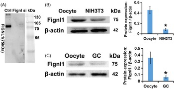
FIGNL1 is predominant in mouse oocytes. To verify the reliability of FIGNL1 antibody, we carried out a western blot experiment. The results showed that mouse monoclonal anti-FIGNL1 (A-4) (cat. no. sc-398264) antibody could display bands in the correct position to prove that the antibody was specific. When Fignl1 was knocked down by a specific siRNA, the band could hardly be detected (Figure 1A). The expression and localization patterns of Fignl1 were examined in mouse oocytes, and western blotting results demonstrated that Fignl1 expression in mouse oocyte was significantly higher compared with that in fibroblasts (Figure 1B) and granulosa cells (Figure 1C). Therefore, it was speculated that Fignl1 may play a role in oocyte function.
FIGNL1 is predominant in mouse oocytes. Western blot shows that mouse monoclonal anti-FIGNL1 (A-4) (cat. no. sc-398264) antibody could display bands in the correct position to prove that the antibody was specific. When Fignl1 was knocked down by specific siRNA, the band intensity was significantly reduced (A). Western blot and related statistical analysis showed that the expression level of Fignl1 in oocytes was two times higher than that in NIH3T3 cells (B) and three times higher than that in granulosa cells (C). β-Actin was used as a control. *P < 0.05 was considered to indicate a statistically significant difference. Here, 100 oocytes were used in each group. Three independent experiments were performed for each result.

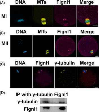
FIGNL1 is located at the spindle poles of metaphase I (MI) and MII oocytes and co-localizes with γ-tubulin. The abundance of FIGNL1 in mouse oocytes indicated that FIGNL1 may participate in germ cell function. Subsequently, the localization of FIGNL1 in mouse oocytes was detected by immunofluorescence, and it was found that FIGNL1 was located at the spindle poles of MI and MII oocytes (Figure 2A,B). Furthermore, the results suggested that FIGNL1 and γ-tubulin co-localized at the spindle poles of oocytes (Figure 2C). Co-immunoprecipitation (Co-IP) experiments showed that both FIGNL1 and γ-tubulin co-precipitated and interacted with each other (Figure. 2D). Here, γ-tubulin is an essential component of the microtubule-organizing centre (MTOC), which plays an important role in spindle assembly. Therefore, the function of FIGNL1 may be related to the MTOC at the two poles of the spindle and the spindle assembly process. Moreover, the present study examined the distribution of FIGNL1 protein in oocytes at other stages. It was demonstrated that FIGNL1 had no specific distribution in GV oocytes (Figure S1); however, FIGNL1 began to gather around chromosomes in germinal vesicle breakdown (GVBD) oocytes. Furthermore, in anaphase I (AI) and telophase I (TI) oocytes, FIGNL1 was clustered on the spindle midzone.
FIGNL1 is located at the spindle poles of MI and MII oocytes and co-localizes with γ-tubulin. Immunofluorescence showed that FIGNL1 was located at the spindle poles of MI (A) and MII (B) mouse oocytes. Tubulin is displayed in yellow, FIGNL1 in magenta and DNA in cyan. (C) At the MI stage, FIGNL1 co-localized with γ-tubulin at the spindle poles. DNA is displayed in cyan, FIGNL1 in magenta and γ-tubulin in yellow. Scale bar, 20 μm. FIGNL1, Fidgetin-like 1. Three independent experiments were performed for each result. (D) Co-IP results showed that the FIGNL1 antibody could precipitate FIGNL1 and γ-tubulin. γ-Tubulin antibody could precipitate γ-tubulin and FIGNL1. Here, 300 oocytes were used in each group. Three independent experiments were performed for each result.

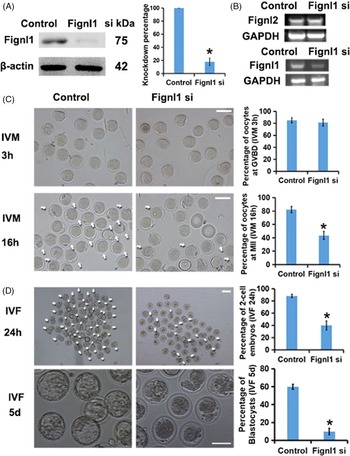
Knockdown of Fignl1 resulted in severe maturation abnormalities in oocytes. To investigate the role of FIGNL1 in oocyte development, specific small interfering RNAs (siRNAs) for Fignl1 were designed and microinjected into GV oocytes. Then, the efficiency and specificity of the siRNA was tested, and reverse transcription PCR (RT-PCR; Figure 3B) and western blotting (Figure 3A) results identified that specific siRNA could reduce Fignl1 mRNA and protein expression levels by 80%. RT-PCR also showed that siRNA could specifically target Fignl1 without knocking down Fignl2, another member of the MTSP family (Figure 3B; upper panel). After knocking down Fignl1, the oocyte was developed in vitro, and the maturity of oocyte was measured. Under the observation of living cells, oocyte development is generally observed in two phenomena: GVBD occurrence and first polar body (1PB) extrusion. 1PB extrusion is generally considered to be a marker of oocyte maturation. The results indicated that, compared with the control group, the rate of germinal vesicle breakdown (GVBD) of Fignl1-deleted oocytes did not decrease significantly after 3 h of in vitro maturation (IVM; Figure 3C; upper panel). However, the percentage of cells exhibiting 1PB extrusion was significantly lower compared with the control group after 16 h of IVM (Figure 3C; lower panel). This shows that FIGNL1 had no significant effect on the occurrence of GVBD in oocytes, but it mainly participates in the process after GVBD and before 1PB extrusion, and finally promotes the maturation of oocytes. In addition, after in vitro fertilization with healthy sperm and culture of the early embryos, the Fignl1-depleted group had a decreased two-cell embryo rate and blastocyst rate compared with the control group (Figure 3D). Collectively, the results suggested that FIGNL1 plays an important role in oocyte development and maturation.
Knockdown of Fignl1 leads to maturation disorder and abnormal fertilization of oocyte. (A) Western blot and related statistical analysis showed that the expression of Fignl1 decreased by ∼80% under the specific siRNA. β-Actin was used as a control. Here, 100 oocytes were used in each group. In the column diagram on the right, % respect to the percentage of FIGNL1 remaining after specific siRNA knockdown. (B) Reverse transcription-quantitative PCR showed that the transcription level of Fign1 was significantly decreased by siRNA targeting Fign1, but there was no significant change in the transcription level of Fignl2. GAPDH was used as a control. Here, 100 oocytes were used in each group. (C) In vitro development of oocytes and related statistical analysis showed that there was no significant change in the ratio of GVBD of oocytes after 3 h of in vitro development, but the ratio of MII of oocytes (indicated by white arrow) decreased significantly after 16 h of in vitro development. Before in vitro culture, 100 oocytes were used in each group. In the upper right column, % respect to percentage of GVBD oocytes in the control and Fignl1 knockdown groups at 3 h of in vitro development. In the lower right column, % respect to percentage of MII oocytes in the control and Fignl1 knockdown groups at 16 h of in vitro development. (D) In vitro fertilization (IVF) of oocytes and related statistical analysis showed that the proportion of 2-cell embryos (indicated by white arrows) formed after the specific siRNA knockdown of Fign1 and 24 h of fertilization and development of normal male sperm was significantly reduced, the proportion of blastocysts formed after 5 days of in vitro development was also significantly reduced. There were 100 MII oocytes in each group before IVF. In the upper right column, % respect to percentage of 2-cell embryos in the control and Fignl1 knockdown groups at 24 h of IVF. In the lower right column, % respect to percentage of blastocysts in the control and Fignl1 knockdown groups at 5 days of IVF. Scale bars, 100 μm. *P < 0.05 is considered to indicate a statistically significant difference. IVF, in vitro fertilization; Fignl, Fidgetin-like; siRNA, small interfering RNA. Three independent experiments were performed for each result.

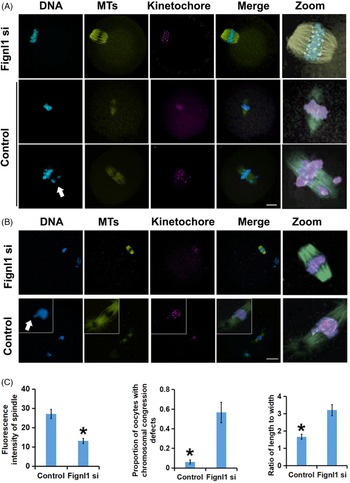
Knockdown of Fignl1 resulted in abnormal pole-correlated spindle organization in metaphase I (MI) or metaphase II (MII) oocytes. To determine how the decrease in FIGNL1 affected oocyte development, we conducted further studies. It was found that, after Fignl1 knockdown, there were some abnormalities in the spindle morphology of meiotic oocytes. The spindle phenotype was characterized after Fignl1 knockdown, and it was identified that this knockdown caused several abnormalities in spindle morphology. However, compared with the control group, the spindle of oocytes lacking FIGNL1 was thinner and longer, and the ratio of length:width was larger (Figure 4A). Moreover, the results suggested that silencing Fignl1 resulted in more astral microtubules and increased multipolar spindles in the cytoplasm compared with the control oocytes (Figure 4B). It was also found that abnormal spindle morphology led to abnormal chromosome arrangement. Furthermore, as shown by the white arrow in Figure 4, there were higher numbers of chromosomal congressional defects in MI and MII oocytes that were not arranged in the equatorial plate, but which were manifested as chromosomal congressional defects. This indicated that oocytes without FIGNL1 may have defects in spindle function due to abnormal spindle morphology. It was also suggested that the abnormal arrangement of chromosomes in oocytes may eventually lead to the failure of oocyte maturation (Figure 4A,B). Abnormality in spindle morphology and function leads to the disorder of chromosome arrangement and separation, and finally leads to the failure of oocyte maturation or fertilization. Measurement and statistical analysis showed that the fluorescence intensity of spindle microtubules in oocytes lacking FIGNL1 decreased apparently, while the spindle length width ratio and the proportion of oocytes with chromosome abnormalities increased significantly (Figure 4C). These results suggested that the reduced expression of Fignl1 may lead to chromosomal abnormalities and aneuploidy in oocyte by affecting spindle assembly, further affecting oocyte maturation and fertilization.
Fignl1 knockdown causes abnormal pole-correlated spindle organization in MI or MII oocytes. (A) When Fignl1 was knocked down by specific siRNA, MI oocytes showed longer and thinner spindles, decreased microtubule density and chromosomal congressional defects. The rightmost column is the image adjusted by each colour channel. The white arrow refers to chromosomal congressional defects. Tubulin is displayed in yellow, kinetochores in magenta and DNA in cyan. (B) When Fignl1 was knocked down by specific siRNA, the MII oocytes showed many stellar structures composed of microtubules independent of the spindle structure. In addition, MII oocytes lacking FIGNL1 still showed decreased microtubule density and chromosomal congressional defects. The rightmost column is the image adjusted by each colour channel. White arrow indicates chromosomal congressional defects. Tubulin is displayed in yellow, kinetochores in magenta and DNA in cyan. (C) Results of Fignl1 knockdown oocyte correlation measurements were as follows: the fluorescence intensity of spindle microtubules was approximately one time lower than that of the control group, the ratio of spindle length to width was approximately one time higher than that of the control group, and the proportion of oocytes with chromosomal abnormalities was ∼10 times higher than that of the control group. Scale bars, 20 μm. *P < 0.05 was considered to indicate a statistically significant difference. Fignl, Fidgetin-like. Here, 100 oocytes were fixed and stained in each group, and the number and proportion of chromosomal abnormalities in each group were counted. Here, 20 oocytes were selected randomly in each group to measure the fluorescence intensity and spindle length:width ratio. Three independent experiments were performed for each result.

Discussion
Normal meiosis ensures oocyte ploidy, followed by normal fertilization and early embryonic development until a healthy neonate is born. During the whole process of meiosis, with the polymerization and depolymerization of microtubules, the spindle morphology changes dynamically, which is the guarantee of normal meiosis. If spindle MT tissue is abnormal, this often causes defective meiosis, chromosome number abnormality and oocyte maturation disorder (Srayko et al., Reference Srayko, O’Toole, Hyman and Müller-Reichert2006; Kogo et al., Reference Kogo, Kowa-Sugiyama, Yamada, Bolor, Tsutsumi, Ohye, Inagaki, Taniguchi, Toda and Kurahashi2010; Smith et al., Reference Smith, Milne, Nelson, Eddie, Brown, Atanassova, O’Bryan, O’Donnell, Rhodes, Wells, Napper, Nolan, Lalanne, Cheeseman and Peters2012; Caburet et al., Reference Caburet, Arboleda, Llano, Overbeek, Barbero, Oka, Harrison, Vaiman, Ben-Neriah, García-Tuñón, Fellous, Pendás, Veitia and Vilain2014; Chen et al., Reference Chen, Zhang, Sun, Kuang, Mao, Wang, Yan, Li, Xu, Yu, Fu, Mu, Zhou, Li, Jin, He, Sang and Wang2017; Nguyen et al., Reference Nguyen, Marin, Zhou, Gentilello, Smoak, Cao, Fedick, Wang, Taylor, Scott, Xing, Treff and Schindler2017). MT-severing activities by MTSPs is one of the mechanisms to ensure the normal assembly of spindles. MTSPs are reported to have three distinct roles during the cell cycle: (i) MT severing can expose the ends of MTs, and microtubule polymerase or depolymerizing enzyme can bind to the ends for continuous aggregation or depolymerization to make MTs elongate or shorten; (ii) long MTs are severed into short fragments, and each fragment will elongate into long MT by polymerization, therefore increasing the density of spindle microtubules, which is called ‘nucleation’; and (iii) MT severing can removing the possible aberrant kinetochore-MT attachments, and ensure the correct separation of chromosomes, to ensure the stability of chromosome number (McNally and Vale, Reference McNally and Vale1993; Hartman et al., Reference Hartman, Mahr, McNally, Okawa, Iwamatsu, Thomas, Cheesman, Heuser, Vale and McNally1998; Cox et al., Reference Cox, Mahaffey, Nystuen, Letts and Frankel2000; Buster et al., Reference Buster, McNally and McNally2002; Mukherjee et al., Reference Mukherjee, Diaz Valencia, Stewman, Metz, Monnier, Rath, Asenjo, Charafeddine, Sosa, Ross, Ma and Sharp2012; Johjima et al., Reference Johjima, Noi, Nishikori, Ogi, Esaki and Ogura2015). In human mitotic U2OS cells, katanin-like 1 (Katnal1) is specifically localized to the spindle poles, and Katnal1 knockdown significantly reduces MT intensity at the poles (Ververis et al., Reference Ververis, Christodoulou, Christoforou, Kamilari, Lederer and Santama2016). Therefore, MT severing within spindle poles can increase the microtubule density of the spindle and promote the assembly of the spindle through ‘nucleation’.
In addition, during meiosis in C. elegans oocytes, the homologues of katanin, a MTSP member, are composed of two subunits meiotic double-stranded break formation protein 1 (MEI-1) and MEI-2, which are located on chromosomes (Srayko et al., Reference Srayko, O’Toole, Hyman and Müller-Reichert2006). Katanin’s severing activity can induce nanoscale damage to the microtubule lattice, leading to the incorporation of GTP tubulin to promote the rescue and the stability of the new positive end formed after severing, which eventually leads to the expansion of microtubules and the increase in spindle density (Schaedel et al., Reference Schaedel, John, Gaillard, Nachury, Blanchoin and Théry2015, Reference Schaedel, Triclin, Chrétien, Abrieu, Aumeier, Gaillard, Blanchoin, Théry and John2019; Vemu et al., Reference Vemu, Szczesna, Zehr, Spector, Grigorieff, Deaconescu and Roll-Mecak2018). Several studies have shown that mei-1/mei-2 mutations lead to a significant reduction in the density of microtubules in the spindle of meiotic oocytes of nematodes (Srayko et al., Reference Srayko, Buster, Bazirgan, McNally and Mains2000, Reference Srayko, O’Toole, Hyman and Müller-Reichert2006; McNally et al., Reference McNally, Berg, Cortes, Hernandez, Mains and McNally2014; Joly et al., Reference Joly, Martino, Gigant, Dumont and Pintard2016). A study combining optical microscope, electron tomography and mathematical model showed that katanin can selectively sever microtubules in close contact with chromosomes in meiotic oocytes of C. elegans, increase microtubule density and maintain the normal morphology of metaphase spindle. The severing effect of katanin decreased significantly in the transition from metaphase to anaphase and in the anaphase chromosome microtubules. In addition, the severing activity of katanin can prevent the microtubules in close contact with chromosomes from growing too long, to effectively improve the turnover rate of spindle microtubules (Lantzsch et al., Reference Lantzsch, Yu, Chen and Redemann2021). These findings indicate that MT severing near chromosomes can also promote spindle assembly through ‘nucleation’.
In mouse meiotic oocytes, we have previously studied KATNAL1 (Gao et al., Reference Gao, Xu, Jin, Ying and Liu2019) and SPASTIN (Jin et al., Reference Jin, Shou, Liu, Jiang, Shen, Cheng and Gao2022), members of MTSPs. KATNAL1 accumulates at the two poles of the spindle, while SPASTIN is dispersed on the microtubules of the spindle. The loss of both can lead to the decline in oocyte maturation rate and fertilization rate.
To the best of our knowledge, the present study was the first to demonstrate that FIGNL1, an MTSP predominantly expressed in mouse oocytes, was distributed at the two poles of the spindle during meiosis of mouse oocytes. In addition, it was found that the reduced expression of Fignl1 resulted in spindle defects characterized by abnormal length:width ratio, reduction of MT density and stellar MT structure. Due to the defects of spindle structure, meiosis cannot proceed normally, and homologous chromosomes (MI) or sister chromatids (MII) cannot be separated accurately, which leads to further aberrant chromosome arrangement and a lagging chromosome, and eventually results in maturation disorder of oocytes, fertilization and abnormal development of the early embryo. There was co-localization between FIGNL1 and γ-tubulin, the key component of MTOC, and co-IP preliminarily proves the interaction between them. These results suggested that the effect of FIGNL1 on meiosis of oocytes may be related to MTOC. It should be noted there is usually 1–3 major MTOC at GV oocytes that then get fragmented upon GVBD. However, the immunofluorescence assay did not show significant aggregation of FIGNL1 in GV oocytes. The reason for this finding needs to be further explored. It may be that MTOCs is too small at this time to visualize the localized proteins.
In HEK293T cells, FIGNL1 is mainly concentrated near the centrosome, which was similar to our results (Zhao et al., Reference Zhao, Jin, Wang, Sun, Hong, Cao and Wang2016). In Drosophila somatic cells, spastin and fidgetin, which are located at the two poles of mitotic spindle, are used to stimulate microtubule terminal depolymerization and flux. Both MTSPs concentrate at centrosomes, where they catalyze the turnover of γ-tubulin, consistent with the hypothesis that they exert their influence by releasing stabilizing γ-tubulin ring complexes (γ-TuRCs) from minus ends (Zhang et al., Reference Zhang, Rogers, Buster and Sharp2007). The γ-TuRCs are believed to cap and stabilize MT ends (Wiese and Zheng, Reference Wiese and Zheng2000), which is closely related to the assembly of the spindle (Rogers et al., Reference Rogers, Rogers, Schwimmer, Ems-McClung, Walczak, Vale, Scholey and Sharp2004). We speculate that, in mouse oocytes, FIGNL1, which accumulates at the two poles of the spindle, may also be affected by γ-tubulin that affects spindle assembly.
It has also been reported that overexpression of Fignl1 results in an incomplete microtubule network. Moreover, by severing MTs, FIGNL1 has been shown to play an important role in the transmission of information in nerve cells (Coralie et al., Reference Coralie, Amélie, Laïla, Christian, Daniel, Stéphanie, Monica, Christophe, Philippe, Céline, Leticia, Susanne, Sylvie, Corinne, Fatiha, Jean-Christophe, Annie and Jamilé2018). Therefore, these results suggested that FIGNL1, as a member of MTSP, has a similar MT-severing activity. Fignl1 has also been reported to be downregulated in the ovaries of ageing rats and has been screened as a possible gene associated with testicular weight loss and moderate malformed spermatozoa, therefore showing the potential role of FIGNL1 in mammalian reproductive function (L’Hôte et al., Reference L’Hôte, Vatin, Auer, Castille, Passet, Montagutelli, Serres and Vaiman2011; Govindaraj and Rao, Reference Govindaraj and Rao2015). Previous studies have revealed that the depletion of the spindle pole matrix causes an abnormal width:length ratio and additional asters when diverse centrosome or pole components are knocked down or depleted (Eot-Houllier et al., Reference Eot-Houllier, Venoux, Vidal-Eychenié, Hoang, Giorgi and Rouquier2010; Kim et al., Reference Kim, Kim, Oh, Park and Hwang2013; Patel et al., Reference Patel, Zich, Serrels, Rickman, Hardwick, Frame and Brunton2013; Connolly et al., Reference Connolly, Osterberg, Christensen, Price, Lu, Chicas-Cruz, Lockery, Mains and Bowerman2014; Pimenta-Marques et al., Reference Pimenta-Marques, Bento, Lopes, Duarte, Jana and Bettencourt-Dias2016). Furthermore, these previous findings are consistent with the present results, suggesting that FIGNL1 plays an important role in the integrity of meiotic spindles by regulating polar tissue. It has also been shown that defective spindle tissue may seriously affect meiosis, subsequent fertilization and early embryonic development. We speculate that FIGNL1, located at the two poles of the spindle, increases microtubule breakage by severing microtubules. The broken ends of microtubules can be elongated into new microtubules by the action of polymerase, therefore increasing the density of microtubules, promoting the formation of spindles and ensuring normal meiosis. In contrast, when FIGNL1 is absent, the density of microtubules in the oocyte spindle decreases and the structure becomes loose, resulting in an increase in the length:width ratio and an increase in the stellar structure composed of microtubules. When abnormal spindles cannot guide the migration of homologous chromosomes or sister chromatids, lagging chromosome will occur, which is also an important reason for aneuploidy. When the oocyte has a developmental disorder or aneuploidy, fertilization and the subsequent early embryo development will obviously be abnormal.
In the present study, it was found that Fignl1 knockdown significantly reduced the first polar body extrusion rate of oocytes and led to a reduction in the fertilization rate and two-cell rate. Therefore, the present results further demonstrated the potential role of FIGNL1 in the meiosis of the oocyte.
In addition, the ligand of RAD51 recombinase, FIGNL1, plays a role in repairing DNA double-stranded breaks in HR, which is essential for maintaining genomic stability and preventing developmental disorders and cancer (Ma et al., Reference Ma, Li, Yao, Lin, Gu, Xu, Deng, Ma and Zhang2017; Matsuzaki et al., Reference Matsuzaki, Kondo, Ishikawa and Shinohara2019). Homologous recombination is essential for ensuring proper segregation of chromosomes in meiotic division (Kim and Mirken, Reference Kim and Mirkin2018). This will particularly focus on cis- and trans-requirements of meiotic DNA double-stranded break formation, a hallmark event during meiosis and a prerequisite for recombination of genetic traits (Edlinger and Schlögelhofer, Reference Edlinger and Schlögelhofer2011). Given the association between Fignl1 and Rad51, we consider that the phenotype of abnormal chromosome arrangement caused by Fignl1 knockdown may be related to the abnormal chromosome HR caused by the failure to correctly create and resolve double-stranded breaks and invasion events in prophase I. This requires more in-depth research to explore further.
According to our previous research, Katnal1, another member of the MTSPs, is also located at the two poles of the meiotic oocyte spindle. Its deletion can also cause oocyte development disorder, fertilization abnormality and spindle morphological abnormality (Gao et al., Reference Gao, Xu, Jin, Ying and Liu2019). However, it is mainly manifested in the increase in spindle length width ratio and a greater proportion of astral microtubules. There was no obvious abnormality in spindle microtubule fluorescence intensity and metaphase chromosome arrangement. We speculated that Katnal1 and FIGNL1 may both play a role at the two poles of the spindle, but that the focus was different. The dynamic distribution characteristics and function coordination mechanism of the two need to be further discussed.
In addition, considering that other MTSPs produce spindle migration defects (Beard et al., Reference Beard, Smit, Chan and Mains2016; Joly et al., Reference Joly, Martino, Gigant, Dumont and Pintard2016), our results illustrate the spindle morphology and congressional defects caused by the loss of FIGNL1, but do not show the spindle position changes relative to the cell cortex. Perhaps spindle migration is not the main way in which FIGNL1, a specific MTSP, affects oocytes meiosis. Further studies and phenotypic validation are needed in this regard.
In conclusion, to the best of our knowledge, the present study is the first to identify that FIGNL1, a member of the MTSP, enriched in oocytes is concentrated on the spindle pole during the meiosis of mouse oocytes, which may be crucial to polar tissue. Furthermore, it was demonstrated that Fignl1 knockdown caused severe spindle defects characterized by an abnormal width:length ratio, reduction in MT density and additional aster MT. It was also indicated that Fignl1 knockdown resulted in abnormal spindle morphology and atypical chromosome arrangement, and that these factors together delay meiosis and reduce fertilization. However, further research is required to identify the underlying mechanisms via which FIGNL1 organizes the spindle pole and the factors that controls its activity.
Supplementary material
To view supplementary material for this article, please visit https://doi.org/10.1017/S0967199422000417
Availability of data and materials
The datasets used during the present study are available from the corresponding author upon reasonable request.
Acknowledgements
We thank Dr Zhang (Hangzhou Medical College) for providing us with specific plasmids.
Author’s contributions
HFS and LLG conceived and designed the experiments. LLG, YY, YCL, QW and ZJ performed the experiments. LLG analyzed the data. HFS and LLG contributed the reagents/materials/analysis tools. HFS and LLG wrote the paper. All authors read and approved the manuscript and agreed to be accountable for all aspects of the research in ensuring that the accuracy or integrity of any part of the work is appropriately investigated and resolved.
Ethics approval and consent to participate
All animal experiments were approved by the Animal Care and Use Committee of Hangzhou Medical College (Hangzhou, China) and performed in accordance with institutional guidelines.
Patient consent for publication
Not applicable.
Competing interests
The authors declare that they have no competing interests.
Declaration of Funding
This research is supported by the youth programme of National Natural Science Foundation of China (programme number: 82001539).