INTRODUCTION
It was a common understanding that female adults of Strongyloides ratti parasitize the small intestine, produce eggs parthenogenetically and are expelled completely in 3–4 weeks from rat hosts by immunity (Mimori et al. Reference Mimori, Nawa, Korenaga and Tada1982; Korenaga et al. Reference Korenaga, Nawa, Mimori and Tada1983). Accordingly, egg excretion in feces after primary infection increases rapidly, making a high peak at days around 7–8 post-infection (p.i.), and reduces to zero in about 1 month. We noticed an additional small peak or a ‘plateau’ (the plateau hereafter) appearing at day around 25 p.i. This finding led to new facts that some adults withstand the worm expulsion and ‘re-settle’ in the large intestine, and that the plateau mostly consists of eggs produced by large intestinal adults (Kimura et al. Reference Kimura, Shintoku, Kadosaka, Fujiwara, Kondo and Itoh1999). The large intestinal parasitism is not a product by chance but a survival strategy of S. ratti, because (i) eggs from the plateau produced more offspring (infective larvae, L3i) than those from the high peak through the heterogonic cycle of development and longer duration of the plateau (Kimura et al. Reference Kimura, Shintoku, Kadosaka, Fujiwara, Kondo and Itoh1999), (ii) adults in the large intestine increased the body length and the number of intrauterine eggs despite mounting host immune responses (Shintoku et al. Reference Shintoku, Takagi, Kadosaka, Nagaoka, Kondo, Itoh, Honda and Kimura2011), and (iii) 32·4% of wild rats captured in Nagoya, Japan, were found to harbour S. ratti adults in the large intestine (Shintoku et al. Reference Shintoku, Kimura, Kadosaka, Hasegawa, Kondo, Itoh and Islam2005).
The mechanisms of S. ratti expulsion from the small intestine have been studied extensively, and a crucial role of mucosal mast cells (MMC) was demonstrated in mice (Nawa et al. Reference Nawa, Kiyota, Korenaga and Kotani1985; Abe et al. Reference Abe, Sugaya, Ishida, Khan, Tasdemir and Yoshimura1993a ; Onah and Nawa, Reference Onah and Nawa2000), or their importance was reported in rats (Olson and Schiller, Reference Olson and Schiller1978; Wilkes et al. Reference Wilkes, Bleay, Paterson and Viney2007). However, there is no study on MMC in the large intestine of a rat, the natural host. Intestinal eosinophilia is also induced by S. ratti infection and Abe et al. (Reference Abe, Yoshimura and Nawa1993b ) related it to worm expulsion in mice. Again, however, no information is available from S. ratti-infected rats. In this paper, we report the kinetics of MMC and eosinophils (Eo) in the small and large intestines of rats, and discuss it in relation to worm expulsion.
MATERIALS AND METHODS
Thirty-eight 6-week-old male Wistar rats, purchased from Japan SLC, Inc. (Shizuoka, Japan), were infected with 3000 L3i of S. ratti subcutaneously. The parasites used and the method of infection were described elsewhere (Kimura et al. Reference Kimura, Shintoku, Kadosaka, Fujiwara, Kondo and Itoh1999). Three animals each were kept overnight without food, killed with an overdose of ether and autopsied at days 5, 11, 14, 18, 21, 25, 28, 32, 35, 40, 60 and 70 p.i. For pathological studies, 4 different parts of the intestine were resected: a segment of upper small intestine 15–18 cm from the pylorus (site A), a segment of lower small intestine 15–18 cm proximal to the ileo-caecal junction (site B), the caecum (site C), and a segment of colon 3–6 cm distal from the caecum (site D). They were fixed in 4% paraformaldehyde at 4 °C for 6 h, embedded in paraffin and sectioned at 4 μm thickness. The simultaneous staining of MMC and Eo was carried out with alcian blue-carbol chromotrope following the method by Newlands et al. (Reference Newlands, Huntley and Miller1984) with some modifications, i.e., alcian blue staining was done overnight instead of for 30 min and then washed with 0·7 M HCl before chromotrope staining. The numbers of MMC and Eo in sites A and B were counted with 10 villus-crypt units (VCU)/rat and those in sites C and D with 10 crypt units (CU)/rat, and expressed as the averages of 3 infected rats. Two non-infected control rats were autopsied at days 14, 28 and 63 and processed as in the infected animals.
A group of 5 infected rats, including the 3 that were autopsied at day 70 p.i., were examined for fecal egg counts (per 12 mg of feces) by the direct smear method at days 6, 13, 17, 26, 34, 41 and 70 p.i.
All experiments were carried out in Aichi Medical University, Japan, with the approval of the University Animal Care and Use Committee.
RESULTS
Egg excretion pattern
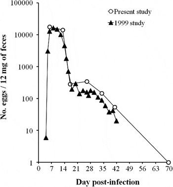
The first peak of egg excretion in this study was observed at day 6 p.i. and the following plateau at around day 26 p.i. This result is compared in Fig. 1 with the other result from our previous study in which fecal eggs were counted under the same experimental conditions except for days p.i. for fecal tests (Kimura et al. Reference Kimura, Shintoku, Kadosaka, Fujiwara, Kondo and Itoh1999). Having an almost identical egg excretion pattern between the two studies, we utilized the adult recovery data of the 1999 study (determined at days 9, 14, 19, 25 and 35 p.i.) to correlate them with the present MMC and Eo data. Considering the proximity of gut position between the two studies, the S-1, S-4, Cc, and C-1 data presented in Table 1 of Kimura et al. (Reference Kimura, Shintoku, Kadosaka, Fujiwara, Kondo and Itoh1999) were superimposed, respectively, on sites A, B, C, and D in the present Figs. 2 and 4.
Egg excretion patterns obtained in the present study and the study by Kimura et al. (Reference Kimura, Shintoku, Kadosaka, Fujiwara, Kondo and Itoh1999). The number of eggs includes eggs and hatched larvae. Both studies used 5 male Wistar rats infected subcutaneously with 3000 infective larvae.

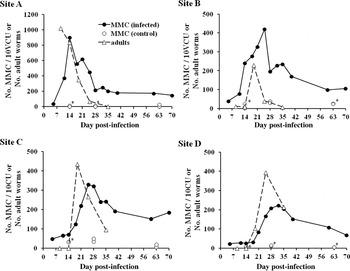
Change in the number of mucosal mast cells (MMC) and adult worms after infection in 4 study sites: (A) upper small intestine, (B) lower small intestine, (C) caecum and (D) colon. VCU, villus-crypt unit; CU, crypt unit. ● Average of 3 infected rats; ○ count in each control rat (asterisk indicates 2 circles).


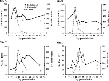
Change in the number of eosinophils (Eo) after infection in 4 study sites. Sites A, B, C, and D; VCU and CU; and ● and ○ are defined as in Fig. 2.

Kinetic analysis of MMC
Without infection, the counts of MMC in the whole intestine (24 data points in total) ranged from 1 to 50, with an average of 18·5 (S.D. = 13·8). The MMC counts in S. ratti-infected animals increased significantly, with different peak times and heights by site of intestine (Fig. 2). The peaks of MMC in sites A, B, C and D were observed at days 14, 25, 25 and 32 p.i. respectively, with the respective counts of 901·3, 421·3, 329·0 and 221·9. In each of sites A and B, a less conspicuous MMC increase was also noticed about 1 week after the high peak. Compared with the peaks of adult recovery, those of MMC count were delayed by 5–7 days in all 4 sites. The number of adults started declining from the peaks when MMC counts reached 261·1, 293·6, 155·5 and 167·2, respectively in sites A, B, C and D (estimated by interpolation).
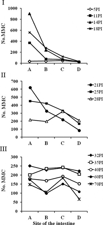
The same MMC data in Fig. 2 were re-analysed according to site of the intestine to overview MMC levels in the whole intestinal canal at different days p.i. (Fig. 3). Between days 11 and 18 p.i., in site A, MMC were overwhelmingly more abundant than in the other sites, whereas no MMC reaction was seen in site D (Fig. 3-I). The MMC increase in site D was recognized at days 21–28 p.i. (Fig. 3-II), while those in site A decreased steadily. On days 32–35 p.i., MMC counts were basically similar in all sites (200–250 level), despite the fact that only a small number of worms (<8 at day 35 p.i.) were found in sites A and B (Kimura et al. Reference Kimura, Shintoku, Kadosaka, Fujiwara, Kondo and Itoh1999). Then a further decrease in the counts was noticed on and after day 40 p.i. (Fig. 3-III).
Kinetic analysis of Eo
The number of Eo varied significantly by site of the intestine in the control animals (Fig. 4). The average of 6 data points in site A was 275·0 (S.D. = 52·9), which decreased to 71·3 (S.D. = 29·1), 20·5 (S.D. = 4·6), and 13·0 (S.D. = 4·9) respectively in sites B, C and D. The infection increased the number of Eo, but the peak count at day 14 p.i. in site A (434·3) was only 1·6 times higher than the control, and the peak at day 18 p.i. in site B (194·3) was 2·7 times higher. In sites C and D, the highest Eo counts were, respectively, 5·1 and 4·2 times higher than the control levels (a peak in site C was not very clear). The time lag between the peaks of adult and Eo counts in sites A, B and D varied from 1 to 5 days: the Eo peak followed the adult peak in sites A and D, but appeared 1 day earlier in site B. The numbers of Eo count when the peak counts of adults started declining were 373·3, 162·6, 72·5 and 27·7 in sites A, B, C, and D respectively (the estimates were made by interpolation).
DISCUSSION
The study showed that MMC buildup (and possible activation) by S. ratti infection could be divided into 2 distinct phases: one is in the upper small intestine (site A) between days 5 and 14 p.i. and the other in the colon (site D) between days 18 and 32 p.i.. Sites B and C were intermediary in terms of MMC buildup and its timing. It is interesting that, after worm expulsion started on day 9 p.i. in site A, very few adults were recovered in sites B, C, and D (0–8·3 at day 14 p.i.; Kimura et al. Reference Kimura, Shintoku, Kadosaka, Fujiwara, Kondo and Itoh1999), and then on day 19 p.i., the recovery increased sharply. We reported that adults obtained from the small intestine on day 7 p.i. were rejected when transplanted into the colon of immune rats, but that adults obtained on day 19 p.i. could parasitize the colon and reproduce (Shintoku et al. Reference Shintoku, Takagi, Kadosaka, Nagaoka, Kondo, Itoh, Honda and Kimura2011). It is likely that the majority of adults was expelled quickly, but some withstood the expulsion and by day 19 PI became immune-resistant in the upper part of small intestine, moved downward and started parasitizing the large intestine, stimulating MMC proliferation there. Thus, we have concluded that the 2 phases of mastocytosis were caused by biologically different adults. Adults in site B could be immune-resistant ones moving on to the caecum.
It has been known that, in the upper small intestine, a peak of S. ratti adults precedes an MMC peak. Olson and Schiller (Reference Olson and Schiller1978) reported a 10-day time lag between the two peaks in rats. Abe and Nawa (Reference Abe and Nawa1988), in mice, reported the peak of MMC 4 days after the peak of fecal larval excretion, which is approximately the time of the adult peak. In a basically similar study, the peak of serum MMCP-I (mouse mast cell protease-I) was reported 3 days after the peak output of S. ratti DNA in feces, an indicator of excreted eggs/larvae (Eschbach et al. Reference Eschbach, Klemm, Kolbaum, Blankenhaus, Brattig and Breloer2010). In the present study, a 5 to 7-day time lag was observed not only in the small intestine but in all sites. The relative consistency of the lag will suggest a direct stimulation by parasitic adults for the buildup of mastocytosis. In site B, however, an increase of MMC was recognized even before adults were recovered (Fig. 2). This could be explained by a widespread MMC reaction in the small intestine which was induced by a large number of adults in site A. In sites C and D, the adult peaks were on days 19 and 25 p.i. respectively, when the hosts’ immune responses had already been activated. As the time lags in these sites were 6–7 days, the immunity did not seem to facilitate MMC buildup. It is interesting to know whether the immune-resistance acquired by adults after day 19 p.i. relates to this finding.
A big variation was observed in the number of MMC at different sites, the peak MMC count varying from 901·3 in site A to 221·9 in site D. Abe et al. (Reference Abe, Ochiai, Minamishima and Nawa1988) with IL-3-treated nude mice observed approximately 10 times stronger mastocytosis in the small intestine than in the caecum, and suggested an innate difference in the distribution of progenitor cells. Meanwhile, Abe et al. (Reference Abe, Nawa and Yoshimura1992) reported the existence of IL-3-stimulating components in excretory-secretory products of S. ratti adults, which implies that the number of parasitic adults can influence the level of mastocytosis. To study the possibility, a simple trial was made: a ratio of the peak MMC count to the peak number of adults was computed at each site, which resulted in 0·88, 1·84, 0·76 and 0·56 respectively in sites A, B, C and D. The relatively small difference (0·56–0·88 MMC/adult) among sites A, C, and D indicates that the number of MMC is dependent on that of parasitic adults, regardless of gut position. A high ratio in site B must be influenced again by upstream worms in site A.
The reduction of adults from the peak count started when the MMC count reached 260–290 in sites A and B and 160–170 in sites C and D. This will support an assumption that a certain level of mastocytosis is required to trigger worm expulsion. The different effector levels between the small and large intestines could be due to 2 different units (VCU and CU) used for MMC enumeration, or 2 biologically distinct adults parasitizing there. They can stimulate MMC differently, or MMC may expel them differentially. Also, the level of MMC activation (Ishiwata et al. Reference Ishiwata, Uchiyama, Maruyama, Kobayashi, Kurokawa, Nawa, Ogra, Mestecky, Lamm, Strober, Bienenstock and McGhee1999) could be different by site of intestine and time after infection, which we could not study. In addition, other effector cells like Eo might be involved (see next paragraph). A very high MMC peak far exceeding the expulsion level in site A probably facilitated ‘washing’ worms away quickly before day 19 p.i.
The reaction of Eo was less explanatory in terms of worm expulsion: the baseline counts in the control rats were so different by site, and the reaction after infection was also variable in each site. A noticeable finding is that the rate of Eo increase (peak count in infected rat/control count) was 4·2–5·1 times in sites C and D, and 1·6–2·7 times in sites A and B, suggesting more activation in the large intestine. A relatively high increase was also reported in the caecum of mice, and related to the expulsion from that site (Abe et al. Reference Abe, Yoshimura and Nawa1993b ). In another study with mice, Eo were implicated in the reduction of worms inside the small intestine, although they did not influence the duration of primary infection (Ovington et al. Reference Ovington, McKie, Matthaei, Young and Behm1998). In sites C and D, Eo might be causing damage on adults facilitating expulsion by MMC.
The present study revealed 2 distinct phases of MMC buildup in S. ratti-infected rats: one in the upper small intestine by stimulation of ‘ordinary’ adults and the other in the colon by immune-resistant adults. Thus, mechanisms of worm expulsion seem to be much more complicated. In addition, it was suggested that mastocytosis is not merely a host reaction to reject parasites but a possible ‘product’ by parasites themselves, suggesting a role of MMC for them. It will be interesting to study the significance of MMC reaction from this viewpoint.