Case report
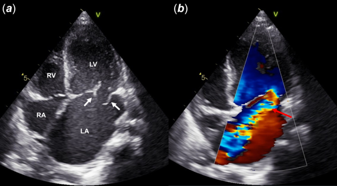
An 8-month-old male infant was referred to our hospital because of progressive coughing for 10 days and severe mitral regurgitation. His parents had not been aware of a heart murmur until then, and he did not have a specific family history. His body temperature was normal. Physical examination showed tachypnea, coarse crackles, and a grade 3/6 systolic regurgitant murmur at the apex with S3 and S4 gallop. Laboratory findings and serial blood cultures did not suggest bacterial infection. The chest radiograph showed cardiomegaly with a cardiothoracic ratio of 0.54 and pulmonary congestion. The electrocardiogram revealed left atrial and ventricular hypertrophy. Echocardiography showed a mobile aneurysmal lesion (8.9 × 7.5 mm) on the posterior mitral leaflet. An isolated hole was found on the aneurysmal lesion, and the Doppler echocardiogram showed severe regurgitation through the hole (Fig 1). No abnormal findings were detected in the subvalvular apparatus. Although the left atrium and ventricle were dilated, the left ventricular pump function was preserved.
Transthoracic apical 4-chamber view of Doppler echocardiography: ( a ) The perforated posterior mitral leaflet aneurysm (white arrow). ( b ) Severe mitral regurgitation through the perforated aneurysm (red arrow).

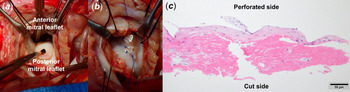
As his cardiorespiratory condition progressively deteriorated despite intensive medical treatment, we performed emergency surgical repair of the mitral valve on the day after admission. During the operation, most of the mitral leaflet appeared intact; however, a barnacle-like aneurysm was noted on the P2 scallop of the posterior mitral leaflet with a 5-mm round hole on its top (Fig 2a). The subvalvular apparatus showed no tendon rupture. The perforation was directly closed and sutured vertical to the leaflet coaptation to avoid coaptation loss of the mitral valve (Fig 2b). The post-operative course was uneventful. Serial echocardiography on post-operative day 1 and that obtained 1-year later showed mild residual mitral regurgitation.
( a ) Intraoperative view of the perforated aneurysm on the P2 segment of the posterior mitral leaflet. ( b ) Direct closure of the perforation with an intentional perpendicular suture. ( c ) Histopathological image of the mitral valve around the perforation (stained with hematoxylin and eosin). Scale bar, 50 µm.

Histopathological examination revealed disruption of the mitral valve leaflet without inflammatory cell infiltration or obvious irregular fibrous thickening, which would suggest infective endocarditis. At the edge of the perforation site, collagen fibre in the valvular leaflet was degenerated, and fibrinous components were attached (Fig 2c).
Discussion
Mitral valve aneurysm is a localised saccular bulging on the mitral leaflet towards the left atrium with systolic expansion and diastolic collapse. Reference Kim, Cho and Jun1 It is most commonly caused by aortic regurgitant jet complicated by infective endocarditis of the aortic valve. Reference Reid, Chandraratna and Harrison2 Perforation of the aneurysm may occasionally occur and lead to significant mitral regurgitation. Reference Tewari, Moorthy and Sinha3 There are other acquired aetiology such as rheumatic diseases and suspected congenital or structural aetiology such as connective tissue diseases including osteogenesis imperfecta, Marfan syndrome, Ehlers–Danlos syndrome type IV, and pseudoxanthoma. Reference Gajjar and Desai4,Reference Rückel, Erbel, Henkel, Kramer and Meyer5 Although these cases are rare, aneurysms caused by these aetiologies commonly occur in the anterior mitral leaflet. Reference Kim, Cho and Jun1
Mitral valve aneurysm in children, excluding acquired aetiologies such as infective endocarditis, rheumatic disease, and iatrogenic disorders, has rarely been reported in the literature. Reference Saxena, Shrivastava and Kumar6 On the other hand, there are a few reports of isolated mitral valve perforation without infective endocarditis in children. Reference Schranz, Oelert and Iversen7,Reference Oztunc, Saltik and Turkoglu8
In our patient, the aneurysmal lesion was on the posterior mitral leaflet with no clinical evidence of infective endocarditis. Other congenital mitral valve diseases, such as prolapse, isolated cleft, double-orifice, tendon rupture, or severely hypoplastic leaflet or acquired mitral valve diseases, such as endocarditis, connective tissue disease, rheumatic fever, or trauma, were excluded. In our case, two mechanisms may have caused mitral aneurysm and regurgitation: congenital isolated mitral valve aneurysm that was subsequently complicated by sudden perforation or a small isolated mitral perforation that may have induced mitral valve aneurysm secondary to a jet lesion, while expansion of the hole eventually caused critical mitral regurgitation. Dilated left ventricle and atrium, as well as left ventricular hypertrophy with preserved pump function on echocardiography, suggested subacute onset. At the same time, the degenerative changes in the collagen fibre of the valvular leaflet and fibrosis on histopathological examination might have been caused by the long-standing jet lesion. Consequently, we believe that a small isolated mitral perforation may have progressed to severe mitral regurgitation complicated by mitral valve aneurysm. It is difficult to conclude with only a single instance. Therefore, there is a need for reporting similar cases with detailed graphics and pathologic information to elucidate the relevant aetiology and pathogenesis.
Mitral perforation and aneurysm are surgically repaired using various techniques. For isolated perforation in children, direct suture or patch closure using autologous pericardium is performed. Reference Schranz, Oelert and Iversen7,Reference Oztunc, Saltik and Turkoglu8 For mitral valve aneurysm complicated by perforation in adults, resection of the aneurysm and mitral valve annuloplasty are done. Reference Kim, Cho and Jun1,Reference Guler, Hatipoglu, Karabay and Kirma9 In our case, the outcome of surgical repair, that is, intentional perpendicular suture to achieve mitral coaptation, was excellent.
In summary, we reported a case of perforated mitral valve aneurysm not complicated by infective endocarditis in an 8-month-old male infant, with surgical and histopathological findings. Although its aetiology remains unclear, we suggest to include mitral valve aneurysm in differential diagnosis as a cause of mitral regurgitation in children.
Supplementary material
To view supplementary material for this article, please visit https://doi.org/10.1017/S1047951120003340
Acknowledgements
We thank H. Tomita and T. Fujii for their thoughtful comments on this manuscript. T. Matsuyama advised about histopathological findings.
Financial support
This research received no specific grant from any funding agency, commercial or not-for-profit sectors.
Conflicts of interest
None.
Ethical Standards
Written informed consent was obtained from parents instead of the patient himself.