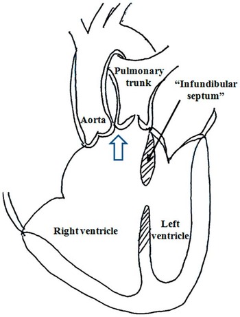
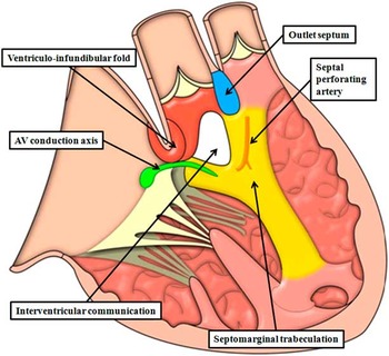
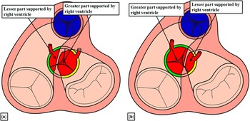
Many lesions, in the past, have been the source of confusion and uncertainty for those seeking to diagnose and treat patients with congenitally malformed hearts. Most of the disagreements related to these entities, such as so-called transposition, best analysed in terms of discordant ventriculo-arterial connections,Reference Becker and Anderson 1 or single ventricle, now recognised as representing a functional rather than an anatomical situation,Reference Jacobs and Anderson 2 have now been satisfactorily resolved. A lesion that remains a source of controversy, however, is the arrangement in which both arterial trunks arise in their greater part from the morphologically right ventricle. Initially described by Witham,Reference Witham 3 the feature underscoring many of the initial arguments was the alleged need for both arterial roots to be supported by bilateral infundibulums so as to justify its diagnosis.Reference Sridaromont, Ritter, Feldt, Davis, Mcgoon and Edwards 4 These polemics stemmed from the additional suggestion that it was necessary to distinguish between double-outlet right ventricle and tetralogy of Fallot.Reference Baron 5 The justification for this premise is itself questionable, as in his original description Fallot explained how, in one of his initial autopsies, the aorta arose in its entirety from the right ventricle, despite the fact that the heart showed the phenotypic features subsequently recognised as being diagnostic for his tetralogy.Reference Fallot 6 We now recognise, furthermore, that using the criterion of “bilateral conuses” to diagnose double outlet contravenes the important principle of the morphological method, which states that one variable feature should not be defined on the basis of another feature that is itself variable.Reference Van Praagh, David, Wright and Van Praagh 7 The real problem in diagnosis reflects the fact that, unlike tetralogy of Fallot, double-outlet right ventricle does not represent a discrete phenotypic entity. On the contrary, various phenotypes can be recognised in the setting of both arterial trunks arising in their greater parts from the morphologically right ventricle.Reference Walters, Mavroudis, Tchervenkov, Jacobs, Lacour-Gayet and Jacobs 8 In a perceptive review, Sidi et alReference Sidi, Vouhé and Ou 9 emphasised these anatomical variations, pointing out that, in terms of diagnosis, the key feature was to determine the optimal strategy for surgical repair. At the same time, these authors suggested that the lesion was best considered in terms of malposition of the arterial trunks, but the argument they used for this suggestion foundered in that they mistook the inner heart curvature for the outlet septum (Fig 1). Their figure is not without its value, however, as it emphasises that, when both arterial trunks arise from the morphologically right ventricle, the outlet from the left ventricle is roofed by the inner heart curvature rather than the outlet septum. In this review, therefore, we build on this crucial difference, showing how, once accurate definitions are provided for the building blocks of the right ventricle (Fig 2), a pragmatic approach can be established for the diagnosis of double-outlet right ventricle, leading in turn to the choice of the most appropriate options for surgical repair.
The figure has been re-drawn from Figure 4c of the review provided by Sidi et al.Reference Sidi, Vouhé and Ou 9 It shows how the structure they identified as the “infundibular septum” is, in reality, interposed between the leaflets of the arterial and atrioventricular valves. The true outlet septum would be positioned between the leaflets of the arterial valves, as shown by the white arrowhead with black borders.

The figure shows both arterial trunks arising from the right ventricle with each arterial trunk supported by a completely muscular infundibulum and a subaortic interventricular communication. The building blocks of the right ventricular outflow tracts are shown. The ventriculo-infundibular fold, or inner heart curvature, is shown in red. The outlet septum is shown in blue, and the septomarginal trabeculation in yellow. See text for further discussion. Note also the location of the atrioventricular (AV) conduction axis and the septal perforating artery.

Definition of the building blocks of the right ventricle
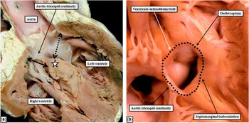
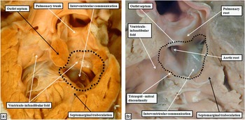
As we will show, the key to differentiating hearts in which both arterial trunks arise from the right ventricle is recognition of the precise nature of the cranial wall of the channel between the ventricles. When there is overriding of an arterial valve, then the key to diagnosis is to determine which plane represents the outflow from the left ventricle, as opposed to the plane representing the area of potential septation to achieve a biventricular surgical repair (Fig 3). As is shown in Figure 3, in the setting of tetralogy of Fallot with concordant ventriculo-arterial connections, it is the leading edge of the muscular outlet septum, which is exclusively a right ventricular structure, that forms the cranial margin of the area closed by the surgeon so as to re-connect the overriding aortic root with the left ventricle. It is this area that is usually taken, in the setting of tetralogy, to represent the “ventricular septal defect”. This is entirely appropriate, as it is also this area that is closed to achieve surgical separation of the circulations.Reference Bailliard, Spicer, Mohun, Henry and Anderson 10 The outlet septum can be readily defined as the structure separating the ventricular outflow tracts. As such it interposes between the hinges of the arterial valves (Fig 2). In most instances, it is a muscular structure; however, as we will show, it can be fibrous when there has been failure of muscularisation, during development, of the subpulmonary infundibulum.Reference Anderson, Brown and Mohun 11 In contrast to the outlet septum, the ventriculo-infundibular fold is the structure that interposes between the leaflets of the atrioventricular and arterial valves (Fig 2). It is a remnant of the initial inner heart curvature. In most instances, its large part remains muscular, as shown in Figure 3b, where the muscular fold separates the leaflets of the aortic and tricuspid valves in the roof of the right ventricle. Its middle component, however, usually becomes fibrous subsequent to the transfer during development of the aorta to the left ventricle.Reference Anderson, Brown and Mohun 11 After the completion of normal ventricular septation, the inner heart curvature of the left ventricle is represented by the fibrous continuity between the leaflets of the aortic and mitral valves. Should the fold retain its initial myocardial structure, then the leaflets of the aortic valve will be separated from those of the mitral valve. This is the essence of the bilateral infundibular support, or the “bilateral conuses” that, for quite some time, were considered a prerequisite for diagnosis of double-outlet right ventricle.Reference Sridaromont, Ritter, Feldt, Davis, Mcgoon and Edwards 4 Such bilateral infundibulums, however, can rarely be found even when the aorta is normally positioned within the left ventricle, in the presence of intact ventricular septal structures.Reference Rosenquist, Clark, Sweeney and McAllister 12 This finding, along with the fact that such bilateral infundibulums are also to be found in some patients with discordant ventriculo-arterial connections, and in most of those with concordant ventriculo-arterial connections and parallel arterial trunks,Reference Cavalle-Garrido, Bernasconi, Perrin and Anderson 13 points to the lack of logic in its use as a diagnostic feature of double-outlet right ventricle.
The images show a “four-chamber” cut ( a ) through the overriding aortic root in a heart obtained from a patient with tetralogy of Fallot, and a similar heart ( b ) viewed from the apex of the right ventricle showing the right ventricular aspect of the channel between the ventricles. The aortic root is overriding the crest of the ventricular septum (white star in a ), but in both hearts it is supported predominantly by the left ventricle, meaning that the ventriculo-arterial connections are concordant. In panel ( a ), the black-dotted, double-headed arrow shows the plane of the left ventricular outflow to the aorta. The solid, double-headed arrow shows the plane that would be closed by the surgeon to re-connect the aorta with the left ventricle. This is the area usually considered to represent the “ventricular septal defect”. As shown in panel ( b ), rather than being truly planar, it is an area bordered by an area of aortic-tricuspid continuity, part of the ventriculo-infundibular fold, the leading edge of the deviated muscular outlet septum, and the limbs of the septomarginal trabeculation. In order to make comparisons with hearts having double-outlet right ventricle, it is crucial to have specific definitions for these components of the right ventricular outflow tract (see Fig 2).

In most hearts with a double-outlet right ventricle, as well as in tetralogy of Fallot, the channel between the ventricles opens to the right ventricle between the limbs of a strap-like mass of myocardium, which reinforces the ventricular septal surface. This is the septomarginal trabeculation (Fig 2), usually described in North America as the septal band. Its body extends towards the apex of the right ventricle, where it gives rise to the anterior papillary muscle of the tricuspid valve, along with one of a series of septoparietal trabeculations – the latter structure described as the moderator band. The caudal limb of the septomarginal trabeculation usually gives rise to the medial papillary muscle, whereas the septal perforating artery typically enters the crest of the muscular ventricular septum beneath its cranial limb (Fig 2).Reference Hosseinpour, Anderson and Ho 14 The relationship between the caudal limb of the septomarginal trabeculation and the ventriculo-infundibular fold is key to determining the vulnerability, during surgical procedures, of the atrioventricular conduction axis. In most cases of tetralogy, as shown in Figure 3b, between the caudal limb of the septomarginal trabeculation and the ventriculo-infundibular fold, there is an area of fibrous continuity between the leaflets of the aortic and tricuspid valves. The fibrous area is continuous with the membranous part of the ventricular septum, through which the atrioventricular conduction axis penetrates as it passes from the apex of the triangle of Koch to reach the left ventricular outflow tract. In those circumstances in which the caudal limb of the septomarginal trabeculation fuses with the ventriculo-infundibular fold, there will be a muscular postero-inferior rim to the channel between the ventricles. This muscular component then protects the atrioventricular conduction axis.Reference Hosseinpour, Jones, Barron, Brawn and Anderson 15
The essence of double-outlet right ventricle
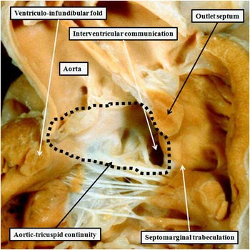
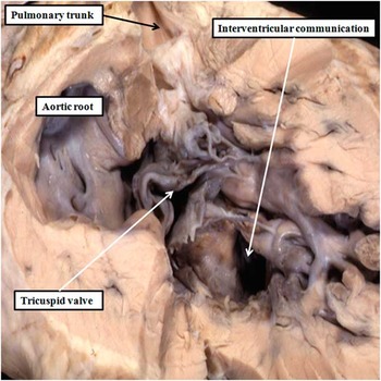
As we have already emphasised, there is no single phenotypic feature of hearts that have in common the origin of both arterial trunks from the right ventricle.Reference Walters, Mavroudis, Tchervenkov, Jacobs, Lacour-Gayet and Jacobs 8 This is because, rather than being an anatomical entity, double-outlet right ventricle is but one type of ventriculo-arterial connection.Reference Wilcox, Ho, Macartney, Becker, Gerlis and Anderson 16 As such, it can be found in its allegedly “classical” variant, in which the leaflets of both arterial valves are exclusively supported by infundibular musculature. It is almost certainly the case, however, that when analysed simply in terms of the ventriculo-arterial connection there is fibrous continuity between the leaflets of an atrioventricular and an arterial valve in the majority of hearts fulfilling the criterion of origin of both arterial trunks from the right ventricle. This is not only the case when one of the arterial roots is positioned astride the crest of the ventricular septum, but with the larger part of its circumference supported by the right ventricle, but also when the entirety of both arterial roots are supported by right ventricular structures. It is this latter feature that is the essence of the so-called “200%” double outlet as described by Lacour-Gayet et al.Reference Lacour-Gayet, Haun and Ntalakoura 17 As shown in Figure 4, such “200%” examples can be found with fibrous continuity between the leaflets of the aortic and mitral valves (Fig 4). Having established that hearts do not need bilateral infundibulums so as to exhibit double outlet from the right ventricle, the question remains as how best to ascribe those hearts with overriding arterial roots to the categories of double-outlet or one-to-one ventriculo-arterial connections. Much has been made, in the past, of the difficulty of making this distinction because of the shift in the long axis of the muscular ventricular septum during the cardiac cycle, with the heart seemingly changing from the situation of double-outlet to that of concordant or discordant ventriculo-arterial connections. This is a fallacious argument. It is not the long axis of the apical ventricular septum that is the determining feature. Rather, it is the cord subtended by the crest of the septum relative to the circumference of the arterial root that determines whether the root is supported predominantly by the right, as opposed to the left, ventricle (Fig 5).
The image shows a heart in which both arterial trunks arise exclusively from the right ventricle, despite the presence of extensive fibrous continuity, in the roof of the interventricular communication, between the leaflets of the aortic and mitral valves. There is also an extensive area of fibrous continuity between the leaflets of the aortic and tricuspid valves. The dotted line shows the area where the surgeon would place a patch so as to tunnel the aortic root into the right ventricle. Comparison with Figure 3 shows that this area, in the setting of tetralogy of Fallot, would be described as the “ventricular septal defect”. It is certainly the area of putative surgical septation to restore biventricular circulations. The exit from the left ventricle to the aortic root is now best described as the interventricular communication. Note that the muscular outlet septum, which is an exclusively right ventricular structure, is attached to the cranial limb of the septomarginal trabeculation. The interventricular communication opens to the right ventricle between the limbs of the septomarginal trabeculation.

The figures, demonstrating the cardiac base as viewed from the ventricular apices, show that it is assessment of an overriding arterial root in its short axis that determines whether or not there is double outlet or concordant ventriculo-arterial connections. Panel A shows the situation when the majority of the overriding root is supported by the left ventricle (yellow component). In Panel B, we show the situation when the greater part of the overriding root is supported by the right ventricle (green component). The illustration shows overriding of the aortic root, but the concept holds equally well for overriding of the pulmonary trunk, or overriding of both arterial trunks when the interventricular communication is doubly committed.

How, then, to define double-outlet right ventricle?
As we have shown in Figures 3 and 4, the area closed by the surgeon so as to reconnect the aortic root to the left ventricle has comparable borders in the setting of tetralogy of Fallot or double-outlet right ventricle with subaortic interventricular communication. In the setting of double outlet, however, it is customary at this time to describe the hole between the ventricles as the “ventricular septal defect”, rather than the area of putative ventricular septation. We suggest that naming the area of putative septation as the “ventricular septal defect” in both situations provides a pragmatic resolution to the problem of defining double-outlet right ventricle. If it is considered possible simply to ask the surgeon to close the ventricular septal defect, then the patient must initially have had concordant ventriculo-arterial connections, or discordant connections in the setting of the Taussig–Bing malformation. If, on the contrary, it is considered necessary to ask the surgeon to tunnel the interventricular communication to one or other of the arterial roots during the procedure performed to close the area of putative ventricular septation, then the patient must initially have had double outlet from the right ventricle. There will also be a difference in the nature of the structure representing the cranial margin of the channel between the ventricles in these two situations. When the overriding arterial root is supported predominantly by the left ventricle, it will be the outlet septum, or its fibrous remnant, that forms the cranial margin of the channel between the ventricles, which will be described as the ventricular septal defect. In situations where the overriding root is supported predominantly by the right ventricle, the channel between the ventricles will be roofed by the inner heart curvature. It will then represent the interventricular communication, or the channel between the ventricles, rather than the area of putative ventricular septation.
What about the location of the interventricular communication?
As Sidi et alReference Sidi, Vouhé and Ou 9 emphasised in their review, the importance of diagnosing the arrangement under discussion, irrespective of how we describe it, is to provide the basis for determining the optimal surgical treatment. When both arterial trunks are supported by the right ventricle, the ideal approach will be to tunnel the interventricular communication, which is the outlet for the left ventricle in this setting, to one or other of the arterial roots, thus producing a biventricular repair. If this can be achieved by incorporating the aorta into the systemic circulation, then no further treatment will be needed. If, however, it is the pulmonary trunk that can most easily be tunnelled to the left ventricle, then the surgeon will also need to undertake redirection of the arterial pathways, usually achieved nowadays by means of an arterial switch. In some circumstances, nonetheless, it is not possible to tunnel either arterial root to the left ventricle. The likely option is then to create the Fontan circulation. Key to determining which of these options is optimal is the location of the interventricular communication relative to the right ventricle. Some figures suggest, in this regard, that the channel between the ventricles itself “moves” relative to the septal landmarks. This is not the case. In the majority of individuals with double-outlet right ventricle, the defect opens to the right ventricle between the limbs of the septomarginal trabeculation (Fig 4). As shown in Figure 4, if the outlet septum is attached to the cranial limb of the trabeculation, then the interventricular communication is positioned in a subaortic location, presuming that the aorta is to the right of the pulmonary trunk and there are concordant atrioventricular connections and usual atrial arrangement. Should, however, the outlet septum be attached to the caudal limb of the trabeculation, then the defect will be subpulmonary (Fig 6a).Reference Macartney, Rigby, Anderson, Stark and Silverman 18 If the outlet septum has attachments neither to the cranial nor to the caudal limbs (Fig 6b), the defect will be doubly committed and juxta-arterial, most usually when the outlet septum itself is fibrous rather than muscular.Reference Macartney, Rigby, Anderson, Stark and Silverman 18
The images show double-outlet right ventricle with subpulmonary interventricular communication ( a ) and with a doubly committed and juxta-arterial communication ( b ). As in the example shown in Figure 4, in which there was a subaortic defect, the channel between the ventricles in both hearts opens between the limbs of the septomarginal trabeculation. Its location relative to the arterial roots depends on the attachment of the outlet septum, which is not to the caudal limb of the septomarginal trabeculation as shown in ( a ), but to the area of atrioventricular-arterial valvar continuity as seen in ( b ). The outlet septum is fibrous rather than muscular in the heart shown as seen in panel ( b ). The black, dotted lines show the areas of putative ventricular septation. In the heart shown in ( a ), the surgeon would also need to perform an arterial switch procedure so as to correct the circulations. Note also that the patient from whom the heart was obtained as shown in ( a ) had bilateral infundibulums (bilateral conuses).

It is not always the case, however, that the interventricular communication opens between the limbs of the septomarginal trabeculation. The location of the embryonic interventricular communication is always between the limbs of the septal band;Reference Anderson, Brown and Mohun 11 thus, if, for example, there is a muscular inlet defect (Fig 7), we must presume that the initial embryonic communication had closed. Inappropriate closure of the embryonic communication also explains why it is rarely possible to find double-outlet right ventricle with intact ventricular septal structures. The defect shown in Figure 7 is obviously non-committed. Patients with double-outlet right ventricle can also be found when the defect is the ventricular component of an atrioventricular septal defect. Such defects also open predominantly to the inlet of the right ventricle, although no longer within the limbs of the septomarginal trabeculartion. In such settings, it is frequently possible for the surgeon to create an unobstructed tunnel from the defect to one or other of the ventricular outflow tracts. It is moot, therefore, as to whether such defects, which in anatomical terms are separated from the limbs of the septomarginal trabeculation, should be described as being surgically “non-committed”.Reference Stellin, Ho, Anderson, Zuberbuhler and Siewers 19 , Reference Devaney, Lee and Gelehrter 20 The basic categorisation of the defect as being subaortic, subpulmonary, doubly committed, or non-committed, as initially proposed by Lev et al,Reference Lev, Bharati, Meng, Liberthson, Paul and Idriss 21 nonetheless, retains its utility as the optimal means of providing the crucial piece of information for therapeutic decision making.
In this patient with double-outlet right ventricle, the interventricular communication is a muscular defect opening to the inlet of the right ventricle. It is obviously non-committed, no longer opening between the limbs of the septomarginal trabeculation, and having the tricuspid valvar apparatus interposed between it and the origins of both arterial roots. There are also bilateral infundibulums.

Additional anatomical features of double-outlet right ventricle
Although the key feature required so as to provide optimal surgical treatment is the location of the interventricular communication, as described above, this in itself is dependent on the inter-relationship of the arterial trunks themselves. In most instances, when the defect is subaortic or doubly committed, the arterial trunks spiral in relatively normal fashion as they extend into the mediastinum. In contrast, when the defect is subpulmonary, the arterial trunks tend to exit the heart in parallel manner, with the aorta to the right of the pulmonary trunk. It is this feature that is the essence of the so-called Taussig–Bing malformation, which is a spectrum of lesions between transposition and double outlet with subpulmonary defect.Reference Stellin, Zuberbuhler, Anderson and Siewers 22 Sometimes, however, the arterial trunks exit from the heart in parallel manner, but the aorta is left sided rather than right sided. In this situation, the defect tends to be located directly beneath the left-sided aorta,Reference Lincoln, Anderson, Shinebourne, English and Wilkinson 23 facilitating anatomical surgical correction, but other arrangements should be anticipated. In our discussions thus far, furthermore, we have presumed the presence of usual atrial arrangement and concordant atrioventricular connections. Double-outlet right ventricle, of course, can be found in manifold segmental combinations, but the rules established in terms of the arrangement of the outflow tract will be applicable in all these settings. The key question that now remains is whether all these features can now be demonstrated during life.
Clinical presentation and investigation of double-outlet right ventricle
The clinical presentation of double-outlet right ventricle depends on the relationship between the interventricular communication and the great arteries, the presence of outflow obstruction, and the relative systemic-to-pulmonary artery resistance, which together determine the haemodynamic situation. The majority of patients with double-outlet right ventricle present either with an antenatal diagnosis or in the first few weeks of life, although those with a subaortic defect and pulmonary stenosis may present later.
Subaortic interventricular communication without pulmonary stenosis
The clinical presentation resembles that of patients with a large ventricular septal defect, with increased pulmonary blood flow and congestive heart failure. If there is associated coarctation or interruption of the aortic arch, the baby may present with heart failure in the first few days of life. If patients present later in life, they are likely to have established pulmonary hypertension and elevated pulmonary resistance. On examination, a hyperactive praecordium, a loud second heart sound, and holosystolic or early systolic murmur are present, as anticipated for the patient with a ventricular septal defect. An apical diastolic rumble may be audible. The electrocardiogram often resembles that of an atrioventricular septal defect with a superior QRS axis. Right or biventricular hypertrophy, as well as left atrial hypertrophy, is common. Occasionally, there may be first-degree atrioventricular block. On the chest radiograph, there will be cardiomegaly with increased pulmonary vascular markings and a prominent pulmonary arterial segment.
Subaortic interventricular communication with pulmonary stenosis
The clinical presentation is similar to that of patients with tetralogy of Fallot, who present with growth retardation and cyanosis. The appearance of severe cyanosis in the neonatal period reflects severe pulmonary stenosis or atresia. A systolic murmur is sometimes present in the neonatal period, but cyanosis may be delayed for a few weeks. A mid-systolic murmur along the left sternal border is audible, often with a systolic thrill. The second heart sound is loud and single. The electrocardiogram shows right axis deviation, right atrial and ventricular hypertrophy, and occasionally right bundle branch block. First-degree atrioventricular block is frequent. Chest radiographs show normal heart size, with an upturned apex, and decreased pulmonary vascularity. A right aortic arch may be evident.
Subpulmonary interventricular communication
The clinical picture resembles that of transposition. Patients develop cyanosis in the neonatal period, although it may not be severe. Cardiac failure may develop later in the first month of life, with tachypnoea, dyspnoea, and failure to thrive. On examination, a loud second heart sound and a systolic murmur at the upper left sternal border are audible. The electrocardiogram shows right-axis deviation, along with right atrial and right ventricular hypertrophy. On the chest radiograph, there is cardiomegaly with increased pulmonary vascular markings and a prominent pulmonary arterial segment.
Doubly committed and non-committed interventricular communications
When the interventricular communication is either doubly committed or non-committed, presentation can vary markedly, as clinical behaviour is dependent on a number of factors, including the precise location of the interventricular communication, possible streaming of deoxygenated blood through the heart, relative outflow stenosis, and other associated lesions. Findings at examination, therefore, will also vary. On the electrocardiogram, a superior QRS axis might be present, with right or biventricular hypertrophy, and left atrial hypertrophy is common. Occasionally, first-degree atrioventricular block is present. On the chest radiograph, cardiomegaly with increased pulmonary vascular markings may be present.
Echocardiographic imaging
The diagnosis of double-outlet right ventricle is frequently made on the basis of echocardiographic interrogation during fetal life, as the key features including the ventriculo-arterial connections and the relationships of the arterial trunks are readily identifiable. In postnatal life, cross-sectional transthoracic echocardiography is now the fundamental tool used in the imaging of patients with double-outlet right ventricle. It provides details of the important anatomy, along with the necessary haemodynamic information, and thus facilitates planning of intervention.Reference Lopez 24 – Reference Smallhorn 26 Sequential segmental analysis is essential in order to identify the anatomical features that will facilitate decision making, in terms of the type of surgical intervention to be planned. The echocardiographic examination, therefore, should include details of the arrangement of the abdominal organs, as all variants are possible. The location of the heart is also of obvious importance, and can readily be identified from the subcostal views. Although it may not be possible using echocardiography to identify and characterise the atrial appendages directly, atrial arrangement can usually be inferred from the positions of the abdominal aorta and inferior caval vein. In our anatomical introduction, we concentrated on the variants of double-outlet right ventricle found with usual atrial arrangement and concordant atrioventricular connections. It should be remembered that double-outlet right ventricle can be found with any atrial arrangement, and with discordant, mixed, or univentricular atrioventricular connections. In a minority of patients, the atrioventricular valves can be found straddling the ventricular septum, or with significant attachments to the ventricular septal crest. These features must be identified, as they can be a significant obstruction to biventricular repair. The most important feature, nonetheless, is the location of the interventricular communication relative to the ventricular origin of the arterial trunks. In the current era, this feature should readily be determined with cross-sectional imaging. The images obtained show the difference between the interventricular communication providing the exit from the left ventricle when both arterial trunks arise exclusively or predominantly from the right ventricles and the area that will be closed by the surgeon so as to connect one of the other arterial trunks with the left ventricle. If possible, of course, the surgeon will seek to connect the subaortic outflow tract with the left ventricle, and the feasibility of this manoeuvre is readily demonstrated by the echocardiographic images (Fig 8).
The image is taken from a patient with a subaortic interventricular communication. The subcostal long-axis view shows the aorta minimally overriding the crest of the septum, but with the majority of its orifice committed to the right ventricle. The image shows how the plane representing the left ventricular outflow tract, currently described most frequently as the “ventricular septal defect”, is readily distinguished from the area that should be closed by the surgeon. The exit from the left ventricle, however, is roofed by the inner heart curvature rather than the muscular outlet septum.

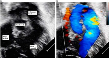
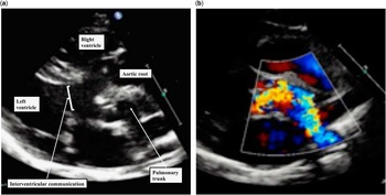
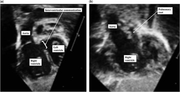
When using multiple imaging planes, and taking advantage of Doppler interrogation (Fig 9), it should also be possible to identify with precision the morphology of the subvalvar outflow tracts, including the presence of subpulmonary or subaortic infundibulums, and any other potential sources of outflow tract obstruction. In addition, the echocardiographic interrogation should identify the aortic origins of the coronary arteries, and any additional abnormalities that might be present, such as interatrial communications, abnormalities of the aortic arch, and in particular the presence of additional ventricular septal defects. The subcostal views are extremely useful in neonates, and may demonstrate almost the entirety of the anatomical features. The parasternal long-axis views are often the most helpful to display the interventricular communication and its relationship to the ventricular origins of the great arteries (Fig 10). It may be necessary, nonetheless, to use multiple imaging planes so as to demonstrate all the borders of the interventricular communication and its relationships with the arterial trunks. Use of multiple planes, for example, can demonstrate when the defect is in non-committed position, and hence distant from the ventricular outlet components (Fig 11). Such defects no longer open to the right ventricle between the limbs of the septomarginal trabeculation. If muscular separation is present between the aortic and mitral valves, this will be evident in long-axis views. It is the parasternal short-axis views, however, that most readily display the spatial arrangement of the great arteries. Long slow sweeps from the subcostal window, and in the parasternal long and short axes, should be used in order to display fully the overall segmental anatomy and the anatomical features of known surgical significance. As we have shown in Figures 9 and 10, color flow Doppler interrogation is used to identify sites of subvalvar or valvar obstruction, as well as to reveal any abnormal features of flow across the atrioventricular valves. Both continuous and pulsed wave Doppler interrogations are used to assess the sites and severity of any identified obstructions. The coronary arterial anatomy is best identified using the parasternal short-axis cuts, as well as with subcostal or apical sweeps. As Pushparajah et alReference Pushparajah, Barlow and Tran 27 have now shown, three-dimensional echocardiography is particularly helpful for planning the optimal surgical strategy.
The images, taken in the subcostal right anterior oblique view, are from a patient with a subaortic interventricular communication and no pulmonary obstruction. As shown by color Doppler interrogation ( b ), there is laminar flow across the interventricular communication and into the pulmonary trunk.

The images, obtained in parasternal long-axis plane, are from patient with double-outlet right ventricle with a subpulmonary interventricular communication. ( a )Location of the aorta anterior to the pulmonary trunk. The addition of color flow Doppler interrogation ( b ) shows the presence of a muscular subpulmonary infundibulum, which is causing obstruction to flow into the pulmonary artery, as demonstrated by the turbulent nature of the flow.

The images shown are from a patient with an interventricular defect considered to be a non-committed defect. As shown in ( a ), taken using the oblique subcostal view, the interventricular communication is distant from the aorta, although it is feasible that a long tunnel could be constructed to channel the left ventricle into the aortic root. When the smaller pulmonary trunk is brought into view ( b ) the interventricular communication is no longer visible, with the aortic root now bisected, suggesting that the defect is not committed directly to either arterial trunk.

Additional three-dimensional interrogation
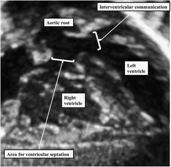
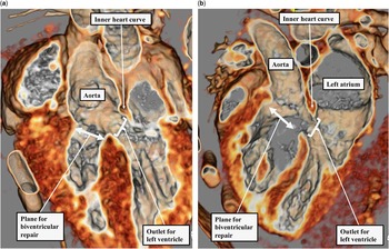
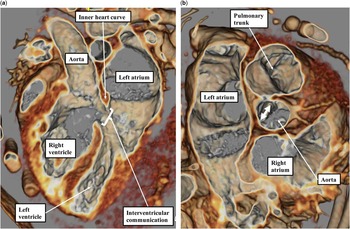
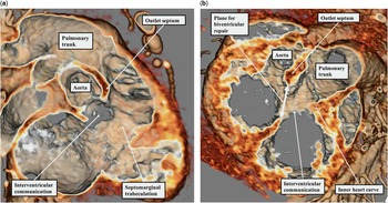
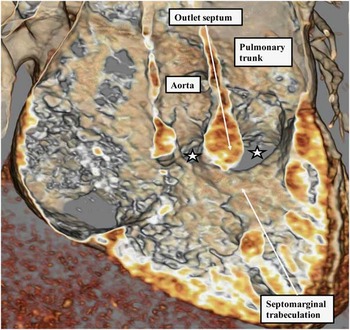
Although cross-sectional echocardiographic interrogation is now the mainstay of the diagnostic work-up, with three-dimensional displays now facilitating decision making,Reference Pushparajah, Barlow and Tran 27 it is the detailed analysis made possible by CT scanning or MRI that now reveal with precision all the required anatomical information. Although these modalities are currently less widely available, particularly for clinical application in small patients, and concerns remain regarding the use of ionising radiation to produce CT images, the images provided show with exquisite accuracy, for example, the fundamental differences between the area through which the ventricular chambers communicate as opposed to the area requiring surgical closure so as to produce biventricular circulations. These images also serve to confirm that it is the area requiring surgical closure that is universally termed the “ventricular septal defect” in the setting of tetralogy of Fallot (Fig 12a). As we have shown, the cranial margin of this area is the muscular outlet septum, or its fibrous remnant. The CT images obtained in the setting of double-outlet right ventricle, in contrast, as was the case with the echocardiographic images, show that the channel currently termed the “ventricular septal defect” is roofed by the inner heart curvature (Fig 12b). The channel, which provided the exit from the body of the left ventricle in both instances, is more accurately termed the interventricular communication when there is double outlet from the right ventricle (Fig 12). The images provided by interrogation of the data sets obtained subsequent to CT scanning also solve the conundrum posed to echocardiographers in the setting of overriding of the arterial roots. As we have already discussed, it can be difficult, when using echocardiographic interrogation, to determine the precise commitment of an overriding outflow tract. The availability of the three-dimensional data set permits relationships between the overriding root and the ventricular septum to be assessed precisely in the short-axis plane (Fig 13). Interrogation of the CT data sets also shows with precision the attachment of the outlet septum, or its fibrous remnant, relative to the interventricular communication. The images confirm that, in the majority of patients, the interventricular communication opens to the right ventricle between the basal limbs of the septomarginal trabeculation or septal band. It is when the outlet septum is attached to the cranial limb of the trabeculation that the interventricular communication is placed in the subaortic location (Fig 14, left-hand pane). In contrast, when the outlet septum is inserted to the rightward margin of the inner heart curvature, cranial to the caudal limb of the septomarginal trabeculation, the interventricular communication is placed in the subpulmonary position (Fig 14b). Interrogation of CT data sets then shows that, although the outlet septum is usually fibrous rather than muscular when the interventricular communication is doubly committed (Fig 6b), this is not always the case. As we have shown in Figure 14, it is the attachment of the outlet septum that is the key feature in committing the defect to one or other arterial root. Thus, when the outlet septum is attached to the midpoint of the inner heart curvature, the defect is doubly committed irrespective of whether the outlet septum itself if fibrous (Fig 6b) or muscular (Fig 15). In the heart shown in Figure 15, in fact, the muscular outlet septum is fused to the crest of the muscular interventricular septum, thus producing discrete subaortic and subpulmonary defects.
The CT images, showing the long axis of the ventricular mass in a patient with tetralogy of Fallot ( a ) and double-outlet right ventricle with subaortic defect ( b ), reveal that the area conventionally termed the “ventricular septal defect” in tetralogy is the plane closed to achieve biventricular repair, whereas in a patient with double outlet it is the outlet for the left ventricle that is currently, in most instances, defined as the “ventricular septal defect”.

( a )The long-axis cut from the patient shown in Figure 12 with a potential double-outlet ventriculo-arterial connection. The aortic root is overriding the crest of the muscular ventricular septum. With the capacity to cut the data set in orthogonal planes, however, it can be shown that the aortic root is committed by at least four-fifths to the cavity of the right ventricle ( b ), confirming that the diagnosis is one of double-outlet right ventricle with subaortic defect.

The cuts from CT data sets show how it is the insertion of the muscular outlet septum relative to the basal limbs of the septomarginal trabeculation that determines the commitment of the interventricular communication to the arterial roots. ( a ) Cranial insertion of the muscular outlet septum in a patient with a subaortic defect. ( b ) Arrangement in a patient with a subpulmonary defect. Note that it is the area roofed by the muscular outlet septum (double-headed arrow) that will be closed by the surgeon to produce a biventricular repair, although in this patient the surgeon will also be required to perform an arterial switch procedure. Note also that the tomographic image shows how the inner heart curvature produces the cranial margin of the interventricular communication, which is not therefore a “ventricular septal defect”.

The CT image, showing the “en face” view of the ventricular septum as seen from its right ventricular aspect, reveals the presence of a defect opening to the right ventricle between the limbs of the septomarginal trabeculation. In this patient, however, the muscular outlet septum is fused to the crest of the muscular interventricular septum, thus dividing a doubly committed interventricular communication into separate subaortic and subpulmonary components (white stars with black borders).

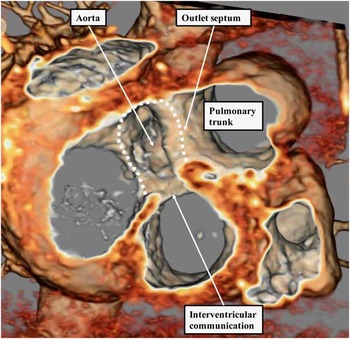
The availability of three-dimensional data sets produced by both CT and magnetic resonance investigation has now opened the way for printing of three-dimensional models to aid in determining the optimal surgical treatment. Use of such three-dimensional models has also been shown to facilitate the understanding of these potentially complicated congenital cardiac malformations.Reference Garekar, Bharati and Chokhandre 28 Printing of such data sets, however, is not without its financial implications, and the information can readily be conveyed simply by display of the images themselves (Fig 16).Reference Riesenkampff, Rietdorf and Wolf 29
The image is a short-axis cut viewed from the cardiac apex from the CT data set obtained from the patient also illustrated in Figure 13. The dotted line shows the borders around which the surgeon will secure the tunnel required to connect the left ventricle, through the interventricular communication, with the subaortic outflow tract. Note that the tunnel will be secured cranially to the muscular outlet septum. Note also that, in this patient, the aortic root is appreciably smaller that the pulmonary root, raising a potential problem in determining the haemodynamic, as opposed to anatomical, feasibility of biventricular repair.

Conclusions
The ability to interrogate the congenitally malformed heart in any desired plane is revolutionising our understanding of so-called “complex” malformations. Such interrogation, for long at the fingertips of the skilled echocardiographer, is now being made available for widespread discussion by the increasing use of techniques such as MRI and CT. The images confirm that the situation in which both arterial trunks take their origin predominantly from the right ventricle is a grouping of individual lesions, rather than a solitary anatomical phenotype. They also confirm that the key feature in determining the diagnosis of those patients appropriately place in this grouping is recognition of the precise borders of the channel providing the outflow from the morphologically left ventricle. Attention to this feature shows that, in the patients with double-outlet right ventricle, the cranial boundary of this channel is the inner heart curvature, be that structure muscular or fibrous, rather than the outlet component of the ventricular septum. When considered literally, therefore, and although indicative of deficient ventricular septation, the channel is more accurately described as an interventricular communication, rather than a ventricular septal defect. It is the space between the crest of the muscular ventricular septum and the outlet component of the ventricular septum that represents the true septal defect. It is also, of course, the margins of this area that are closed by the surgeon so as to restore biventricular circulations, presuming that such a manoeuvre is haemodynamically feasible.
Financial Support
This research received no specific grant from any funding agency, commercial or not-for-profit sectors.
Conflicts of Interest
None.