Milrinone and levosimendan are both used post-operatively following congenital cardiac surgery to increase cardiac output by reducing systemic and pulmonary afterload and increasing myocardial contractility.Reference Hoffman, Wernovsky and Atz1–Reference Follath, Cleland and Just4 Synthetic catecholamines, such as epinephrine and dobutamine, offer an alternative option, but their use has several drawbacks including increased heart rate, increased myocardial oxygen consumption, and increased risk of myocardial damage.Reference Caspi, Coles and Benson5, Reference Fowler, Alderman and Oesterle6
Milrinone, a phosphodiesterase 3 inhibitor, reduces the hydrolysation of cyclic adenosine monophosphate, and consequently increases the intracellular concentration of Ca2+, thereby increasing cardiac muscle contractility and vascular muscle relaxation. Levosimendan produces potent inotropic actions by sensitising myocardial troponin C to calcium. Vasodilatation is mediated by stimulating the adenosine triphosphate-sensitive potassium channels in vascular smooth muscle cells.
The newborn heart has a higher β-adrenoceptor density but a lower receptor affinity than the mature heart.Reference Wittnich, Belanger and Bandali7 This is the underlying reason for a diminished ability to raise cyclic adenosine monophosphateReference Tanaka and Shigenobu8 and increase cardiac output in response to β-adrenergic stimulation.Reference Shaddy, Tyndall, Teitel, Li and Rudolph9 A high number of maturational changes in the myocytes have been discovered, including expression rates of phosphodiesterases and calcium handling, and thus an age-dependent difference in the effect of inodilators could be expected.
Although data on the effects of milrinone and levosimendan in the paediatric population have been presented, a study focused on the effects of the two inodilators in newborns had not been published before Paradisis et al in 2009 reported that milrinone did not increase blood flow and did not prevent low systemic blood flow during the first 24 hours in very pre-term infants.Reference Paradisis, Evans, Kluckow and Osborn10
To this point, evidence on the effect of levosimendan in the newborn is solely based on case reports.Reference Egan, Clarke and Williams11, Reference Namachivayam, Crossland, Butt and Shekerdemian12 The purpose of this study was to evaluate possible age-dependent effects of milrinone and levosimendan in newborn and mature pigs, especially focusing on inotropic and vasorelaxing effects.
Materials and methods
The study was approved by the Danish National Board on Animal Care and Use, and experiments were conducted in accordance with the Danish Law on animal experiments.
Subjects
We chose 48 healthy Danish landrace piglets and divided them into two age groups: newborns with a mean age of 5.6 days plus or minus 1.7, with a weight of 2.2 plus or minus 0.4, and mature animals with a mean age of 5 weeks, with a weight of 18.9 plus or minus 1.9.
Anaesthesia
All animals were pre-medicated with an intramuscular injection of ketamine at a doze of 0.5 milligrams per kilogram and midazolam at 5 milligrams per kilogram. Mature piglets of 5 weeks were anaesthetised with an intravenous injection of etomidate at a doze of 0.5 milligrams per kilogram. Newborn piglets of 5–6 days were anaesthetised with an intravenous injection of midazolam at 0.5 milligrams per kilogram and ketamine at 1.25 milligrams per kilogram. Anaesthesia was maintained throughout the experiment with inhaled sevoflurane and morphine at 0.5 milligrams per kilogram per hour. The animals were intubated and ventilated with a tidal volume of 8–10 millilitres per kilogram, and a respiratory rate of 14 per minute in the mature animals and 45–55 per minute in the newborn group. The newborns were kept on pressure-controlled ventilation, and mature animals on volume-controlled ventilation. Arterial blood gases were analysed (ABL, Radiometer, Copenhagen, Denmark) at baseline and during the experiment, and ventilator settings adjusted to obtain normoventilation and normal oxygenation during the study. Intravenous infusion of Ringer's acetate was administered (that is, 2 millilitres per kilogram per hour).
Study protocol
Piglets were randomised to receive milrinone, levosimendan, or saline after instrumentation, by drawing identical sealed envelopes. The intervention was not blinded to the researchers during the study period, but the investigators were blinded to the actual treatment given in connection with all analyses of haemodynamics and conductance measurements. Owing to long half-lives of milrinone and levosimendan, loading doses were used to reach two different serum levels of each drug. For both drugs, loading doses were administered during 10-minute periods followed by an infusion period of 50 minutes. For milrinone (Sanofi-Aventis, Hoersholm, Denmark), a loading dose of 50 micrograms per kilogram was administered, followed by an infusion of 0.3 micrograms per kilogram per minute. After 60 minutes, a second bolus of 75 micrograms per kilogram was administered, followed by an infusion of 0.6 micrograms per kilogram per minute. Levosimendan (Simdax, Abbott, Copenhagen, Denmark) was administered with a loading dose of 12 micrograms per kilogram, followed by an infusion of 0.1 micrograms per kilogram per minute. After 60 minutes, a second loading dose of 24 micrograms per kilogram was followed by an infusion of 0.2 micrograms per kilogram per minute. In the control group, an equivalent amount of saline was infused. Measurements of cardiac function and pressures were registered every 30 minutes starting at baseline – 0, 30, 60, 90, and 120 minutes (Fig 1). Ringer's acetate was administered to re-establish a mean arterial pressure according to baseline measurements. At the end of the experiment, animals were sacrificed with an overdose of pentobarbital.
Infusion protocol, timeline, and timing of measurements. B = bolus; IVI = I.V. infusion. *Measurements.

Surgical procedure and instrumentation
Bilateral throat cut-down incisions were made and five valved sheaths (William Cook, Ireland and St. Jude Medical Europe, Belgium) were advanced through two arterial and three venous accesses. Two appropriately sized conductance catheters (Millar Instruments, Dallas, Texas, United States of America) were positioned with the tip at the apex of the right ventricle and left ventricle, respectively. Mature animals 5 Fr., 12 segments, newborn 3 Fr., 8 segments. A Fogarty occlusion catheter (Boston Scientific, Denmark) was positioned in the inferior caval vein. A bipolar pacing wire was positioned at the right atrium appendage. A Swan–Ganz catheter (Edwards Lifesciences, Horw, Switzerland) was positioned in the pulmonary artery. Correct placement of all catheters and wires was confirmed by X-ray flouroscopy before data collection.
Ventricular pressure–volume relationship
The conductance and micromanometer-tipped catheter was connected to a signal conditioner (Sigma-5-DF, CD Leycom, The Netherlands). After calibrating the conductance catheter, we recorded pressure–volume measurements at baseline during steady-state conditions and pre-load reduction by gradual occlusion of the inferior caval vein. All conductance volumes were corrected for parallel conductance, blood resistivity, and the gain constant. Parallel wall conductance and total ventricular conductance were calculated as described elsewhere.Reference Baan, van der Velde and de Bruin13 One condition processor had the excitation frequency altered to avoid electrical interference between the two systems, thereby enabling simultaneous biventricular volume measurements. Analyses of the parallel conductance, steady-state, and occlusion parameters were performed using PVAN 3.3 (Millar instruments, Dallas, Texas, United States of America).
Physiological data
Cardiac output (litre per minute) was calculated by a dedicated processing computer (Vigilance Baxter, Irvine, California, United States of America) as a continuous cardiac output in the mature group and by injection of 3 millilitres of tempered saline in the superior caval vein in the newborn group. Three injections were administered at each measurement and the median value was used in the analysis. Cardiac output measurements were only recorded three times during the experiment in order to prevent fluid overload. Cardiac output was indexed to body weight (litre per minute per kilogram). Pulmonary arterial pressure (millimetres of mercury) and mean pulmonary arterial pressures (millimetres of mercury) were measured from the tip of the pulmonary artery catheter. Systolic artery pressure and mean arterial pressure were measured from the valved introducer in the left carotid, and heart rates were obtained from the arterial pressure waveform. Systemic vascular resistance (dyne times second per centimetre to the fifth) was derived by the equation: 80 times mean arterial pressure minus central venous pressure divided by the cardiac output. Arterial elastance (millimetres of mercury per millilitre) was derived by dividing the end-systolic pressure by the stroke volume.
Pressure–volume measurements were recorded under steady-state conditions and during occlusion of the inferior caval vein. The atrium was paced at a rate of 100 per minute in the mature group and 200 per minute in the newborns during each measurement to ensure comparability of the time-dependent parameters within the groups. The ventilator was disconnected when calculating steady-state measurements of the maximum pressure development over time, maximum negative development of pressure over time, and the time constant of isovolumetric relaxation (millisecond). Time constant of isovolumetric relaxation was calculated using a monoexponential model of pressure decay with a zero asymptote.Reference Weiss, Frederiksen and Weisfeldt14 Pre-load reduction was performed subsequently during the same period of apnoea, to allow off-line calculation of load-independent indices of contractility, that is, pre-load recruitable stroke work (millimetres of mercury times millilitres per millilitre) and end-systolic elastance (millilitres of mercury per millilitre). Pre-load recruitable stroke work was calculated by regression of the stroke work, defined as the area of the pressure–volume loop, and the end-diastolic pressure, during pre-load reduction.Reference Glower, Spratt and Snow15 End-systolic elastance was defined as the slope of the end-systolic pressure–volume relationship.
Statistical analyses
Statistical analyses were carried out using Stata Statistical Software, 10.0 (Stata Corporation, College Station, Texas, United States of America) using multiple variance regression analyses. All data are presented as percentage changes in relation to placebo plus or minus standard deviation. An unpaired Student's t-test was used to determine differences in parameters between the two age groups at baseline, after assumptions of normal distributions were confirmed. A p-value of 0.05 or lower was considered statistically significant.
Results
Age group differences
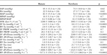
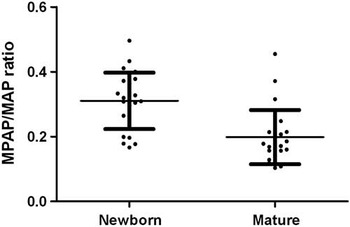
At baseline, the age groups displayed haemodynamic differences in systolic artery pressure, pulmonary arterial pressure, mean arterial pressure, arterial elastance, systemic vascular resistance, maximum pressure development over time, end-systolic elastance, time constant of isovolumetric relaxation, and maximum negative development of pressure over time (Table 1). As expected, the ratio of mean pulmonary arterial pressures/mean arterial pressure was higher in the newborns (p < 0.0001), indicating that the relatively increased pulmonary pressures after birth were still present (Fig 2). At baseline, no differences were found between the treatment groups in either of the age groups.
Comparisons of haemodynamics and ventricular performance variables in newborn and mature piglets.

Ea = arterial elastance; Ees = end-systolic elastance; dP/dTmax = maximum development of pressure over time; dP/dTmin = minimum development of pressure over time; LV = left ventricle; MAP = mean arterial output; MPAP = mean pulmonary arterial pressure; PRSW = pre-load recruitable stroke work; RV = right ventricle; SAP = systolic arterial pressure; SVR = systemic vascular resistance; Tau = time constant of isovolumetric relaxation.
Values given are mean of the entire age group at baseline and standard deviation using two-tailed Student's t-test.
Maximum pressure development over time; maximum negative pressure development over time, and the time constant of isovolumetric contraction are all time-dependent variables, and differences between groups may be partly affected by pulse rate
Mean pulmonary arterial pressure/mean arterial pressure ratio for mature (n = 24) and newborn animals (n = 24). Plus or minus standard deviation indicated by bars. The groups were statistically different, p < 0.0001. Two-tailed Student's t-test.

Mature animals
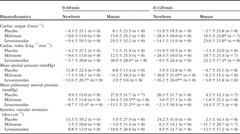
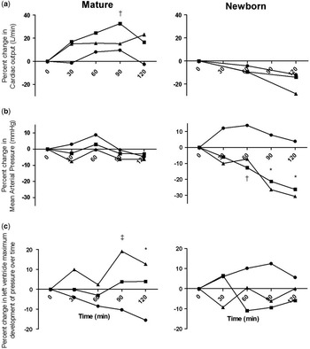
Both milrinone and levosimendan increased cardiac output and cardiac index compared with the control group during the 120-minute observation period (levosimendan increased cardiac output by 23% (p < 0.05), and milrinone caused a 16.5% increase (p = 0.05) after 120 minutes; Table 2). These changes were not associated with any decrease in mean arterial pressure, which remained stable throughout the entire period (Fig 3, Table 2). Mean pulmonary arterial pressures decreased significantly during the first 60 minutes for both levosimendan (p = 0.002) and milrinone (p = 0.004) compared with the control group. However, mean pulmonary arterial pressures returned to similar values as in the control group after 120 minutes. Systemic vascular resistance decreased by 13–30%, although not statistically significant, in both levosimendan- and milrinone-treated animals (Table 2).
Changes in haemodynamics during the infusion protocol.

*Significant change compared to placebo, p < 0.05
**Significant change compared to placebo, p < 0.01
Values given are percentage change from baseline at 60 and 120 min and their standard deviations
Changes in cardiac output (a), mean arterial pressure (b), and left ventricle maximum pressure development over time (c) during infusions milrinone (▪), levosimendan (▴), and controls (●). The number of animals in each group is shown in Table 1. Left column = mature animals; right column = newborn animals. *Both milrinone and Levosimendan are significantly different from controls = p < 0.05. ‡Milrinone is significantly different from controls, p < 0.05. †Levosimendan is significantly different from controls, p < 0.05.

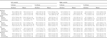
In the left ventricle, a significant increase in maximum pressure development over time was observed for both levosimendan and milrinone after 120 minutes compared with the control group (Table 3). At the 60 min mark, milrinone had significantly decreased the time constant of isovolumetric relaxation by −10.7%. At 120 min, a decrease was found in both groups – milrinone 16% and levosimendan 9.6% – although not statistically significant. In accordance, pre-load recruitable stroke work was increased by 73% and 37% for levosimendan (p = 0.005) and milrinone (p = 0.07) after 90 minutes, respectively. At 60 min, only milrinone produced significant increases in PRSW, but neither milrinone nor levosimendan produced significant increases at 120 min. The time constant of isovolumetric relaxation remained unchanged in the right ventricle (Table 3).
Changes in left and right ventricular systolic and diastolic performance during the infusion protocol.

dP/dTmax = maximum development of pressure over time; dP/dTmin = minimum development of pressure over time; Tau = time constant of isovolumetric relaxation; PRSW = pre-load recruitable stroke work; Ees = end-systolic elastance
Values given are percentage change from baseline at 60 and 120 min and their standard deviations
*Significant change compared to placebo, p < 0.05
**Significant change compared to placebo, p < 0.01
Newborn animals
In contrast to the changes in cardiac output in the mature animals, neither levosimendan nor milrinone was able to increase cardiac output or cardiac index in newborn piglets (Table 2). In the milrinone-treated animals, cardiac output decreased, although numbers did not reach statistical significance. Both inodilators, however, caused a decrease in mean arterial pressure. In the levosimendan-treated animals mean arterial pressure decreased by 26.2% (p < 0.01) and in the milrinone-treated animals by 31.6% (p < 0.01). Mean pulmonary arterial pressures decreased by 8.7% in the levosimendan-treated group (p = 0.03). Despite the decrease in mean arterial pressure in both intervention groups, no significant changes were observed in systemic vascular resistance in either group (Table 2). Neither maximum pressure development over time, pre-load recruitable stroke work nor time constant of isovolumetric relaxation showed any significant changes in any of the intervention groups in the left ventricle (Table 3). Milrinone increased pre-load recruitable stroke work by 82% in the right ventricle after 90 minutes (p < 0.05) and 96.2% after 120 minutes. Neither maximum pressure development over time nor time constant of isovolumetric relaxation was changed in any of the intervention groups in the right ventricle.
Discussion
In mature pigs, both milrinone and levosimendan increased cardiac output, as previously shown in both animalReference Stocker, Shekerdemian and Norgaard3, Reference Segreti, Marsh, Polakowski and Fryer16, Reference Banfor, Preusser and Campbell17 and clinical studies.Reference Hoffman, Wernovsky and Atz1, Reference Bailey, Hoffman and Wessel18 Quite surprisingly, however, this effect was not observed in newborn pigs, where a vasodilatory response was much more pronounced.
Age group differences
We found highly significant differences in baseline haemodynamics and ventricular performance parameters between the two age groups. Most markedly, the ratio of mean pulmonary arterial pressures/mean arterial pressure was more than 50% higher in the newborn animals. We therefore believe that this study of vascular physiology and myocardial performance has been carried out at two different stages of maturation in the cardiovascular system, equivalent to the transition period immediately after foetal circulation.
Pulmonary artery pressure
It has previously been described how milrinoneReference Feneck19, Reference Chang, Atz, Wernovsky, Burke and Wessel20 and levosimendanReference Stocker, Shekerdemian and Norgaard3, Reference Follath, Cleland and Just4 reduced pulmonary arterial pressure in the mature vascular system in humans and animals with elevated pulmonary pressures. In this study, only levosimendan produced a significant decrease in mean pulmonary arterial pressures in the newborn group during the infusion period. This is most likely explained by the low basal tone of the vessel smooth muscle during the experiment. In the mature group, we found a decrease in pulmonary pressure at 60 minutes, but no significant difference was observed after the second infusion period.
Arterial pressures
The marked decrease in mean arterial pressure in the newborn animals could hypothetically be related to the expression or activity of phosphodiesterase 3 in the systemic vessels, with high expression and activity increasing the impact of milrinone. However, there are currently no data on age-related changes in activity and expression of phosphodiesterase 3 in arterial smooth muscle.
Systemic vascular resistance
We could not demonstrate any significant changes in systemic vascular resistance. In newborns, this was due to a simultaneous decrease in cardiac output and mean arterial pressure. In a recent study, levosimendan in high doses decreased systemic vascular resistance significantly, whereas milrinone caused no significant changes.Reference Stocker, Shekerdemian and Norgaard3 In agreement with these findings, we observed marked but inconsistent changes in systemic vascular resistance in the levosimendan-treated mature animals. A similar tendency was observed for milrinone after 120 minutes, but changes were not statistically significant.
Age-dependent responses
Animal studies have shown that in the mature circulation, the positive inotropic effect seems to predominate over the vasodilatory effect of both milrinoneReference Sys, Goenen, Chalant and Brutsaert21 and levosimendan.Reference Leather, Ver and Segers22 This is in accordance with the findings of the present study. However, we found the exact opposite to be true for the newborn animals, where no increase in ventricular performance was observed. In newborns, β-stimulation with catecholamines requires much higher weight-adjusted doses compared with adultsReference Tanaka and Shigenobu8 to obtain a similar haemodynamic response because of low affinity to β-receptors in spite of high density.Reference Wittnich, Belanger and Bandali7 The same could be the case for inodilators, and it could be argued that higher concentrations should have been administered. However, as both inodilators caused a marked decrease in mean arterial pressure of approximately 30%, a further increase in dose is unlikely to be acceptable in a clinical setting. Although metabolism of both inodilators may be different in newborn animals requiring higher doses to sustain a sufficient concentration, the vasodilatory response progressed throughout the experiment indicated that this is an unlikely explanation.
Ventricular performance in the newborns
Milrinone is effective in the prevention of low cardiac output syndrome in children after congenital cardiac surgery.Reference Hoffman, Wernovsky and Atz1 This, however, has not been established specifically for the neonatal patient population. It has been reported that milrinone increases the cardiac index in patients aged between 3 and 27 days, with a mean of 5, 6 hours after hypothermic CPB.Reference Chang, Atz, Wernovsky, Burke and Wessel20 However, all patients received dopamine, and because of a possible synergistic effect the actual contribution of milrinone to improve ventricular performance is unclear. Our findings are in conflict with previous in vitro studies in which milrinone has been reported to increase myocardial performance in myocytes of the newborn. Although amrinone, another phosphodiesterase 3 inhibitor, has been shown to decrease contractility in the newborn myocardium,Reference Binah, Legato, Danilo and Rosen23–Reference Ross-Ascuitto, Ascuitto, Chen and Downing25 evidence of a positive inotropic effect of milrinone in the newborn piglet heart has been published.Reference Ross-Ascuitto, Ascuitto, Ramage and McDonough26 However, all reports on a positive effect of milrinone on ventricular performance in the newborn heart originate from in vitro studies using concentrations that are 10–100 times higherReference Ross-Ascuitto, Ascuitto, Ramage and McDonough26, Reference Binah, Sodowick, Vulliemoz, Danilo and Rosen27 than what is clinically relevant (100–300 nanograms per millilitres). A possible cause of the age-dependent differences in the effect of milrinone could be the developmental changes in the expression rate of phosphodiesterase 3 in the myocardium. Perinatal changes in the expression of phosphodiesterase 3 would be highly likely to alter the effect of milrinone. High expression rates of phosphodiesterase 3 would make milrinone in normal doses insufficient, whereas high doses, as used previously,Reference Ross-Ascuitto, Ascuitto, Ramage and McDonough26, Reference Binah, Sodowick, Vulliemoz, Danilo and Rosen27 would be effective. Another possible cause of the age-dependent differences in the effect of milrinone is probably rooted in the array of developmental changes in the myocardium. Among those are changes in Ca2+ flow over the cell membraneReference Akita, Joyner, Lu, Kumar and Hartzell28 and sarcoplasmatic reticulum.Reference Seguchi, Harding and Jarmakani29 Both are activated by cyclic adenosine monophosphate-dependent protein kinases, and their activity can therefore be manipulated indirectly by milrinone.
Cardiac output
In our study, cardiac output was increased in the mature animals because of increased contractility. In the newborns, both milrinone and levosimendan failed to increase cardiac output despite a significantly decreased mean arterial pressure. In accordance with these observations, a recent clinical study demonstrated that milrinone was unable to increase systemic blood flow in pre-term neonates.Reference Paradisis, Evans, Kluckow and Osborn10 Our findings suggest that the same could apply for levosimendan. There are currently only casuistic data in the literature on the use of levosimendan in neonates and only in combination with other inotropes.Reference Egan, Clarke and Williams11, Reference Namachivayam, Crossland, Butt and Shekerdemian12, Reference Lechner, Moosbauer, Pinter, Mair and Tulzer30
Model
Pacing rates were adjusted for each age group to ensure that contractility was recorded at a frequency relevant from a clinical and physiological perspective. Comparison of changes after an intervention was only carried out within the specific age group and compared with the control group. As newborns were paced at a higher frequency, it could be assumed that the lack of response to inodilators was due to an absent inotropic reserve potential. However, it has previously been described how non-clinical doses of milrinone can increase peak systolic pressure in newborn pig hearts paced at 180 beats per minute.Reference Ross-Ascuitto, Ascuitto, Ramage and McDonough26 Furthermore, the authors found that dobutamine in clinically relevant doses could significantly increase contractility expressed as maximum pressure development over time and pre-load recruitable stroke work in newborn hearts when paced at 200 beats per minute (data not shown). As both age groups were only paced prior and during but not between the measurements, a pace-induced decrease in contractility is unlikely to occur.
Limitations
Our findings are based on an animal model and may therefore not necessarily apply to human physiology. Nevertheless, the haemodynamic and ventricular response in the mature animals is very similar to what has been observed in adults and children.Reference Hoffman, Wernovsky and Atz1, Reference Bailey, Miller and Lu2, Reference Follath, Cleland and Just4, Reference Feneck, Sherry, Withington and Oduro-Dominah31
Comparisons between human beings and pigs on maturation of the cardiovascular system in terms of a normalised mean pulmonary arterial pressures/mean arterial pressure ratio and changes in metabolism are sparse, and the findings of this study are therefore not directly related to the equivalent age groups in humans.
Recent reports from piglet studies regarding the use of milrinone in newborn piglets found that infusion rates cumulating to a total infusion of 165 microgram per kilogram over 135 minutes – present study 170 microgram per kilogram over 120 minutes – produced serum values just below the low end of the therapeutic window.Reference Joynt, Bigam and Charrois32 Serum concentrations were measured 120 minutes after bolus infusion, compared with the present study in which last measurements were recorded 50 minutes after loading doses. Considering this, we believe that the infusion protocol with two boluses administered within 2 hours is sufficient to achieve a therapeutic concentration.
Conclusion
This study is the first to report data on the age-dependent effect of levosimendan and milrinone in an experimental in vivo, closed-chest model. Both milrinone and levosimendan failed to increase cardiac output in the newborn pigs, whereas the vasodilatory response was pronounced. If our findings would translate to the post-operative setting, the inodilators milrinone and levosimendan only contribute to the prevention of low cardiac output syndrome in the newborns by decreasing afterload. Future studies in the neonatal population addressing these issues specifically are warranted. Additional animal studies are needed in order to elucidate the change in response to two inodilators in combination with catecholamines, as well as the effect during a low cardiac output syndrome situation.
Acknowledgements
We would like to thank the technical staff of The Institute of Clinical Medicine, Aarhus University Hospital, Skejby. Financial support: The Danish Council for Independent Research – Medical Sciences (FSS), Institute of Clinical Medicine, Aarhus University Hospital, Skejby, The Danish Heart Foundation and The Jens Anker Andersen Foundation.