In recent years, early complete repair of congenitally malformed hearts has become a common practice in neonates, and has also been advocated for infants born with low weight.Reference Reddy and Hanley1, Reference Kopf and Mello2 In some patients with functionally univentricular arrangements, nonetheless, or others with complex biventricular anatomy, the preferred initial surgical approach is a palliative construction of a systemic-to-pulmonary arterial shunt.Reference Tamisier, Vouhe, Vernant, Leca, Massot and Neveux3
In patients with the flow of blood to the lungs dependent on patency of the arterial duct, establishing a stable and adequate flow using a systemic-to-pulmonary arterial shunt, early after birth, is critical for survival. In other patients with small pulmonary arteries, or multiple aortopulmonary collateral arteries, this palliative procedure may be an important factor for promoting growth of the pulmonary arteries, thus increasing the potential for later biventricular repair.Reference Duncan, Mee and Prieto4, Reference Ishikawa, Takahashi and Sato5
Many studies recognized weight less than 3 kilograms at the time of placement of the shunt as a significant risk factor for survival.Reference Tamisier, Vouhe, Vernant, Leca, Massot and Neveux3, Reference de Leval, McKay, Jones, Stark and Macartney6 In an attempt to identify other predicting factors of survival, we reviewed our experience with this procedure in patients weighing less than 3 kilograms in whom we used a venous homograft to create the shunt. We studied the differences between patients with a functionally univentricular heart as opposed to patients with biventricular circulations.
Methods
We reviewed retrospectively our computerized data base to identify all children weighing less than 3 kilograms who had undergone placement of a Blalock-Taussig shunt using a saphenous venous homograft at Cook Children’s Medical Center between June, 2002, and May, 2007. Medical records for each patient were then reviewed to obtain information regarding baseline demographics, operative, post-operative and follow-up factors. Our Institutional Review Board approved this study on March 28, 2006, and individual consent for the study was waived.
We identified 32 infants, all weighing less than 3 kilograms at the time of surgery, and all with complex cyanotic congenitally malformed hearts, who underwent construction of the shunt during the period of study. Their weights ranged from 1.8 to 2.86 kg, with a median of 2.51 kg. The median age at time of placement of the shunt was 8 days, with a range from 2 to 70 days. Of the patients, 29 (91%) were neonates, while 15 (47%) were born prematurely, and 13 (41%) were less than one week old at the time of surgery. In 17 patients (53%), the anatomic defects produced a functionally univentricular arrangement, while 15 (47%) had defects which permitted staging to biventricular repair.
Patients staged to univentricular palliation were much more likely to have physiology dependent on patency of the arterial duct as compared with those staged to biventricular palliation (p < 0.001). The diameters of the pulmonary arteries were measured by transthoracic echocardiography. The size of the left pulmonary artery was significantly smaller at presentation in those staged to biventricular repair than in those with functionally univentricular arrangements, at a median of 2 mm as opposed to 3.6 mm (p = 0.01 – Table 1).
Comparison of pre-operative characteristics stratified by intended strategy for surgical palliation.

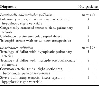
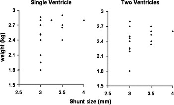
In Table 2, we show a comparison of the preoperative diagnoses between patients with functionally univentricular as opposed to biventricular physiology. As indicated, in all patients we used a saphenous venous homograft as the graft, its diameter ranging in size from 2.5 to 4 mm, with a median of 3 mm. These allografts were cryopreserved homografts from donors, and were matched to the blood groups of the patients (Cryolife, Inc. Kennesaw, GA). The chosen sizes were generally closer to the weight of patients with functionally univentricular hearts (r 2 = 0.33) as compared to those having biventricular circulations (r 2 = 0.22) (Fig. 1). In those with functionally univentricular hearts, the chosen size was calculated on the basis of 1 mm equaling 1 kilogram of body weight. In those with biventricular circulations, shunts of 1 mm larger were usually chosen to encourage increased flow of blood to the lungs, and thus promote pulmonary arterial growth.
Primary diagnosis in patients stratified by intended strategy for palliation.

Scatter plot showing the size of the shunt according to the weight of the patients stratified according to the strategy for palliation.

Description of surgical technique
The operation was performed through a sternotomy in 28 out of the 32 patients (87%). Only the patients undergoing unifocalisation of aortopulmonary collateral arteries were approached through a thoracotomy.
The shunts were anastomosed to an arterial branch on the opposite side to the aortic arch in all patients. Typically, the shunt was placed between the brachiocephalic or subclavian artery and the pulmonary artery on the same side. We administered 100 units of heparin per kilograms prior to creation of the shunt, reversing the dose at the end of the operation.
An arteriotomy was made on the inferior aspect of the inflow artery, and an anastomosis made with the saphenous venous homograft using a continuous 7-0 monofilament polypropylene suture. A haemoclip was then applied proximally across the graft, permitting removal of the C-clamp from the inflow arteries. The graft was then cut to its final length. A vascular C-clamp is applied to the pulmonary artery ensuring that flow into the pulmonary artery on the opposite side from the arterial duct or pulmonary trunk was not compromised. The pulmonary artery was opened longitudinally, and the distal anastomosis completed, again using a continuous 7-0 monofilament polypropylene suture. The vascular clamp was released to allow back-bleeding from the pulmonary artery. Haemostasis was accomplished, and antegrade flow was permitted by removing the haemoclip from the graft. Once we were satisfied with the creation of the shunt, the patent arterial duct, if present, was always ligated.
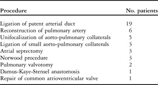
In 6 patients, we used cardiopulmonary bypass while creating the shunt due to the concomitant performance of additional intracardiac procedures, with 3 patients undergoing a modified Norwood procedure. The additional procedures are listed in Table 3. All patients were discharged from the hospital on aspirin.
Additional procedures at time of shunting.

Comparison and analysis of data
Baseline factors were compared for the entire cohort, as well as being stratified on the intended strategy for functionally univentricular or biventricular palliation. Continuous variables were expressed as medians, with range, due to the low number of patients involved. Dichotomous variables were expressed as percentages or proportions. Statistical significance of univariate comparisons was made using the Kruskal-Wallace test of equality for continuous variables and the Fisher Exact test for dichotomous variables. We calculated and tested correlation coefficients (r 2) between continuous variables using the Spearman rank correlation coefficient method. The life table method was used to construct Kaplan-Meier plots for survival of both the patients and the shunts. In the former comparison, patients were censored at the time of last follow-up, while for the latter, patients were censored either at the time of last follow-up or at take-down of the shunt, whichever came first. Statistical comparison between groups when the life table method was employed used the log rank test. Computation of statistical values was aided by use of Intercooled Stata 6.0 for Windows.
Results
Of the 32 patients, 2 died (6.25%), both having functionally univentricular hearts (p = 0.2). Of these, 1 patient had pulmonary atresia with intact ventricular septum, a severely hypoplastic right ventricle, and no right coronary artery. The second patient had congenital heart block, isomerism of the right atrial appendages, pulmonary atresia, unbalanced atrioventricular septal defect with hypoplastic left ventricle, and severe atrioventricular valvar regurgitation. In both patients, poor cardiac function preoperatively and postoperatively was the primary cause of death.
The stay in intensive care ranged from 2 to 58 days, with a median of 5 days, and in hospital ranged from 6 to 98 days, with a median of 19 days. There was no significant difference in these periods for the two groups. Postoperative infections occurred twice, once in a patient with tetralogy of Fallot with pulmonary atresia and multiple aortopulmonary collateral arteries, who suffered thrombosis of the shunt following unifocalization on the left side, and needed creation of a new shunt.
Follow-up ranged from 3 months to 4.7 years, with a mean of 2.1 years. During the period of follow-up, there were 4 late deaths, 3 in patients with functionally univentricular physiology (p = 0.3). All deaths occurred before takedown of the shunt. Of these 4 late deaths, 2 patients died suddenly at home, both having pulmonary atresia and intact ventricular septum. The precise cause of death is unknown. A high rate of death among patients with pulmonary atresia and intact ventricular septum during the period of shunting has been noted previously.Reference Tamisier, Vouhe, Vernant, Leca, Massot and Neveux3 Another patient died during cardiac catheterization in preparation for a bidirectional Glenn anastomosis, while the final patient died due to sepsis.
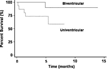
A Kaplan-Meier survival plot stratified by the strategy for palliation is shown in Figure 2. A trend toward longer survival was noted in those with biventricular as compared with functionally univentricular physiology (p = 0.06). At six months, 90% of those with biventricular physiology were still alive, as compared with 58% of those with functionally univentricular physiology (p = 0.16).
Kaplan-Meier plot of survival stratified according to the strategy for palliation.

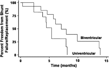
In Figure 3, we show a Kaplan-Meier plot of the time to failure or replacement of the shunt as stratified by the strategy for palliation. A statistically significant difference was found between the two groups (p = 0.05). The median time to failure or replacement of the shunt in those with functionally univentricular hearts was 5 months, as compared with 6.6 months in those with biventricular arrangements. Of 12 surviving patients with functionally univentricular hearts, 10 have proceeded to their bidirectional Glenn anastomosis, which was created as early as 3 months, and no later than 4 months of age.
Kaplan-Meier plot of time to take-down of the shunt stratified according to the strategy for palliation.

Of the 14 surviving patients with biventricular physiology, 10 have proceeded to definitive repair.
Discussion
Construction of a systemic-to-pulmonary arterial shunt is an important palliative procedure in newborns with cyanotic congenital cardiac defects. The most common technique for creation of such shunts is the modified approach using a polytetrafluoroethylene tube as graft between the subclavian artery and the pulmonary artery on the same side.Reference de Leval, McKay, Jones, Stark and Macartney6 This technique offers several advantages, allowing some degree of regulation of blood flow through the shunt determined by the size and length of the tube and the origin of flow, and preservation of flow in the subclavian artery, with lower rates of distortion of the inflow and outflow arteries. Early and late results with the modified Blalock-Taussig shunt have been widely reported. Shunting in the neonatal period is considered to be one of the important risk factors for increased perioperative mortality. Several reports suggest that a birth weight less then 3 kilograms may be an additional significant factor affecting survival.Reference Tamisier, Vouhe, Vernant, Leca, Massot and Neveux3, Reference Al Jubair, Al Fagih and Al Jarallah7–Reference Sivakumar, Shivaprakasha, Rao and Kumar9 Specific reports about the creation of a systemic-to-pulmonary arterial shunt in patients born with low weight, however, are rare.Reference Bove, Francois and De Groote10
The main goal in this subgroup of patients is to avoid over circulation to the lungs, which may cause congestive heart failure, prolonged ventilation, and need for inotropic support, leading to prolonged stays in intensive care, and increased morbidity and mortality. In an attempt to identify the predicting factors for survival, we reviewed our experience in patients born weighing less than 3 kilograms and undergoing placement of a modified Blalock-Taussig shunt.
Most of our patients were neonates, and almost half were born prematurely, earlier than 37 weeks of gestation. It is our preference to perform an early primary repair in patients with a biventricular heart defect and favourable anatomy. Our group was comprised by approximately equal halves of patients with functionally univentricular and potentially biventricular hearts, the latter patients deemed to be at high risk for early primary repair (see Table 2).
In the newborns, we always attempted to wait for the pulmonary resistance to decrease before constructing the shunts. Pulmonary vascular resistance was estimated clinically by elevation in systemic saturations of oxygen, with or without the development of congestive heart failure. This approach may assist with a better prediction of flow of blood to the lungs after creation of the shunt. In preterm patients, the immaturity of the lungs may cause the flow to be less predictable following the operation.
Based on previous studies, we recognized the difference between patients having functionally univentricular as opposed to potentially biventricular hearts.Reference Tamisier, Vouhe, Vernant, Leca, Massot and Neveux3, Reference Fermanis, Ekangaki and Salmon11 Those with functionally univentricular arrangements had a wider diversity in their basic diagnosis, albeit that most had circulations dependent on the arterial duct. Those with potentially biventricular arrangements tended to have a form of Tetralogy of Fallot as their principal diagnosis, with a higher risk for early primary repair. This has led, not surprisingly, to the observation that those with functionally univentricular hearts had larger pulmonary arteries, significantly so for the left pulmonary artery. Interestingly, the presence of smaller pulmonary arteries did not express itself in a significantly higher rate of failure of the shunt in those with biventricular hearts. This as opposed to previous studies that reported increased failure with pulmonary arteries of smaller size.Reference Al Jubair, Al Fagih and Al Jarallah7 Only 1 patient with the diagnosis of tetralogy of Fallot and pulmonary atresia with multiple aortopulmonary collateral arteries suffered thrombosis of the shunt following a unifocalization procedure.
In addition, there have been reports regarding a high incidence of pulmonary arterial distortion following creation of the shunt, leading to late failure.Reference Tamisier, Vouhe, Vernant, Leca, Massot and Neveux3 Review of our data from follow-up cardiac catheterization has not confirmed this observation. We noticed some patients with mild stenosis of an anastomotic site. If deemed necessary, this was reconstructed during takedown of the shunt. The relatively low incidence of postoperative complications may be attributed to the use of the saphenous venous homograft as the shunt. This technique was also used successfully in the past, albeit mainly in the era preceding the introduction of polytetrafluoroethylene grafts.Reference Danilowicz, Ishmael, Doyle, Isom, Colvin and Greco12, Reference Bogats, Kertesz, Katona, Toszegi and Kovacs13 We use the saphenous venous homograft for all our patients needing a Blalock-Taussig shunt. We consider it to be more useful in small patients. Technically, the saphenous venous homograft is easier to use, more haemostatic, and less thrombogenic than artificial grafts. The homograft is more pliable as opposed to the stiff polytetrafluoroethylene grafts, minimizing the risk for distortion of the pulmonary arteries. It is available in smaller sizes, which allows use of grafts of 2.5 or 3 mm diameter without increasing the risk for thrombosis. This reduces the possibility of these small patients having excessive pulmonary blood flow.
The use of a homograft to create the shunt may increase immunosensitization, with the elevation of panel reactive antibodies. It may lead to increased morbidity and mortality associated with cardiac transplantation, if heart transplantation is ever considered for these patients.Reference Jacobs, Quintessenza and Boucek14, Reference Jacobs, Quintessenza and Chai15 Even with this potential weakness associated with using a homograft, we believe that it has increased the overall survival of these patients.
In all 28 patients who did not need unifocalization, our preferred surgical approach was through a sternotomy. The advantages of this approach have been described previously.Reference Odim, Portzky and Zurakowski16 Specifically, for those born with low weight, the use of the proximal part of the subclavian or brachiocephalic artery results in a better inflow.
We observed a better survival for the shunt in patients with biventricular physiology. This is similar to previous studies, that reported mortality rates as low as 0.6% for neonates with tetralogy of FallotReference Ilbawi, Grieco and DeLeon17 as opposed to mortalities of 10 and 21% in studies that included both functionally univentricular and biventricular arrangements.Reference Tamisier, Vouhe, Vernant, Leca, Massot and Neveux3, Reference Sivakumar, Shivaprakasha, Rao and Kumar9 Given these findings, our approach has been to minimize the period of shunting in those with functionally univentricular hearts, proceeding to the bidirectional Glenn anastomosis as early as three months of age.
In conclusion, construction of a systemic-to-pulmonary arterial shunt can be performed safely in patients born weighing less than 3 kilograms and having biventricular physiology. For those with functionally univentricular physiology, the procedure carries an increased rate of mortality for the period of shunting. The saphenous venous homograft allows the use of smaller grafts, which does not significantly increase the risk for thrombosis or death when compared to previous studies using polytetrafluoroethylene grafts.