In tetralogy of Fallot with pulmonary atresia, arterial supply to the lungs is provided by the arterial duct, systemic-to-pulmonary collateral arteries, or rarely by an alternative source such as an aortopulmonary window or a coronary arterial fistulous communication. Accurate identification of the source and distribution of such flow is essential for clinical management.Reference Haworth and Macartney1, Reference Baker2 Postnatally, this is achieved by echocardiography, angiography, or magnetic resonance scanning. Prenatally, all diagnostic information is derived from ultrasonic examination, on which counselling is based, and decisions are made regarding continuation or termination of pregnancy. The question remains, however, as to how much can be discovered about the pulmonary arterial supply prenatally?
Fetal echocardiography often identifies the arterial duct and demonstrates the intrapericardial pulmonary arteries.Reference Hornberger, Sanders and Sahn3–Reference Pepa, Savis, Jones, Sharland, Tulloh and Simpson7 Prenatal identification of systemic-to-pulmonary collateral arteries, in contrast, has rarely been documented.Reference Volpe, Campobasso and Stanziano8–Reference Miyashita and Chiba10 Our aim in this study, therefore, was to review the diagnostic features of systemic-to-pulmonary collaterals during fetal life, and to assess their impact on counselling, antenatal, and postnatal management.
Methods
Using computerised databases, we identified all antenatal diagnoses of significant structural congenital cardiac disease seen by a single fetal cardiologist working in three tertiary referral centres between January, 1997, and April, 2006. Scans were performed using various ultrasound systems and curvilinear probes of appropriate frequencies from 3.5 to 7.5 MHz, namely the Hewlett-Packard 5500 machine (Hewlett-Packard, Palo Alto, California, USA), The ATL 5000 platform (Advanced Technology Laboratories, Bothell, Washington, USA) and the Aloka Prosound PHD 5000/5500 and α-10 systems (Aloka, Tokyo, Japan). We then identified all diagnoses of tetralogy of Fallot with pulmonary atresia, subsequently reviewing the reports of the fetal echocardiographic interrogations and, whenever appropriate, the S-VHS and digital tapes. We established the outcomes of all the pregnancies, seeking postmortem and postnatal data whenever available. Findings from the prenatal echocardiogram were then compared to the postnatal and autopsy findings. Ethical approval was obtained to assess the data.
Results
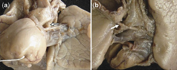
A total of 6587 fetuses had been seen during the period. Amongst these, we identified 11 cases of tetralogy of Fallot with pulmonary atresia. Of the 11 cases, in 5 pulmonary arterial supply had been considered to be via an arterial duct, while in 6 the fetal echocardiograms had revealed the presence of systemic-to-pulmonary collateral arteries. In the latter group, the arterial duct was considered to be absent in 5. In the sixth case, in addition to systemic-to-pulmonary collaterals from descending thoracic aorta, a vessel was seen in the region of the aortic arch in which a duct might have been anticipated, but it was unclear during the fetal examination whether the vessel represented a further collateral artery, or whether it was indeed a small arterial duct. Pregnancy was terminated, and post-mortem confirmed presence of both systemic-to-pulmonary collateral arteries and a small, almost occluded, arterial duct (Fig. 1).
The view of left side (a) shows the aortic arch, pulmonary trunk and the left arterial duct(arrow). The view of the back of the heart (b) shows a systemic-to-pulmonary collateral artery (arrow) arising from the anterior aspect of the descending aorta. Its branches follow the main bronchuses to enter the hilums of the right and left lungs.

Systemic-to-pulmonary collateral arteries
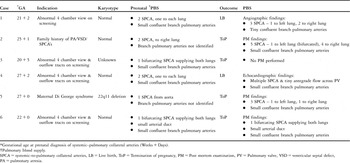
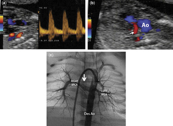
The features and outcomes of all cases diagnosed prenatally with systemic-to-pulmonary collateral arteries are shown in the Table 1. The gestational age at which the collateral arteries were identified varied from 20 weeks plus 5 days to 27 weeks plus 2 days, with a median of 23 weeks plus 5 days. The presence of a major systemic-to-pulmonary collateral artery was initially suspected on colour flow mapping as a vessel originating from the descending thoracic aorta. Settings for colour Doppler interrogation were then optimised to enhance the visualisation of such arteries, and to track their pathway towards the pulmonary parenchyma. Additional use of power Doppler also facilitated imaging of the collateral arteries, whereas cross-sectional imaging alone failed to delineate their proximal parenchymal course. Pulsed wave Doppler waveforms showed a saw-toothed trace, with continuation of flow throughout diastole (Fig. 2a). This pattern was distinct from the Doppler signal obtained in the descending aorta. In some cases, the maximal pulsed-wave Doppler velocity recorded in the collateral artery was greater than 1 metre per second. This possibly indicated stenosis in the vessel.
Details of cases of tetralogy of Fallot with pulmonary atresia and prenatal diagnosis of systemic to pulmonary collateral arteries (SPCA).

*Gestational age at prenatal diagnosis of systemic–pulmonary collateral arteries (Weeks + Days).
†Pulmonary blood supply.
SPCA = systemic-to-pulmonary collateral arteries, LB = Live birth, ToP = Termination of pregnancy, PM = Post mortem examination, PV = Pulmonary valve, VSD = ventricular septal defect, PA = pulmonary atresia.
The prenatal and postnatal images of our first case. Panel (a) shows a sagittal view of the prenatal scan, with 2 systemic-to-pulmonary collateral arteries (red flow) arising from the descending aorta. Pulsed wave Doppler flow in these arteries is of relatively high velocity, and has a saw-toothed appearance. Panel (b) shows retrograde flow to the pulmonary trunk (white arrow). The descending aortogram (c) taken postnatally shows systemic-to-pulmonary collateral arteries and diminutive intrapericardial pulmonary arteries (white arrow). Abbreviations: Ao: aorta, SPCA: systemic-to-pulmonary collateral arteries, Des Ao: descending aorta.

Confirmation of pulmonary arterial supply
In 10 cases, either at post-mortem examination, or in cases where pregnancy was continued, the pattern of pulmonary arterial supply identified prenatally was confirmed. In the remaining case, in which the pregnancy was terminated, postmortem study was not performed.
In 2 of the fetuses thought initially to have pulmonary atresia, a tiny amount of forward flow was demonstrated across the pulmonary valve on postnatal investigation. Of these patients, 1 had systemic-to-pulmonary collateral arteries and the other an arterial duct.
Intrapericardial pulmonary arteries
In 3 of the 5 cases with pulmonary supply through an arterial duct, intrapericardial pulmonary arteries of adequate size were visualised antenatally. Acoustic windows were inadequate in one to assess the pulmonary arteries, while in the other there was an associated left diaphragmatic hernia, and the left pulmonary artery was not visualised. Confluent intrapericardial pulmonary arteries of adequate size were subsequently confirmed in 4 cases, either by postmortem study in 2, or by postnatal echocardiography in the other 2. In the case with associated diaphragmatic hernia, postmortem revealed a hypoplastic left lung, with a very small but confluent left pulmonary artery supplied by an arterial duct.
In the cases where the pulmonary arterial flow was derived from systemic-to-pulmonary collaterals, intrapericardial pulmonary arteries could be identified prenatally in 4 of the 6 cases. All were considered to be abnormally small, and in one case (Fetus #1) failed to grow as gestation progressed. The presence of confluent but hypoplastic intrapericardial pulmonary arteries was confirmed in 3 instances, by post-mortem examination, postnatal echocardiography, and angiography, respectively. In one case, the pregnancy was terminated and no postmortem examination was performed. In the remaining 2 cases, grossly hypoplastic but confluent intrapericardial pulmonary arteries were identified only at autopsy.
Outcome
Pregnancy was terminated in 4 out of the 6 cases (66%) in whom flow to the lungs had been demonstrated through collateral arteries. In the two liveborn infants, prostaglandin was not started, as the arterial duct was known to be absent. A Blalock Taussig shunt was constructed in one when aged 2.5 months, and a patch placed across the ventriculo-pulmonary junction in the other at the age of 1.5 months. Only one of the cases, however, was still alive at the age of 1 year.
In the 5 cases with flow to the lungs through an arterial duct, pregnancy was terminated in 3 (60%). In the two liveborn infants, prostaglandin was started, and a modified Blalock Taussig shunt was constructed on the third day in one, and the sixteenth day in the other. Cardiac surgery was delayed in the second case to permit surgical repair of oesophageal atresia and a tracheosophageal fistula. Both children are alive at 1 year.
Differential diagnosis of systemic-to-pulmonary collateral arteries
In 1 fetus diagnosed at 23 weeks gestation, a vessel could be seen originating from the descending aorta that had features similar to a systemic-to-pulmonary collateral artery. The intrapericardial pulmonary arteries in this instance, however, were of relatively good size, and the course of the vessel was unusual for a collateral artery, as it did not seem to enter the lung parenchyma. In addition, it was possible to identify the arterial duct. The diagnosis made was that of a right aortic arch with aberrant origin of the left subclavian artery. This was confirmed at postmortem examination when the pregnancy was terminated at 23 weeks. There was no evidence of a systemic-to-pulmonary collateral artery, and pulmonary arterial supply was via the arterial duct.
Discussion
In tetralogy of Fallot with pulmonary atresia, the ventricular anatomy is usually suitable for biventricular repair, but the success of this procedure depends also on the pattern of pulmonary arterial supply, in particular the presence or absence of intrapericardial pulmonary arteries and their size. For fetuses with this diagnosis, identification of the pulmonary arterial supply, and description of the presence and size of the intrapericardial pulmonary arteries, is the key to counselling, treatment, and prognosis. In this study, we were able to define the main source of pulmonary arterial supply, and the anatomy of the pulmonary arteries, in most cases.
Antenatal cross sectional echocardiography has been used to evaluate the presence and size of the intrapericardial pulmonary arteries,Reference Hornberger, Sanders and Sahn3–Reference Pepa, Savis, Jones, Sharland, Tulloh and Simpson7 but few studies have assessed systemic-to-pulmonary collateral arteries.Reference Volpe, Campobasso and Stanziano8–Reference Miyashita and Chiba10 In one investigation, four-dimensional sonography with B-flow imaging and spatio-temporal correlation was used to image such vessels, showing that cross-sectional and colour Doppler echocardiography had failed to assess the anatomy of the intrapericardial pulmonary arteries in one-third of cases, and the source of arterial supply in one-quarter.Reference Volpe, Campobasso and Stanziano8 These findings were echoed in another study,Reference Vesel, Rollings, Jones, Callaghan, Simpson and Sharland9 where the intrapericardial pulmonary arteries were correctly identified in four-fifths, the source of arterial supply in three-fifths, and systemic-to-pulmonary collateral arteries in under half.
In our study, we were able in all fetuses to define the main source of flow of blood to the lungs, whether it was from systemic-to-pulmonary collateral arteries or from an arterial duct. Our findings were confirmed in all cases when postmortem or postnatal data was available. This included all cases with important systemic-to-pulmonary collateral arteries. Our increased rate of detection of these collateral arteries, when compared to other studies,Reference Volpe, Campobasso and Stanziano8, Reference Vesel, Rollings, Jones, Callaghan, Simpson and Sharland9 may be due to targeted fetal echocardiography searching for systemic-to-pulmonary collateral arteries, particularly in cases with small intrapericardial pulmonary arteries, and whenever the arterial duct was not promptly identified. Colour flow mapping should be used in the search for abnormal vessels arising from the descending thoracic aorta in all cases of pulmonary atresia with a ventricular septal defect identified in the fetus. Although we have shown that the presence of systemic-to-pulmonary collateral arteries can be confirmed prenatally, delineation of the exact course and distribution remains problematic. This may become possible with evolving spatio-temporal image correlation and other colour flow techniques involving three-dimensional reconstruction.
We were able to identify confluent intrapericardial pulmonary arteries in 7 cases, including 4 with systemic-to-pulmonary collateral arteries, as had previous investigators.Reference Volpe, Campobasso and Stanziano8, Reference Vesel, Rollings, Jones, Callaghan, Simpson and Sharland9 This was not possible in 4 cases. Failure to demonstrate the intrapericardial pulmonary arteries in these cases was due to their very small size in 3. In 2 with systemic-to-pulmonary collateral arteries, miniscule intrapericardial pulmonary arteries were found at postmortem. In a further case, there was left diaphragmatic hernia with very hypoplastic pulmonary arteries. It is possible that, with the advent of newer imaging techniques such as four-dimensional sonography, as well as improving resolution of the ultrasonic equipment, our ability to identify even minute pulmonary arteries will improve.
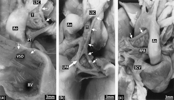
In our second case, identification of collateral arteries helped in prenatal decision-making. Having had one previous child, who had died in the first days of life with tetralogy of Fallot, pulmonary atresia, and absent intrapericardial pulmonary arteries, identification of the collateral vessels facilitated the decision of the mother to terminate the pregnancy. Had the second fetus been shown to have good-sized central pulmonary arteries with a duct-dependent pulmonary circulation, this would not have been her choice. The postmortem examination confirmed the prenatal findings of multiple collateral arteries, and the presence of grossly hypoplastic intrapericardial pulmonary arteries (Fig. 3).
Postmortem findings in our second case. The white arrows point to systemic-to-pulmonary collateral arteries. Abbreviations: Ao: aorta, LPA: left pulmonary artery, LSC: left subclavian artery, PT: pulmonary trunk, RPA: right pulmonary artery, RV: right ventricle, SCV: superior caval vein, T: trachea, VSD: ventricular septal defect.

There was no significant difference in the rate of termination of pregnancy between those with a duct-dependent circulation and those with collateral arteries, although the numbers of patients are small. Identification of systemic-to-pulmonary collateral arteries introduces uncertainty as to feasibility of achieving repair with continuity from the right ventricle to the pulmonary arteries, as the precise pulmonary arterial anatomy, and particularly that of the intraparenchymal vessels, is less predictable.Reference Dinarevic, Redington, Rigby and Shinebourne11 Patients with systemic-to-pulmonary collateral arteries are likely to require staged surgery, which can be a daunting prospect for a family, although life expectancy without intervention may well be longer than for those with a duct-dependent pulmonary circulation.
In all cases, prenatal identification of the main source of pulmonary arterial supply assisted postnatal decision-making regarding use of prostaglandin E. This was commenced electively if retrograde flow in the arterial duct was seen prenatally, but was not started in cases with absence of the arterial duct. Our advice in these latter cases had been to monitor neonatal saturations of oxygen. Providing these remained at greater than 80%, prostaglandin was avoided. Unnecessary use of prostaglandin can cause significant complications, such as apnoea leading to the need for ventilation.
Our data do not allow comments regarding fetal growth of intrapericardial pulmonary arteries. In our first case, however, there was evidence of confluent pulmonary arteries at 21 weeks gestation. Flow could be seen extending up to a thin atretic pulmonary valve (Fig. 2b). Serial echocardiograms showed that the pulmonary trunk and its branches failed to grow with time, and there was progressive obstruction within the right ventricular outflow tract, with obliteration of the infundibulum with muscular overgrowth. At birth, all that remained were the grossly hypoplastic intrapericardial pulmonary arteries (Fig. 2c). It is possible there may have been benefit in performing pulmonary valvotomy at 21 weeks gestation,Reference Tulzer, Artz, Franklin, Loughnan, Mair and Gardiner12 which may have established forward flow across the atretic pulmonary valve, promoting growth of the intrapericardial pulmonary arteries, and possibly allowing them to be used in postnatal life. In comparison, our fourth case, although considered to have pulmonary atresia in prenatal life, was found to have some antegrade flow across the pulmonary valve at birth. The pulmonary arteries in this case, although small, were comparatively bigger at term. The child later proceeded to have relief of the obstructed right ventricular outflow tract in the hope of promoting forward flow and pulmonary arterial growth prior to eventual repair. We speculate that the obstructive process occurred later in gestation in the second child, enabling more growth of the intrapericardial pulmonary arteries.
Systemic-to-pulmonary collateral arteries can anastomose with a lobar pulmonary artery, with a segmental intrapulmonary artery, or with a pulmonary artery at the hilum.Reference Haworth and Macartney1, Reference Haworth13, Reference Boshoff and Gewillig14 Prenatal echocardiography in our first case showed blood entering the pulmonary trunk retrogradely from two collateral arteries. There was suspicion that the collateral arteries were stenosed due to the high velocity observed on the pulsed wave Doppler trace. This was confirmed by postnatal angiography. Stenosis of the centrally connected systemic-to-pulmonary collateral arteries may account for the grossly hypoplastic intrapericardial pulmonary arteries seen at term, with obstruction of the collateral artery preventing forward flow and growth of the pulmonary arteries. There was no suspicion of stenosis in the collateral arteries of our fourth case, where the intrapericardial pulmonary arteries were larger at term. In our 2 liveborn cases where pulmonary arterial supply was from an arterial duct, both children had pulmonary arteries of adequate size, and a modified Blalock Taussig shunt was subsequently constructed.
Our study is limited by its retrospective nature, being dependent upon the data already collected. The numbers are also small for statistical analysis. Our study is essentially descriptive, but does show the feasibility of identifying systemic-to-pulmonary collateral arteries in the fetus. We have found a learning curve in our ability to diagnose such collateral vessels, and therefore caution the reader that counselling should reflect the experience of their particular centre.
In conclusion, prenatal diagnosis of systemic-to-pulmonary collateral arteries is possible, and is important in establishing likely outcomes and planning management. Currently it should be possible to evaluate these arteries, as well as the presence and size of the intrapericardial arteries, in most cases of tetralogy of Fallot with pulmonary atresia diagnosed prenatally. Newer imaging techniques may further aid prenatal diagnosis of systemic-to-pulmonary collateral arteries, as well as the anatomy of even the very smallest intrapericardial pulmonary arteries.
Acknowledgements
During the course of this research, Dr Anna Seale was a research fellow at the Royal Brompton Hospital supported by grants from the Harrison Heart Foundation and the Joe Gandon Memorial Trust. She is now a consultant at the hospital.