Case report
A 27-year-old woman with uncomplicated pregnancy was referred to our fetal center for possible tetralogy of Fallot at 23 weeks gestation. Over several fetal echocardiograms, the anatomy was defined as tetralogy with pulmonary atresia and absent pulmonary arteries. A large maligned ventricular septal defect with an overriding aortic valve was seen as expected. Furthermore, we were unable to identify a ductus arteriosus or collateral vessels feeding the lungs. The parents were counseled about the concerns given the inability to identify a source of pulmonary blood flow and the possibility of early postnatal demise. After counseling, the family elected for comfort care.
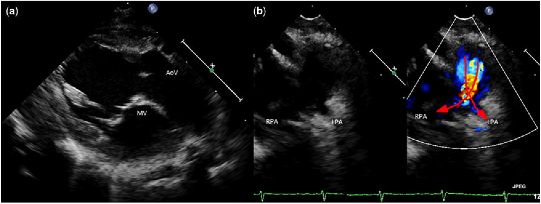
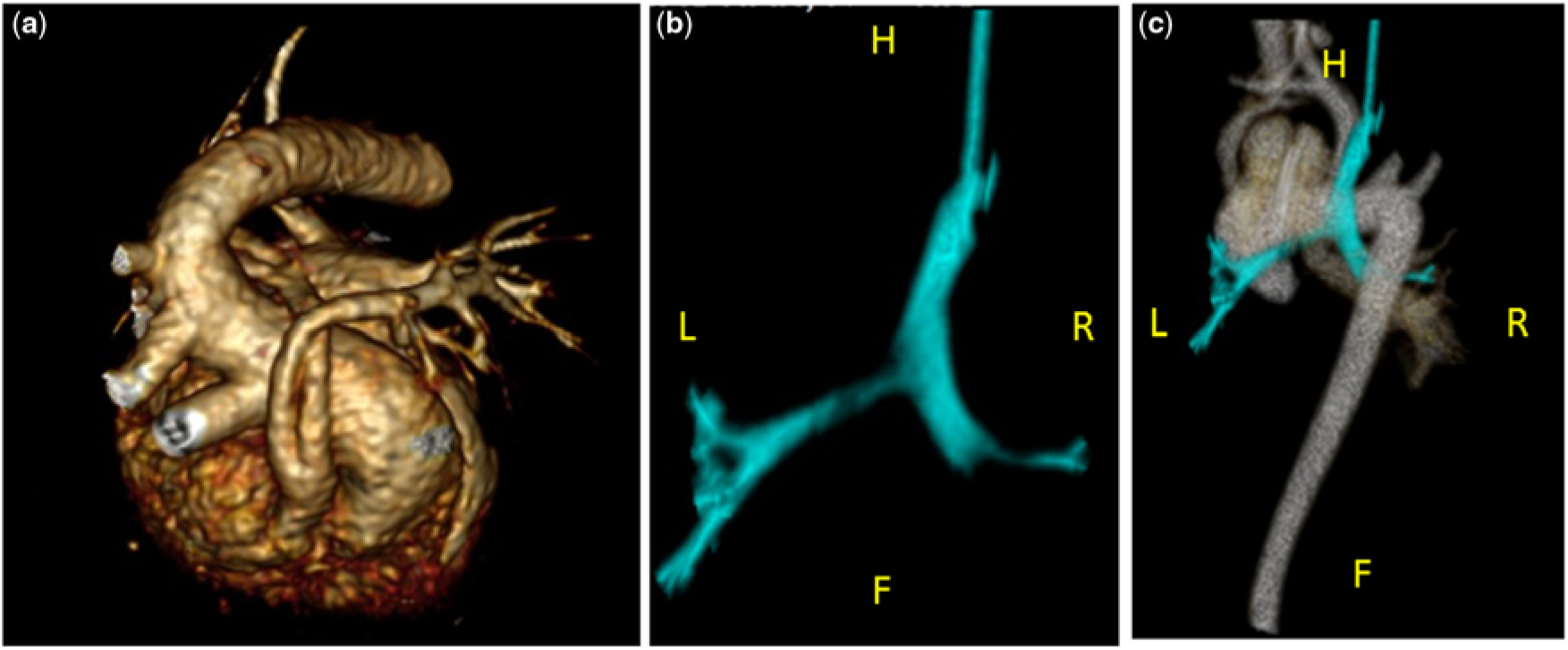
At 37 weeks gestation, a 2.3 kg male infant was born via cesarean section secondary to fetal decelerations. The family then opted to continue with medical management. Oxygen saturations were <60% at 3 minutes, but eventually rose to the 80’s to 90’s on CPAP respiratory support in the Cardiac Intensive Care Unit. Postnatal transthoracic echocardiography confirmed levocardia, normal abdominal, and suggestion of normal atrial situs with D-ventricular looping and atrioventricular concordance. Also, noted was bilateral superior vena cava, with the right-sided vessel draining to the right-sided atrium, the left draining to the left-sided atrium, and a small bridging vein. There were four pulmonary veins seen entering the left-sided atrium. There were a large anterior malalignment ventricular septal defect and a moderate-sized secundum atrial septal defect. Unlike the prenatal study, postnatally both great arteries could be identified. There was malposition of the great arteries with the pulmonary valve anterior and rightward of the aorta, but with ventriculoarterial concordance, giving the segmental designation of {S, D, I}. The right ventricular outflow tract was narrow, but without significant obstruction, whereas the pulmonic valve appeared bicuspid, thickened, and doming. The branch pulmonary arteries were identified; they were hypoplastic with a crisscross pattern that coursed in front of the distal ascending aorta in an anterior to posterior fashion with the right pulmonary artery running under the dilated, winding rightward aortic arch with mirror image branching. There was aortic to mitral valve fibrous continuity (Fig 1). There was no patent ductus arteriosus. Left coronary origin was difficult to visualize. Computed tomography at two days of age confirmed the anatomic diagnosis (Fig 2). Cardiac catheterization defined separate left and right coronary arteries. Computed tomography confirmed abdominal and atrial situs, in addition to giving more information regarding pulmonary artery anatomy and the relationship to the aortic arch. Distal tracheobronchomalacia caused by vascular compression was also noted on this first CT; however, the patient was weaned to room air, breathing spontaneously, and discharged prior to cardiac surgery.
Transthoracic echocardiography in the parasternal long-axis with a view of the aortic and mitral valve continuity (a), and with superior angulation showing the crossover of the pulmonary arteries just prior to their bifurcation (b). AoV = aortic valve; LPA = left pulmonary artery; PV = pulmonic valve; RPA = right pulmonary artery.

Three-dimensional CT reconstruction of the heart with crisscross pulmonary arteries, arching caudally over the dilated aortic arch (a), and a CT reconstruction of the trachea and mainstem bronchus viewed from the rear showing the branching into the mainstem bronchi (b), and with the dilated aorta superimposed (c). Airway reconstructions reveal distal tracheal and bilateral bronchial hypoplasia secondary to vascular compression from the dilated aorta.

Chromosomal microarray revealed 22q11.2 deletion. He underwent right ventricular outflow tract transannular patch and ventricular septal defect patch closure at 3.5 months of age. Though he weaned from mechanical ventilation postoperatively, his respiratory insufficiency continued, requiring CPAP and high flow nasal cannula. Further evaluation with bronchoscopy revealed severe distal tracheal compression, and computed tomography angiography revealed right mainstem bronchus hypoplasia, likely from the vascular compression of the dilated transverse aorta. After in depth conversation with the cardiothoracic and pulmonary teams, the patient underwent aortopexy with the hope of relieving the distal tracheal compression (Fig 2). Unfortunately, he had persistent malacia of his distal trachea that required tracheostomy placement and currently requires mechanical ventilation, now at 8 months age.
Discussion
This case highlights the difficulty in both diagnosing and naming these rare variants of complex congenital heart disease.Reference Bravo-Valenzuela, Carrilho, Peixoto, Bezerra and Araujo Júnior1 In addition, we describe the pitfalls in prenatal diagnosis, which was difficult due to the severe tortuosity of the pulmonary arteries. Finally, finding an accurate, complete, and non-ambiguous description for this arrangement of arterial trunks has been challenging using almost any method of nomenclature.
Initially, this conotruncal anomaly was thought to be embryologically impossible.Reference Vanmierop and Wiglesworth2 However, in 1971, Van Praagh et al. described four patients with D-transposition of the great arteries with near normal appearing external positioning.Reference Van Praagh, Perez-Trevino and Lõpez-Cuellar3 They challenged the previous notions of conotruncal development in transposition and the definition of the term “transposition” itself.
Instead, they proposed the term “malposition” to describe all abnormal conotruncal relationships except when the aorta arises from the morphologic right ventricle and the pulmonary artery arises from the morphologic left ventricle in, otherwise, normal situs, true “transposition.” Two of the cases had two ventricles with ventriculoarterial concordance but malposition of the great arteries (levocardia with {S, D, L} and dextrocardia with {I, L, D}). These cases expounded upon the Van Praagh precedent and argued that “the term anatomically corrected malposition” describes not a discrete anomaly but only a ventriculo-arterial relation, which is one of ventricular-arterial concordances.Reference Van Praagh, Perez-Trevino and Lõpez-Cuellar3
In the current literature, anatomically corrected malposition describes concordant atrioventricular and ventriculoarterial connections where flow pathways through the heart are as expected but the arrangement of the arterial trunks – relative to each other – is not oriented in their anticipated manner.Reference Gupta, Ramakrishnan and Gulati4 This definition encompasses our patient’s anatomy; however, the nomenclature anatomically corrected malposition alone does not convey a complete description of the arterial trunk arrangement.
One of the characteristics that makes our case interesting is the presence of aortic-mitral continuity and the lack of subaortic conus, which, for some anatomists, would disqualify this anatomy from the definition of “anatomically corrected malposition.”Reference Kirklin, Pacifico, Bargeronjr and Soto5 The aortic mitral continuity seen in this case has been described previously by Cavalle et al. in several of their series of eight patients, and they argued that this should not disqualify them from this designation.Reference Cavalle-Garrido, Bernasconi, Perrin and Anderson6 Also, the absence of bilateral infundibulums does not disqualify the case as anatomically corrected malposition as it has also been discussed in previous publications.Reference Gupta, Ramakrishnan and Gulati4 In a schema proposed by Foran et al., our patient’s anatomy most closely fits with what they described as isolated infundibuloarterial discordance;Reference Foran, Belcourt and Nanton7 however, it can be a confusing descriptor for those unfamiliar with the article. Using a segmental sequential analysis, we can describe our aortic valve as being posterior and leftward of the pulmonary valve, Reference Bernasconi, Cavalle-Garrido, Perrin and Anderson8 with an “aortic arch” that is oriented horizontally in the transverse body plane.
Our case is also unusual in the clinical presentation and highlights the broad array of anatomic possibilities, illustrating the difficulty of prenatal diagnosis and range of postnatal complications.
Our patient’s moderately hypoplastic pulmonary arteries with such tortuosity are unusual and made prenatal diagnosis extraordinarily difficult. Fortunately, postnatal echocardiography was able to elucidate patent pulmonary arteries supplying pulmonary blood flow. Unfortunately, the degree of respiratory distress experienced by our patient is also unusual. Vascular compression from the low-lying, horizontal, and dilated transverse arch caused such significant obstruction, resulting in significant bronchopulmonary dysplasia necessitating tracheostomy, a complication not previously described in the literature. This case and cases like it highlight the difficulty in prenatal echocardiography, and how conotruncal anomalies can lead to complicated post-operative courses.
Supplementary Material
To view supplementary material for this article, please visit https://doi.org/10.1017/S1047951119000684.
Acknowledgements
None.
Financial Support
This research received no specific grant from any funding agency, commercial or not-for-profit sectors.
Conflicts of Interest
None.