Implications
Nutritional stressors affect energy balance imposing detrimental effects on growth and immune status. With the discovery of leptin, adipose tissue was recognized as a highly active metabolic and endocrine organ: adipokines, specific hormones and cytokines secreted by adipocytes, modulate energy balance and link growth and immune axis. The regulation of energy balance is complex: it is important to determine the main metabolic and signaling networks. Our in vitro model can provide insights into the adipocyte response to nutritional inputs and into the relationship between adipose tissue and growth hormone.
Introduction
The discovery of leptin over two decades ago (Zhang et al., Reference Zhang, Proenca, Maffei, Barone, Leopold and Friedman1994) has led to a radical change of perspective on the role of white adipose tissue (WAT). WAT has been studied for its role as an endocrine organ able to produce a variety of proinflammatory and antiinflammatory cytokines, mediators and hormones (Trayhurn and Wood, Reference Trayhurn and Wood2004; Borghetti et al., Reference Borghetti, Saleri, Mocchegiani, Corradi and Martelli2009). Growth and growth-related processes are regulated by nutritional (energy reserves and/or food availability) and hormonal status. A cross-talk mediated by numerous cytokines and hormones is believed to be responsible for integrating energy balance and immune function (Bernotiene et al., Reference Bernotiene, Palmer and Gabay2006; Borghetti et al., Reference Borghetti, Saleri, Mocchegiani, Corradi and Martelli2009). In particular, interleukin-6 (IL-6) and tumor necrosis factor-α (TNF-α) have been indicated as biomarkers of white and subcutaneous adipose tissue and body mass index (Pou et al., Reference Pou, Massaro, Hoffmann, Vasan, Maurovich-Horvat, Larson, Keaney, Meigs, Lipinska, Kathiresan, Murabito, O’Donnell, Benjamin and Fox2007). It is well documented that proinflammatory cytokines increase muscle protein degradation, reduce muscle protein synthesis and divert nutrients to the synthesis of components in the immune system (Frost and Lang, Reference Frost and Lang2004; Elsasser et al., Reference Elsasser, Caperna, Li, Kahl and Sartin2004). In addition, a direct effect of growth hormone (GH) on TNF-α and IL-6 secretion has been shown by in vitro and in vivo studies (Uronen-Hansson et al., Reference Uronen-Hansson, Allen, Lichtarowicz-Krynska, Aynsley-Green, Cole, Hoiden-Guthenberg, Fryklund and Klein2003; Pagani et al., Reference Pagani, Meazza, Travaglino, De Benedetti, Tinelli and Bozzola2005; Hochdorfer et al., Reference Hochdorfer, Kuhny, Zorn, Hendriks, Vanhaesebroeck, Bohnacker, Krystal and Huber2011). In the pig, adipose tissue plays a fundamental role in productive and reproductive performance (Hausman et al., Reference Hausman, Barb and Dean2008). Environmental and disease stressors cause negative effects on food intake, muscle growth and animal health: immune challenges may slow growth rate and feed intake with a significant economic impact (Hevener et al., Reference Hevener, Routh and Almond1999). Studies are needed to elucidate the adipose tissue physiology, before evaluating its real impact and importance in pig metabolism and energy regulation. Therefore, the aims of the present study were first to determine leptin, IL-6 and TNF-α expression and secretion by pig adipocytes in response to different culture conditions, second to evaluate the ability of GH to modulate adipokine production under varying conditions. Finally, we valuated whether nitric oxide (NO) pathways are involved in modulating adipokine expression and secretion in our experimental conditions. We used a previous-described experimental model (Saleri et al., Reference Saleri, Grasselli and Tamanini2005) and cells were incubated using DMEM/Ham’s F12 salts, a DMEM/Ham’s F12 formulation deprived of nutritional components, to verify whether and how nutritional conditions can influence the adipocyte cytokine expression and secretion. A system of co-culture with anterior pituitary was chosen to evaluate the direct effect of GH on the WAT.
Material and methods
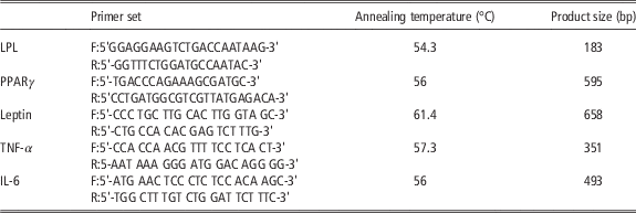
Omental adipose tissue was obtained at slaughter from 15 healthy gilts (aged 8 months) and immediately carried to the laboratory in phosphate buffered saline. Connective tissue and visible blood vessels were removed before the isolation of adipocytes. Approximately 1 g of adipose tissue was minced and digested in a water bath at 37°C for 1 h in Dulbecco’s Modified Eagle Medium/F12 (DMEM/F12) containing 1 mg/ml of collagenase. Digested tissue was filtered through two layers of polyester chiffon (up with 250 μm mesh and down with 75 μm mesh) and the cell suspension was centrifuged at 800×g for 10 min. Cell culture was performed as previously described by Qu et al. (Reference Qu, Zhang, Zhang and Gong2007). Cells were cultured in 15 ml flask in growth medium (DMEM/Ham’s F12+10% FCS) at 37°C and 5% CO2. Cells were examined daily and were kept in growth medium until confluence and medium was replaced every 3 days. At confluence, cells were detached with 0.25% trypsin and transferred into six well plates (200 000 cells/well) to induce adipogenic differentiation. Cells were cultured for 2 weeks in medium supplemented with 5% fetal calf serum (FCS), 10−6 M dexamethasone and 5 μg/ml insulin. At day 14, adipocytes were identified morphologically under phase-light microscope, and lipid droplets in cells were examined with Oil Red O staining. To confirm adipocyte differentiation, total RNA was isolated, from duplicate-treated plates and mRNA abundance for the peroxisome proliferator-activated receptor (PPARγ) and lipoprotein lipase (LPL) was measured using reverse-transcription PCR. Primer sequences, the respective annealing temperatures and amplified products are summarized in Table 1. From day 14, medium was removed and adipocytes (200 000 cells/well) were cultured for 24 h (T0) in DMEM/Ham’s F12+10% FCS (group A) or in DMEM/Ham’s F12 salts+10% FCS (group B). After 24 h, media were collected and replaced with DMEM/Ham’s F12 (group A) or DMEM/Ham’s F12 salts (group B): both experimental groups (A and B) were co-cultured for 4 h (T+4) or 12 h (T+12) with anterior pituitary slices: group A1+4 or +12 (control without pituitary), group A2+4 or +12 (coincubation with pituitary); group B1+4 or +12 (control without pituitary), group B2+4 or +12 (coincubation with pituitary). Anterior pituitaries (from 8-month-old sows) were separated from the neurohypophysis, sliced by a Thomas-Stadie-Riggs tissue slicer (Thomas Scientific, Swedesboro, NJ, USA) (section thickness 500 µm on average) and placed into transwell permeable support plates (Corning Incorporated Life Sciences, Chelmsford St. Lowell, MA, USA). To stimulate GH secretion by pituitary cells, growth hormone releasing hormone (GHRH) 10−8 M (Geref; Serono International S.A., Geneva, Switzerland) was added at the start of the incubation times (T+4 or T+12).
Sequences of PCR primers, annealing temperature and PCR product length

LPL=lipoprotein lipase; F=forward; R=reverse; PPARγ=peroxisome proliferator-activated receptor; IL-6=interleukin-6; TNF-α=tumor necrosis factor-α.
RNA isolation and RT-PCR
At the end of incubation, medium was removed and RNA was extracted by TRI Reagent® solution (Applied Biosystems, Monza, Italy) according to the manufacturer’s instructions. RNA quantification was carried out by spectrophotometer (GENEQUANT pro®; Amersham Pharmacia Biotech, Chalfont, Buckinghamshire, England). RT-PCR was carried out using a Ready-to-goTM You-Prime First-Strand Beads kit (Amersham Pharmacia Biotech, Uppsala, Sweden) as described by the manufacturer. A total of 2 µg of total RNA was used in the RT. Aliquots (5 µl) from the generated cDNA were used for subsequent PCR amplification in the reaction buffer containing 1.5 µl MgCl2 (50 mM), 1 µl dNTPs (12.5 mM) and 1 µl Taq DNA polymerase (1 µg/µl), to a final volume of 50 µl. Amplification was carried out for 27 cycles, when the reaction was in the middle of the linear range (before reaching the amplification plateau). Each cycle consisted of denaturation at 94°C for 1 min, annealing at specific temperature for each primer set for 1 min, extension at 72°C for 1 min; at the end of 27th cycle, an additional extension was carried out for 5 min. Specific primer set used, their annealing temperature and amplified product weight (expressed in base pairs, bp) are summarized in Table 1. QuantumRNA 18S was used (no. 1717, Ambion Inc., Austin, Texas, USA) as an internal positive control for a relative quantitative PCR. To amplify the 18S fragment (323 bp) without reaching the plateau phase, 18S PCR alternate primer pairs (5 µM) were coincubated with 18S PCR competimers (5 µM; ratio, 2 : 8) according to manufacturer’s instructions. PCR products were visualized after electrophoresis on 2% agarose gel added with SYBR® Safe DNA gel stain (Invitrogen S.R.L., San Giuliano Milanese, Italy). The gel was analyzed on a computerized densitometry program (Scion Capture Driver 1.2 for Image-Pro Plus; Scion Corp., Walnut, CA, USA); the values represented the ratio of the band intensities of the PCR products over those of the corresponding ribosomal 18S RT-PCR product and were expressed in relative arbitrary units.
Assays
To test the effectiveness of GHRH treatment, we assayed GH concentration in culture media by a competitive ELISA previously validated (Baratta et al., Reference Baratta, Saleri, Mainardi, Valle, Giustina and Tamanini2002). The intra- and interassay CV were 4.5% and 6.7%, respectively. We evaluate also prolactin (PRL), structurally very similar to GH, by a validated ELISA (Borghetti et al., 2006). The intra- and interassay CV were 6.9% and 9.3%, respectively. NO production was assessed at T0, T+4 and T+12 by measuring the amount of nitrite (NO2 −), a stable metabolic product of NO, in culture medium, as previously described (Baratta et al., Reference Baratta, Saleri, Mainardi, Valle, Giustina and Tamanini2002). Interassay variability was <2%.
The concentration of leptin, IL-6 and TNF-α were evaluated at T0, T+4 and T+12. A commercial kit was used to evaluate leptin concentrations (porcine ELISA Kit for Leptin; Uscn Life Science Inc., D.B.A Italia, Milano, Italy) . Sensitivity was 0.114 ng/ml; intra- and interassay CV were 4.5% and 6.2%, respectively. TNF-α levels were analyzed by using a commercial kit (Quantikine porcine TNF-α; R&D Systems, Abingdon, UK). The intra- and interassay CV were 5% and 7%, respectively. The sensitivity was 4 pg/ml. IL-6 was measured by Quantikine porcine IL-6 (R&D Systems). The intra- and interassay CV were 6.9% and 8%, respectively. The sensitivity was 10 pg/ml.
Statistical analysis
Data were analyzed by multifactorial ANOVA (SPSS), by applying a model with treatment group (two levels), incubation time (three levels), interaction between group and incubation time (2×2×3) and replicates (four levels) as fixed factors. Experimental data are presented as mean±SD.
When significant differences were found, means were compared by Scheffé’s F test.
Results
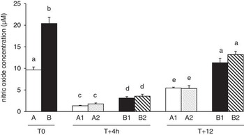
GH release was significantly (P<0.05) affected by GHRH: at +4 and +12 h, mean GH concentrations were 400±35 and 900±56 ng/ml, respectively. In absence of GHRH, GH concentrations are undetectable after 4 and 12 h. GHRH does not effect on PRL levels. At T0, T+4 and T+12, NO increased (P<0.05) up to twofold in groups B, B1 and B2 as compared with group A, A1 and A2, respectively (Figure 1). The coincubation with anterior pituitary did not induce significant effect on NO accumulation.
Nitrite release in adipocyte cultures. Adipocytes were cultured in DMEM/Ham’s F12+10% FCS (group A) or in DMEM/Ham’s F12 salts+10% FCS (group B) for 24 h (T0). After 24 h, adipocytes were cultured for 4 h (T+4) or 12 h (T+12) with or without anterior pituitary slices: A1=control without pituitary; A2=coincubation with pituitary; B1=control without pituitary; B2=coincubation with pituitary. Each data point represents mean±RSD of six separate wells in four different replicates. Significant differences (at least P<0.05) among the groups and treatments are labeled with different letters.

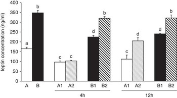
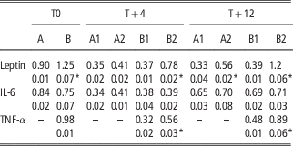
At T0, cells cultured in deprived condition (group B) showed a significantly (P<0.05) higher leptin expression as compared with group A (Table 2). In group A2, the co-culture with anterior pituitary slices, increased leptin expression at T+12, but not at T+4 as compared with the respective control group (A1 at T+4 or T+12). A significant increase (P<0.01) of leptin expression was observed in group B2, as compared with B1, after 4 and 12 h of coincubation with anterior pituitary (Table 2). Leptin concentrations are summarized in Figure 2. Briefly, at T0, leptin concentrations were 165±8 and 348±12 ng/ml in groups A and B, respectively. At T+4 and T+12, group A1 did not show significant difference (97.5±0.6 and 102.4±3 ng/ml, respectively). Group A2 showed a significant rise in leptin secretion after 12 h of pituitary co-culture (205.4±5 ng/ml) as compared with A1 at T+12 (102.4±3 ng/ml). Group B2 showed a significant (P<0.05) increase in leptin production at both incubation time as compared with controls (T+4: B1=225.6±8 ng/ml, B2=320±9 ng/ml; T+12: B1=241.5±3 ng/ml, B2=322±15 ng/ml). No significant differences were found between levels at T+4 and T+12 in B1 and B2 groups, respectively.
Leptin levels (mean±RSD) in adipocyte cultures. Adipocytes were cultured in DMEM/Ham’s F12+10% FCS (group A) or in DMEM/Ham’s F12 salts+10% FCS (group B) for 24 h (T0). After 24 h, adipocytes were cultured for 4 h (T+4) or 12 h (T+12) with or without anterior pituitary slices: A1=control without pituitary; A2=coincubation with pituitary; B1=control without pituitary; B2=coincubation with pituitary. Different letters mean at least P<0.05.

Relative amount of leptin, TNF-α and IL-6 mRNA in adipocytes

IL-6=interleukin-6; TNF-α=tumor necrosis factor-α.
Adipocytes were cultured in DMEM/Ham’s F12+10% FCS (A) or in DMEM/Ham’s F12 salts+10% FCS (B) for 24 h (T0). After 24 h, adipocytes were cultured for 4 h (T+4) or 12 h (T+12) with or without anterior pituitary slices: A1=control without pituitary; A2=coincubation with pituitary; B1=control without pituitary; B2=coincubation with pituitary. Each data point represents mean±RSD of six separate wells in four different replicates.
*P<0.05.
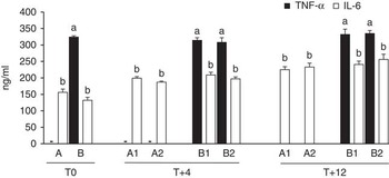
Table 2 reports results of TNF-α expression. TNF-α was expressed only in deprived culture condition groups (B, B1 and B2). The co-culture with anterior pituitary significantly (P<0.05) affected the expression at T+4 (B2: 0.56±0.02 v. B1: 0.32±0.01) and at T+12 (B2: 0.89±0.02 v. B1: 0.48±0.01). Basal TNF-α output was 324±4 pg/ml (group B). Anterior pituitary co-culture did not influence TNF-α secretion. At T+4, levels were: 314±8 (B1) v. 308±14 pg/ml (B2); at T+12, levels were 332±16 (B1) v. 335±9 pg/ml (B2). Results are shown in Figure 3. Nutrient availability and pituitary co-culture did not affected IL-6 expression (Table 2). At T0, IL-6 secretion was 156±10 and 132±9 pg/ml in A and B groups, respectively. Co-culture with anterior pituitary did not modulate IL-6 levels after 4 or 12 h of incubation in experimental groups A (T+4: A1=199±5 pg/ml, A2=187±3 pg/ml; T+12: A1=225±9 pg/ml, A2=233±12 pg/ml) and B (T+4: B1=209±8 pg/ml, B2=197±6 pg/ml; T+12: B1=241±10 pg/ml, B2=256±16 pg/ml). Results are summarized in Figure 3.
TNF-α and IL-6 levels (mean±RSD) in adipocyte cultures. Adipocytes were cultured in DMEM/Ham’s F12+10% FCS (group A) or in DMEM/Ham’s F12 salts+10% FCS (group B) for 24 h (T0). After 24 h, adipocytes were cultured for 4 h (T+4) or 12 h (T+12) with or without anterior pituitary slices: A1=control without pituitary; A2=coincubation with pituitary; B1=control without pituitary; B2=coincubation with pituitary. Different letters mean at least P<0.05. For each cytokine, significant differences (at least P<0.05) among the groups and treatments are labeled with different letters. IL-6=interleukin-6; TNF-α=tumor necrosis factor-α.

Discussion
The role of adipose tissue in the production and secretion of cytokines has been repeatedly demonstrated over the past 20 years (Tilg and Moschen, Reference Tilg and Moschen2006). Many studies have also focused on adipose tissue biology in swine. The function of mature adipocytes as a major cell type in adipose tissue extends beyond lipid storage. These cells actively modulate energy metabolism by secreting bioactive molecules called adipokines, which can act in an autocrine, paracrine and/or endocrine fashion. The reduction or absence of nutrients and other essential ‘ingredients’ available to cells, is a source of nutritional stress. Our results demonstrate a direct connection between nutrient availability and adipocyte capacity to produce NO and cytokines. We showed an increase in NO levels in absence of nutrients. NO is a key regulator of metabolic homeostasis and regulates metabolic lipid and carbohydrate metabolism (Andersson et al., Reference Andersson, Gaudiot, Ribiere, Elizalde, Giudicelli and Arner1999). In particular, Mehebik et al. (Reference Mehebik, Jaubert, Sabourault, Giudicelli and Ribière2005) reported that NO production, which is required for the lipolytic activity of adipocytes, seems to be related to the antioxidant protective action against oxidant effects of superoxide anions. NO production is also strongly influenced by leptin (Joffin et al., Reference Kern, Ranganathan, Li, Wood and Ranganathan2012): in the present study, leptin expression and secretion were higher in cells cultured in DMEM/Ham’s F12 salts (group B) as compared with cells cultured in complete medium (group A). Nutritional status and nutrient availability have been identified as an important determinant in the regulation of leptin expression (Friedman and Halaas, Reference Friedman and Halaas1998). Here we show an increase in leptin when nutrient availability was reduced, similarly to NO. Our findings suggest that NO production is modulated by leptin to correct energy balance, probably by a lipolytic activity, which also protect cells against oxidant damage. Leptin secretion was also positively influenced following 12 h of coincubation with anterior pituitary slices in both groups. Although the relationship between GH and leptin in adipose tissue is still unclear, the expression of GH and leptin receptor in adipose tissue has long been demonstrated in pigs (Sørensen et al., Reference Sørensen, Chaudhuri, Louveau, Coleman and Etherton1992; Barb et al., Reference Barb, Hausman and Houseknecht2001) and the effects of GH on lipid metabolism are well documented in porcine adipose cells. GH decreases fat mass, and this effect is thought to be mediated by the stimulation of lipolysis and the inhibition of lipogenesis (Louveau and Gondret, Reference Jensen2004). Taken together, we may hypothesize that GH directly regulates and modulates cell metabolism by influencing leptin and also by acting on cell metabolism to counteract the shortage necessary of nutritional factors. Another important factor that directly acts on adipocyte metabolism is TNF-α. It stimulates lipolysis, increases free fatty acid production (Chen et al., Reference Chen, Xun, Chen and Wang2009) and promotes leptin release from adipocytes (Simons et al., Reference Simons, van den Pangaart, van Roomen and Aerts2005). TNF-α can be secreted by the adipose tissue itself and it can metabolically promote negative energy balance (Galic et al., Reference Galic, Oakhill and Steinberg2010) by paracrine functions. In our model, it was expressed only in DMEM/Ham’s F12 salts (group B) suggesting that TNF-α expression can be stimulated by the reduction of nutrient to mobilize triglycerides for cell needs. In fact, evidence supporting a key role for TNF-α in homeostasis has been reported in both in vitro and in vivo studies. In particular, in porcine adipocytes (Shan et al., Reference Shan, Wu, Reng and Wang2009), TNF-α is a promoter of hydrolysis of triglycerides and of gene expression of the adipose triglyceride lipase and of the hormone sensitive lipase, that are the novel major lipase in animals (Chen et al., Reference Chen, Xun, Chen and Wang2009). TNF-α is also strongly correlated to IL-6 (Kern et al., Reference Louveau and Gondret2004) and previous studies demonstrated an increase of IL-6 expression in response to TNF-α (Berg et al., Reference Berg, Fraker and Alexander1994; Jensen, Reference Joffin, Niang, Forest and Jaubert2003). Several recent studies point out the importance of both TNF-α and IL-6, the most widely studied cytokines produced by adipose tissue, in the regulation of adipose tissue with regard to obesity and insulin resistance, even if the role of adipocyte IL-6 has yet to be clarified. Even though ~1/3 of the IL-6 detected in plasma is attributed to the production from WAT, most of the adipose-derived IL-6 comes from cells of the stromal vascular fraction and not from adipocyte (Galic et al., Reference Galic, Oakhill and Steinberg2010). In our study, no differences in IL-6 expression and secretion were found in the two groups, supporting the hypothesis of a limited role of adipocyte cell derived IL-6. It should also be remembered that IL-6 has traditionally been known for its immunomodulatory effects, and only recently have studies focused on its metabolic role. IL-6 has been demonstrated to induce lipolysis and fatty acid oxidation in humans in vivo (Petersen et al., Reference Petersen, Carey, Sacchetti, Steinberg, Macaulay, Febbraio and Pedersen2005), but the effect appears to be centrally mediated (Wallenius et al., Reference Wallenius, Wallenius, Sunter, Dickson and Jansson2002; Green et al., Reference Green, Rumberger, Stuart and Ruhoff2004).
In conclusion, our study showed an important endocrine role for porcine adipocytes. In our model, adipocytes readily responded to nutritional inputs by secretion of molecules affecting energy balance. This secretion was strongly influenced by GH. It would be necessary to develop quantitative models of metabolic and signaling networks to achieve a more complete understanding of adipocyte biology in swine.
Acknowledgment
All the authors equally contributed to the paper.