Introduction
Flexible endoscopic evaluation of swallowing is a diagnostic tool used to investigate the oropharyngeal phase of deglutition in dysphagic patients. This procedure has been shown by multiple investigators to be safe and accurate.Reference Langmore, Schatz and Olsen1–Reference Cohen, Setzen, Perlman, Ditkoff, Mattucci and Guss3 Oesophageal phase dysphagia may be suspected during flexible endoscopic evaluation of swallowing, for example due to regurgitation, but this procedure does not provide definitive information about oesophageal swallowing. The oesophageal phase of swallowing is typically evaluated separately, with fluoroscopy, manometry and/or endoscopy. The ideal diagnostic investigation of the oesophageal phase of deglutition could be performed in the same setting as flexible endoscopic evaluation of swallowing and would not involve sedation, radiation exposure or additional patient discomfort. Fluoroscopic studies generally use barium products which do not necessarily simulate a real meal. In addition, relatively small amounts of solid food are used in these studies. During manometry with or without impedance testing, only saline and/or viscous substances are evaluated. Traditional oesophagoscopy is performed transorally in a sedated patient, making oral feeding impossible.
Thin calibre endoscopes enable transnasal oesophagoscopy in the unsedated patient in a clinic setting.Reference Postma, Cohen, Belafsky, Halum, Gupta and Bach4, Reference Amin, Postma, Setzen and Koufman5 This technology gives the endoscopist a rapid method of assessing the oesophageal phase of deglutition, as part of flexible endoscopic evaluation of swallowing. Guided observation of oesophageal swallowing represents an extended version of the flexible endoscopic evaluation of swallowing procedure. The oropharyngeal phase of swallowing is first examined using the traditional flexible endoscopic evaluation of swallowing procedure. If this procedure does not reveal a problem with the oropharyngeal phase of swallowing, or if a concomitant oesophageal phase problem is suspected, guided observation of oesophageal swallowing is then performed in the same setting. The objective of this study was to evaluate the safety and efficacy of guided observation of oesophageal swallowing in the evaluation of oesophageal phase dysphagia.
Methods
Permission to conduct this study was granted by the institutional board of the University of California, Davis. Data from all patients who had undergone guided observation of oesophageal swallowing and who had also undergone a secondary oesophageal evaluation with oesophageal videofluoroscopy and/or manometry between 1 January 2005 and 10 October 2007 were retrospectively retrieved from a clinical database. Information regarding patient demographics, diagnoses, procedure safety, and fluoroscopic and manometric findings was abstracted. Findings for guided observation of oesophageal swallowing were compared with those for oesophageal manometry and fluoroscopy.
Protocol
Our protocol for performing guided observation of oesophageal swallowing is as follows.
Firstly, informed consent for the oesophagoscopy is obtained.
The patient is seated upright in an otolaryngology examination chair.
The most patent nasal cavity is sprayed twice, using a standard atomiser (Wolfe Tory Medical, Salt Lake City, Utah, USA), with 1:1 oxymetazoline 0.05 per cent and lidocaine 4 per cent.
The oesophagoscope (VE-1530, Pentax Precision Instrument Corp, Orangeburg, New York, USA) is placed through the nare and positioned in the oropharynx just below the tip of the uvula.
Flexible endoscopic evaluation of swallowing is then performed according to an established protocol. The anatomy of the tongue base, hypopharynx, larynx and vocal folds are assessed. Velopharyngeal closure and vocal fold mobility are evaluated. Pharyngeal motor function is assessed using the pharyngeal squeeze manoeuvre. Pooling of secretions is noted. The patient is administered 10 mm3 of purée (applesauce mixed with green food colouring). If the patient is safely able to tolerate this bolus, 50 and then 150 mm3 of purée are administered. The patient is then given 10 mm3 of water mixed with green food colouring. If the patient is safely able to tolerate this small bolus, 50 and then 150 mm3 of water are administered. If the 150 mm3 bolus is safely consumed, the patient is asked to take sequential, unmetered swallows through a straw. If deemed safe, the patient is then given food the consistency of a dry cracker, and the swallowing mechanics are observed.
If deep penetration to the level of the vocal folds or tracheal aspiration is visualised during flexible endoscopic evaluation of swallowing, oesophagoscopy alone will be performed, without guided observation of oesophageal swallowing.
Assuming that no such deep penetration or tracheal aspiration is visualised, the oesophagoscope is then passed through the upper oesophageal sphincter into the cervical oesophagus. Any pooled food or liquid from the recently completed flexible endoscopic evaluation of swallowing is noted. The normal oesophageal transit time is approximately 13 seconds; thus, any bolus in the oesophagus that is present 13 seconds after completion of flexible endoscopic evaluation of swallowing is considered abnormal.
The entire length of the oesophagus is evaluated, and the presence of any oesophagitis, web, ring, stricture or neoplasm is noted.
The endoscope is then positioned 2 cm above the aortic compression in the cervical oesophagus. The patient is given a 150 mm3 bolus of purée. Bolus transit and oesophageal motility are evaluated. With the endoscope in this position, the patient is then administered 150 mm3 of water and finally a bolus of cracker consistency. The endoscope is then advanced to the distal oesophagus 6 cm above the gastroesophageal junction. Further 150 mm3 boluses of purée, thin liquid and cracker are then administered. Distal oesophageal motility and bolus passage through the lower oesophageal sphincter into the stomach are noted. The endoscope is then passed into the stomach and a retroflex view of the fundus and gastric cardia is obtained. The patient is again administered 150 mm3 of purée, thin liquid and cracker. Bolus passage into the stomach is visualised. The time required from the initiation of the swallow to transit into the stomach is noted.
Any apparently abnormal mucosa is biopsied at the conclusion of the procedure.
The stomach is then suctioned free of air and the endoscope is carefully withdrawn.
Results
Twenty-one patients were identified who had undergone guided observation of oesophageal swallowing and oesophageal videofluoroscopy and/or manometry. Fifty-two per cent of patients were male and 48 per cent were female. The mean age was 63 years (range 18–85). No complications related to guided observation of oesophageal swallowing were identified.
All 21 subjects had videofluoroscopic oesophagrams available, enabling comparison with guided observation of oesophageal swallowing. The results of guided observation of oesophageal swallowing were in agreement with those of fluoroscopy regarding localising the site of dysphagia in 15/21 (71 per cent) patients. Impaired oesophageal motility was suggested by guided observation of oesophageal swallowing but not by fluoroscopy in two of 21 (9 per cent) patients. Videofluoroscopy suggested oesophageal dysmotility not appreciated on guided observation of oesophageal swallowing in four of 21 (18 per cent) patients. Simultaneous oesophageal impedance manometry was performed in six of the 21 subjects. In these subjects, guided observation of oesophageal swallowing results were in concurrence with manometric findings in five of six (83 per cent) patients.
Guided observation of oesophageal swallowing included a full endoscopic examination of the oesophagus. Anatomic pathology likely to contribute to dysphagia was seen in 15/21 (71 per cent) guided observation of oesophageal swallowing examinations. In 13/21 (62 per cent) cases, guided observation of oesophageal swallowing identified pathology not appreciated on fluoroscopy and manometry, such as hiatal hernia, pill-induced oesophagitis, reflux oesophagitis, candida oesophagitis and stricture.
Discussion
The advent of thin calibre endoscopes suitable for oesophagoscopy in a clinic setting has dramatically changed the safety of and access to oesophageal screening.Reference Postma, Cohen, Belafsky, Halum, Gupta and Bach4, Reference Amin, Postma, Setzen and Koufman5 Transnasal oesophagoscopy can be performed in most patients in just a few minutes using only topical nasal anaesthesia. This technique provides an opportunity to evaluate the oesophagus in the upright, unsedated patient. Adding food and beverages to the examination enables a unique evaluation of the oesophageal phase of deglutition.
Videofluoroscopy, impedance manometry and oesophagoscopy are the primary diagnostic investigations used to assess the oesophageal phase of deglutition. However, none of these studies in isolation can provide complete information about the oesophagus. The videofluoroscopic oesophagram, or barium swallow, may be performed in the same setting as an oropharyngeal videofluoroscopic swallowing assessment, if the provider has had the foresight to request the examination. We are fortunate to practise in settings in which we can perform our own oesophagrams, but this is a relatively uncommon occurrence. In most settings, clinicians do not perform fluoroscopic studies themselves, but rather rely upon the technique and interpretation of a radiologist. The barium swallow is an excellent tool for evaluating diverticulae, strictures and obstructing lesions, but its sensitivity decreases when evaluating oesophagitis, reflux disease and smaller neoplasms. The sensitivity of barium swallow for the evaluation of achalasia is quite good, but its sensitivity decreases to about 50 per cent for more subtle motility disorders, such as ineffective oesophageal motility.Reference Fuller, Huprich, Theisen, Hagen, Crookes and Demeester6, Reference Shakespear, Blom and Huprich7 Exposure to radiation is small but not insignificant,Reference Livingstone, Augustine, Aparna and Raj8 especially if multiple evaluations are performed over time.
Manometry, particularly with impedance testing, is an excellent diagnostic tool for assessing oesophageal motility and bolus transit.Reference Tutuian and Castell9 However, this investigation is somewhat limited by the semi-recumbent positioning of the patient and the inability to test solid food swallowing. Manometry and/or impedance testing is not available in all areas and involves an additional, uncomfortable procedure for the patient.
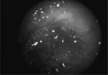
Oesophagoscopy is the ‘gold standard’ for evaluating the oesophageal mucosa. Traditional large calibre, transoral, procedures requiring sedation do not allow for oral feeding during the examination. However, when undergoing transnasal oesophagoscopy the patient is awake and cooperative, so food and pill challenges are easily achieved. During guided observation of oesophageal swallowing, the oesophagus can first be examined for abnormalities or oesophageal residue left after the flexible endoscopic evaluation of swallowing examination (see Figure 1).
Pooling of ingested food in the oesophagus, viewed during guided observation of oesophageal swallowing.

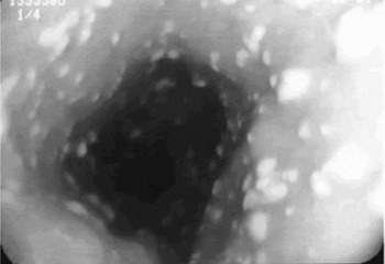
Oesophagitis, either reflux-induced or infectious (Figure 2), is not uncommonly seen during guided observation of oesophageal swallowing.
Candida oesophagitis viewed during guided observation of oesophageal swallowing.

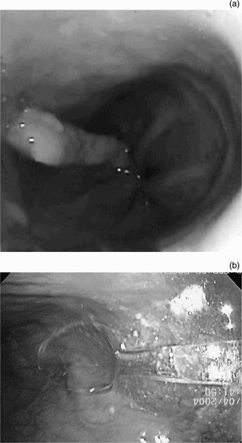
Biopsies to investigate suspicious lesions or to rule out eosinophilic oesophagitis can be performed at any time during guided observation of oesophageal swallowing.Reference Prasad, Talley, Romero, Arora, Kryzer and Smyrck10 Strictures, rings and diverticulae can also be carefully examined during this procedure. On occasion, large oesophageal foreign bodies have been seen (Figure 3).
Retained oesophageal foreign bodies seen on guided observation of oesophageal swallowing: (a) potato chip, (b) plastic spoon.

The patient can be fed orally while the oesophagoscope is retroflexed in the stomach toward the lower oesophageal sphincter, allowing for assessment of transit times with real food, liquid and pills. Gastric emptying may also be assessed with transnasal oesophagoscopy, eliminating the radiation exposure associated with scintigraphy.Reference Attila, Hellman, Krasnow, Hofmann, Saeian and Dua11 Even when transnasal oesophagoscopy is not available, the flexible nasopharyngoscope used for flexible endoscopic evaluation of swallowing can be used to evaluate the cervical oesophagus. Such cervical oesophagoscopy is obviously limited, but may provide information about oesophageal pooling, diverticulae or infectious oesophagitis.
The current study was small and retrospective in nature. The flaws of the study design include non-blinded evaluation of the oesophagrams and manometry and endoscopy procedures by the authors. However, the manometric and fluoroscopic results were not generally available when the guided observation of oesophageal swallowing procedure was performed. Guided observation of oesophageal swallowing was typically performed as part of the initial evaluation of dysphagia, in conjunction with flexible endoscopic evaluation of swallowing. We believe that guided observation of oesophageal swallowing is a valuable tool in assessing oesophageal deglutition. However, further, prospective studies are needed to clarify its clinical role.
Conclusion
Guided observation of oesophageal swallowing appears to be a safe and efficacious diagnostic tool with which to evaluate the oesophageal phase of deglutition during flexible endoscopic evaluation of swallowing. In this series, the results of guided observation of oesophageal swallowing agreed with those of fluoroscopy in 71 per cent of cases and with those of manometry in 83 per cent of cases. The guided observation of oesophageal swallowing procedure identified pathology not detected by fluoroscopy or manometry in 62 per cent of cases. This study emphasises the importance of oesophagoscopy in the evaluation of dysphagia, particularly in the setting of a normal oropharyngeal phase of deglutition. Transnasal oesophagoscopy can be performed in nonsedated patients, allowing real-time visualisation of food and liquid bolus transport through the oesophagus. Like flexible endoscopic evaluation of swallowing, guided observation of oesophageal swallowing uses real food which may more closely simulate a meal than the barium products used during fluoroscopy.