Introduction
Nutrition is a crucial aspect of health and well-being.Reference Gopalan and Khanna1 Optimising nutritional status is particularly important for head and neck cancer patients as it will result in a better response to oncologic or surgical treatment and will improve recuperation.Reference Sobol, Conoyer and Sessions2 In these patients, and all patients with swallowing problems,Reference Sobol, Conoyer and Sessions2, Reference Lin, Liu, Lee, Tang, Wei and Yang3 enteral feeding can be achieved using a nasogastric tube.Reference Gopalan and Khanna1 Nasogastric tubes are well accepted by most patients; however, others are intolerant of the feeding tube.
Placement of a nasogastric tube requires adequate training, and in general it is a requirement that a chest X-ray is performed to confirm correct positioning of the tube prior to commencement of feeding. Incorrectly placed or displaced feeding tubes can result in serious complications such as aspiration,Reference Metheny5 persistent chest infection, pneumothorax and even be fatel.Reference Chang, Melnick, Bedger and Bleyaert6, Reference Fisman and Ward7
We present a case that highlights the benefits of carrying out an X-ray to confirm the position of a nasogastric tube.
Case report
A 50-year-old Caucasian man, undergoing radiotherapy for squamous cell carcinoma of the tongue base, was admitted for pain control and nasogastric tube feeding. This patient required multiple nasogastric tubes over a two-week period. The patient denied pulling the nasogastric tube out on each occasion and we were unable to establish the exact mode of nasogastric tube removal.
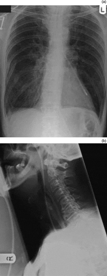
On one such occasion, the nasogastric tube was missing and the patient claimed it had accidentally fallen out whilst he was sleeping. However, the tube was not found. Following reinsertion of a new tube, a check X-ray was carried out (Figure 1). This not only revealed that the new tube was lying within the left main bronchus, but also showed the ‘lost’ tube, which was located partially within the hypopharynx and extended into the stomach. These findings were confirmed with flexible laryngoscopy.
(a) Chest X-ray showing two nasogastric tubes: the newly inserted one in the left bronchus and the tip of the ‘lost’ severed tube extending into the stomach. (b) X-ray of the lateral soft tissue of the neck showing the two nasogastric tubes, with the lost severed tube in the hypopharynx and the new tube entering the trachea.

The newly placed nasogastric tube (which had entered the lung) was simply removed. The lost tube was removed by the gastroenterologist using a flexible oesophagoscope under local anaesthesia with mild sedation.
The nasogastric tube removed from the hypopharynx (extending into the stomach) was found to have been obliquely severed at 50 cm. The patient denied any knowledge of pulling or cutting it and swallowing it.
Discussion
Nasogastric tube insertion is a routine procedure carried out by both medical and nursing staff. Although simple, the procedure is not without its complications. The most common complication observed is epistaxis due to direct trauma from the tube insertion. In addition, prolonged tube placement can result in infection, sinusitisReference Baskin8 and erosion of the surrounding structures. There are even reported incidences of tube misplacement in the lungs and brain with adverse outcomes.Reference Wang, Tseng, Yang, Chou and Chen4, Reference Baskin8–Reference Galloway and Grudis11 Misplaced tubes are associated with other risks such as pneumothoraxReference Ciocon12–Reference Kolbitsch, Pomaroli, Lorenz, Gassner and Luger14 and aspiration.Reference Baskin8, Reference Gomes, Pisani, Macedo and Campos15, Reference Scolapio16 The literature also reports isolated cases of: mediastinitis due to perforation of the cervical oesophagus,Reference Fisman and Ward7 massive fatal haemorrhage due to tearing of the large intrathoracic blood vessels in a patient with mediastinitisReference Wu, Kang, Hui, Hung, Sun and Chan17 and a knotted nasogastric tube in the posterior pharynx, which was removed under endoscopic guidance.Reference Dinsmore and Benson18
In some units it is routine practice to check the position of the nasogastric tube by aspirating gastric secretions for pH testing,Reference May19 without conducting a check X-ray. Although pH testing is useful, there is a definite place for the direct visualisationReference Chang, Melnick, Bedger and Bleyaert6 of a nasogastric tube in order to check the position of the tube. This might entail using a flexible nasendoscope,Reference Karagama, Lancaster and Karkanevatos20 air insufflation in the stomach or X-ray techniques.Reference Chang, Melnick, Bedger and Bleyaert6, Reference Fisman and Ward7 There are instances where difficult tube insertion may need to be assisted with endoscopic controlReference Karagama, Lancaster and Karkanevatos20 or radiological guidance. In addition, the vocalisation technique can be used while advancing the tube in order to adduct the vocal folds,Reference Takwoingi and Demspter21 which can be helpful for correct tube placement.
In certain circumstances, particularly if a nasogastric tube cannot be located, we would recommend the use of flexible nasendoscopy to avoid missing any retained tubes. This examination should precede any imaging and the reinsertion of another nasogastric tube.
In the case reported here, the nasogastric tube removed from the patient was obliquely severed at 50 cm. It appeared that the patient had cut the tube as a result of his intolerance, and had failed to alert the medical staff either because of acute confusion or the fear of retribution from staff.
• Flexible nasendoscopy is recommended when a nasogastric tube cannot be located
• Chest X-rays should be carried out to confirm correct tube placement
• Some patients are intolerant of nasogastric tubes
• Percutaneous endoscopic gastrostomy should be considered for such patients if long-term enteral feeding is anticipated
This patient was unfit for general anaesthetic for the removal of the retained tube from his stomach. However, as this report highlights, such cases can be safely and successfully managed using a flexible oesophagoscope under local anaesthetic.
For patients with poor tolerance of nasogastric tubes, percutaneous endoscopic gastrostomy tube insertion may be required for comfort and convenience.
Conclusion
Clinical tests such as pH testing with litmus paper or auscultations over the stomach during quick air insufflation are helpful to confirm the position of nasogastric tubes in the stomach. However, the present case stresses the importance of direct visualisation or imaging of any inserted nasogastric tube. This is especially vital in confused patients or patients with an unclear history of previous tube dislodgement. Early consideration should be given to percutaneous gastrostomy tube placement in patients requiring long-term enteral feeding and who are intolerant of nasogastric tubes.