Introduction
Breast cancer is classified into several subtypes based on the presence or absence of oestrogen receptor (ER), progesterone receptor (PR) and human epidermal growth factor receptor 2 (HER-2) and, lately, by KI67 expression (Ref. Reference Hergueta-Redondo1). Based on molecular expression of these important receptors in tumour tissue, the cancer can be classified into distinct subtypes, which demand different stratagems for clinical management (Ref. Reference Yanagawa2). Perou et al. conducted the first study assessing molecular classifications of breast cancer (Ref. Reference Perou3). Currently, based on global gene expression evaluations using microarray technology, breast cancer is divided into six subtypes: luminal A (ER+, PR+, HER-2-, low KI67), luminal B (ER+ and/or PR+, HER-2+ or HER-2-, with high KI67), basal-like (BL) (ER-, PR-, HER-2-, any KI67, CK5/6+ and/or EGFR+) (Ref. Reference Ribelles4), ‘normal-like’ breast tumours, HER-2-enriched (HER-2-E) and claudin low (ER-, PR-, low basal cytokeratin and low expression of claudin 3, 4 and 7) (Refs Reference Perou3, Reference Peddi, Ellis and Ma5).
The term ‘triple-negative breast cancer’ (TNBC) defined by the lack of expression of ER, PR and HER-2 (Ref. Reference Ademuyiwa, Ellis and Ma6) and ‘basal-like breast cancer’ (BL) are frequently used interchangeably but they do not overlap completely; BL accounts for between 70 and 90% of TNBC cases.
TNBC that overlaps with the BL subtype is characterised by the overexpression of keratins 5, 6 and 17 and overexpression of the genes implicated in cell proliferation (Ref. Reference Liu7). In addition, overexpression of KI67 is a characteristic of the BL subtype and the overexpression of KI67 is a characteristic of the BL subtype (Ref. Reference Turner and Reis-Filho8).
Worldwide, over one million women are diagnosed each year with breast cancer, and the number of breast cancer-related deaths has risen to almost 400 000 annually according to data from the USA (Ref. 9). About 170 000 of the newly diagnosed patients with breast cancer will be expected to be classified as having TNBC. Since the publication of the GLOBOCAN 2012 cancer statistics, the incidence of breast cancers has risen by over 20% globally (Ref. Reference Brewster, Chavez-MacGregor and Brown10); these statistical data are also reflected in the number of incident cases of TNBC
Compared with hormone-dependent breast cancer, TNBC appears to affect younger patients and the disease is overrepresented in African-American women. TNBC is also associated with a poorer prognosis and significantly higher rates of metastasis and relapse than hormone-dependent breast cancer (Ref. Reference Masuda11). The absence of well-characterised molecular targets that are susceptible to intervention moreover renders TNBC very challenging to treat (Refs Reference Pegram12, Reference Carey13) and it remains the most aggressive subtype of human breast cancer. However, on account of its marked genetic heterogeneity, TNBC has become a central focus of research into the molecular epidemiology of human breast cancer and with this comes the ultimate hope of improved and specific therapy (Refs Reference Hicks14, Reference Dent15).
Since TNBC became a well-defined subgroup in breast cancer management with, many TNBC studies have been initiated in the past 10 years (Ref. Reference Turner and Reis-Filho8). Based on this molecular classification, patients can be stratified in an attempt to define their optimal therapy. Presently only a few clinical trials in which angiogenesis have been targeted have shown encouraging outcomes (Refs Reference Dent15, Reference Minami, Chung and Chang16, Reference Lehmann17, Reference Zhang18).
In addition, improved knowledge of angiogenic processes has brought about the discovery of molecules such as semaphorins and their receptors; transcription factors. Despite the slow progress in scientific discovery related to this cancer, early studies in the field have given rise to therapeutic innovation and new options for exploration in clinical trials. One relevant example is the new clinical trial that is under observation for the use of olaparib, an efficient poly(ADP-ribose) polymerase (PARP) inhibitor which is used in combination with paclitaxel in TNBC patients (Ref. Reference Dent15). More generally, identification of particular cellular characteristics, including epithelial–mesenchymal transition, downregulation of particular noncoding RNAs (ncRNAs) and the relationship between angiogenesis and metabolic syndrome has opened up fresh areas of investigation.
In sum, TNBC has emerged as a critically important field in the management of breast cancer: its poor prognosis and predilection for young women has stimulated much research over the last decade. The latest investigations depict a complex molecular portrait of this disease and it thus poses a particular challenge for research designed to translate the extensive documentation of molecular cytopathology to improved clinical practice. Here we review the latest information about this disease with an emphasis on the role of angiogenesis and the utility of anti-angiogenic therapy (Ref. Reference Fox, Generali and Harris19), We present a conspectus of the principal cellular pathways in the development of TNBC and candidate targets for future therapeutic exploration; we also summarise preclinical studies of candidate agents and some clinical trials involving drugs designed to intervene at key points in the specific molecular pathogenesis of this virulent cancer.
Molecular classification of TNBC
TNBC encompasses a complex spectrum of subtypes that includes different types of malignant cells with different cellular microenvironments and different patterns of development, progression, invasion and metastasis. Multiple complex molecular pathways orchestrate all of these processes (Ref. Reference Garraway and Lander20). Owing to a lack of understanding regarding these complex and intricate molecular pathways, most treatments are focused on restoration of the wild-type phenotype and not on modification of the initial alterations in the genotype. In most cases, this results in treatment failure (Ref. Reference Garraway and Lander20). The relevant altered pathways with therapeutic potential in TNBC consist of Notch signalling, the Wnt/β-catenin Hedgehog pathways, PARP1, mechanistic target of rapamycin (mTOR) and angiogenesis (e.g. RTK, transforming growth factor (TGF)-β, etc.) (Ref. Reference Jamdade21).
TNBCs are classified as BL (70–88%) and non-BL (20–30%) (Ref. Reference Prat22). Non-BL breast cancers are classified as luminal A and B, and HER-2-enriched. Based on PAM50 classification, a new subtype of BL cancer, defined as claudin low (approximately 10% of TNBC tumours), has been identified (Ref. Reference Cheang23). In order to facilitate personalised treatment for patients with this particular subtype of breast cancer, the claudin low classification will remain dynamic until it has been validated in a large number of patients.
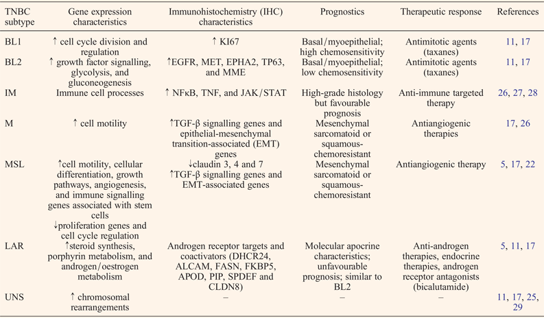
Gene-expression profiling has significantly contributed to the understanding of TNBC biology, with the resulting significant impacts in clinical care (Ref. Reference Prat22). Lehmann et al. reported the identification of seven molecular subtypes in TNBC, based on the microarray evaluation patterns of 224 TNBC cases, which lead to the identification of hierarchical gene clustering (Refs Reference Masuda11, Reference Lehmann17). Based on the gene ontology classifications of the altered gene patterns, the researchers were able to identify seven distinct TNBC subtypes: BL1, BL2, immunomodulatory (IM), mesenchymal (M), mesenchymal stem-like (MSL), luminal androgen receptor (LAR) and unstable (UNS) (Refs Reference Masuda11, Reference Chen24). Table 1 presents an updated Lehmann classification for the main features of the TNBC subtypes.
Characteristic features of TNBC subtypes

The BL1 subtype exhibits overexpression of genes involved in DNA damage repair and cell cycle regulation and is characterised by a positive therapeutic response to platinum salts and PARP inhibitors (Refs Reference Dent15, Reference Zhang18). The BL2 subtype displays overexpression of growth factor receptor genes and a positive response to cetuximab. The IM and M subtypes show upregulation of genes participating in epithelial-to-mesenchymal transitions, and the MSL subtype is further characterised by the upregulation of angiogenic genes. These three subtypes all respond to dasatinib (an SRC/ABL inhibitor). Androgen receptor (AR) signalling characterises the LAR subtype, which is uniquely sensitive to the action of bicalutamide, an AR antagonist (Refs Reference Masuda11, Reference Chen24, Reference Abramson25).
Based on this molecular profile of TNBC, multiple therapies are already being investigated and are at various stages in clinical trials. Treatment examples include the use of PARP inhibitors with BRCA-mutated TNBC or antiandrogens for AR-positive TNBC (Ref. Reference Lehmann, Pietenpol and Tan30). Novel therapeutic strategies are currently being tested in phase II and III trials and will likely require patient stratification before therapy. Examples of these tailored approaches also include the targeting of overexpressed mutated receptors, such as epidermal growth factor receptor (EGFR), vascular endothelial growth factor receptor (VEGFR) and fibroblast growth factor receptor (FGFR) (Ref. Reference Lehmann and Pietenpol31).
Molecular pathways of angiogenesis in TNBC
Angiogenesis, defined as the formation of new blood vessels to supply a tumour with oxygen and nutrients, is a major mechanism in tumour development (Refs Reference Murakami, Elfenbein and Simons32, Reference Jain33). Although the importance of angiogenesis in cancer progression has already been established, it is still unclear whether antiangiogenic therapies should also be applied in TNBC; this is because of the yet unidentified carcinogenic pathways operating as a result of the hormone receptor-free nature of TNBC and the lack of specific targets (Ref. Reference Papa34). Angiogenesis inhibition has received particular attention in TNBCs, primarily because of lymphatic vessel density, vascular invasion and microvessel density related to vascular endothelial growth factor (VEGF) family factors, which are often more pronounced in TNBCs than in other breast cancer subtypes (Refs Reference Bender and Mac Gabhann35, Reference Li36, Reference Tolaney, Boucher and Duda37, Reference Mohammed38). Antiangiogenic agents, such as novel generations of monoclonal antibodies (bevacizumab, sunitinib, pazopanib, erlotinib, cetuximab and panitumumab), have also raised hopes for effective TNBC treatment (Ref. Reference Hurvitz and Mead39). A wide range of factors stimulate angiogenesis; VEGF (Refs Reference Ribeiro-Silva, Ribeiro do Vale and Zucoloto40, Reference Li41) is one of the most relevant examples. VEGF-A is the most important and well-studied proangiogenic factor (Ref. Reference Murakami, Elfenbein and Simons32) and plays an important role in the development of TNBC (Ref. Reference Bender and Mac Gabhann35). A prognostic role for VEGF-A gene expression levels have been demonstrated in TNBC evolution (Refs Reference Ortiz-Martinez42, Reference Bahnassy43). Increased expression of this molecule has also been observed in hypoxic and inflammatory conditions (Ref. Reference De Laurentiis44). A higher expression for VEGF-C has been demonstrated in the MSL subtype, compared with other TNBCs (Ref. Reference Bender and Mac Gabhann35). VEGF-A, TGF-β1 and insulin-like growth factor (IGF)-I/IGF-IR overexpression have been connected to more aggressive cancer progression (Ref. Reference Bahhnassy45). In addition, VEGF receptor signalling is clearly related to invasive and metastatic processes and VEGFR receptor 2 (VEGFR2) is more highly expressed in TNBC than in other breast cancer subtypes (Ref. Reference Ryden46).
Another research direction concerns the inhibition of overexpressed hypoxic factors. Hypoxia plays a significant role in multiple cellular outcomes (Refs Reference Tanaka47, Reference O'Reilly48) apart from angiogenesis; therefore, it may represent a relevant therapeutic target in TNBC (Ref. Reference O'Reilly48). Hypoxia-inducible factor-1α (HIF1α) is key factor for activation angiogenesis (Ref. Reference Yehia49). HIF1α plays an important role in hereditary breast cancer. It has important clinical significance because most TNBC cases involve BRCA1/2 mutations (Refs Reference van der Groep50, Reference van der Groep51). In spite of the fact that HIF1α is not a specific marker for TNBC, it appears to be related to tumour aggressiveness and unfavourable prognosis for TNBC patients (Ref. Reference Tanaka47). HIF1α can thus be used in tandem with other angiogenic markers to support diagnostics.
Vascular components play an important role in physiological and pathological conditions. With regards to cancer inhibition, vascular components represent a crucial research direction, primarily in efforts to develop multiple anti-angiogenic therapies (Refs Reference Tolaney, Boucher and Duda37, Reference Mohammed38). A study conducted on a cohort of patients that included 197 BL breast cancer tumours, specifically 99 TNBCs and 200 non-BL tumours, evaluated CD34 and CD31 as markers for lymphatic and microvessel density. This study revealed a higher microvessel density in the BL and TNBC cases than in non-BL and non-TNBC cancers (Ref. Reference Mohammed38). Li et al. demonstrated a correlation among microvascular density and clinicopathological factors, including survival rates (Ref. Reference Li36).
Osteopontin (OPN) is a multifunctional cytokine that has been proven to have increased expression levels in TNBC (Ref. Reference Wang52). Moreover, ICAM-1 and VEGF-A expression levels can be regulated by osteopontin in TNBC cases (Ref. Reference Ortiz-Martinez42). In addition, proinflammatory cytokines, including tumour necrosis factor α (TNFα), can promote tumour progression through death receptor pathways and activation of c-Jun and AP-1 (Refs Reference Ji53, Reference Qiao54). This highlights the utility of targeting TNFα-TNFR1-related pathways in TNBC (Refs Reference Ji53, Reference Pileczki55).
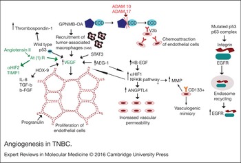
Progranulin is a recently discovered 88-kDa glycoprotein which stimulates proliferation of endothelial cells and was originally purified from a mouse teratoma-derived cell line (Ref. Reference Li41). It has been suggested that progranulin plays an important role in angiogenesis during placental development (Ref. Reference Desmarais56). TNBC shows increased expression of progranulin, VEGF and CD105, especially in patients with negative lymph nodes. These increased expression levels may explain the aggressive behaviour of lymph node negative TNBC. The key pathways and intermolecular interactions involved in angiogenesis are shown in Figure 1.
Angiogenesis in TNBC. Proliferation of endothelial cells is stimulated by VEGF and progranulin. Vascular permeability is increased by ANGPTL4. Expression of VEGF is increased by glycoprotein nonmetastatic melanoma protein B (GPNMB-OA), STAT3, AEG-1, HB-EGF, HOX9 and angiotensin II. GPNBM-OA stimulates recruitment of TAM, results in release of VEGF. ADAM-10 and ADAM 17 are proteases that cleave the ECD of GPNMB-OA, which acts as a chemoattractant for endothelial cells through interaction with the V3b receptor. It has been observed that HB-EGF upregulates the expression of VEGF and ANGPTL4 via upregulation of HIF1α and nuclear factor kappa B (NFκB). Angiotensin II also increases VEGF expression, via AT(1) R receptor. It also induces the expression of αHIF2 and TIMP1. The HOX9 gene also stimulates expression of IL-8, TGF-β and bFGF. AEG-1 has been demonstrated to increase MMPs expression, via the NFκB pathway. Expression of CD133 was associated with VM and upregulation of MMPs expression. Wild-type p53 inhibits angiogenesis via inhibition of VEGF and stimulation of thrombospondin-1. The mutated p53 protein binds to p63 protein (member of p53 family). Lack of a functional p63 initiates endosome recycling processes and upregulation of EGFR and integrin, leading to activation of the invasive capacity of the tumoural cells.

VEGF expression is increased by glycoprotein nonmetastatic melanoma protein B (GPNMB), STAT3, astrocyte elevated gene-1 (AEG1), heparin-binding epidermal growth factor-like growth factor (HB-EGF), home box (HOX) 9 and angiotensin II (Refs Reference Ribeiro-Silva, Ribeiro do Vale and Zucoloto40, Reference De Laurentiis44, Reference Yotsumoto57). In addition, multiple studies have demonstrated an association between VEGF expression and the upregulation of HIF1α (Refs Reference van der Groep50, Reference van der Groep51, Reference Saponaro58).
GPNMB, also known as dendritic cell-heparin integrin ligand (DC-HIL) (Ref. Reference Maric59), osteoactivin (OA), and haematopoietic growth factor-inducible neurokinin-1 (HGFIN), is a type I transmembrane protein expressed in normal tissues, including bone, skin and the haematopoietic system. It is part of the osteomimetic protein family and mediates the differentiation of osteoblasts and osteoclasts. GPNMB is overexpressed in bone pathologies, such as fracture repair and osteoarthritis (Refs Reference Karlsson60, Reference Abdelmagid61). Upregulation of GPNMB in 4T1 cell subpopulations has also been shown to induce bone, liver, and lung metastases. It is also involved in angiogenesis through stimulation of VEGF, tumour-associated macrophages (TAM) recruitment and tumour growth (Ref. Reference Rose62).
ADAM10 and ADAM17 are proteases that cleave the extracellular domain (ECD) of GPNMB, which then acts as a chemoattractant and induces the migration of endothelial cells. The ECD of GPNBM-OA interacts with a Vb3 receptor located on the surface of endothelial cells (Refs Reference Rose62, Reference Duffy, Crown and Mullooly63).
AEG1, also known as metadherin (MTDH), has been identified in primary human foetal astrocytes. It plays an important role in HRas-mediated angiogenesis via the PI3K/AKT pathway. It also regulates the expression of matrix metalloproteinase (MMP) through nuclear factor kappa B (NFκB) signalling. Increased expression of AEG1 has been shown in patients with TNBC and is positively correlated with VEGF level, tumour size, clinical stage and lymph node metastasis (Refs Reference Li36, Reference Danza64, Reference Danza65). This supports Folkman's theory that, in the absence of neovascularisation, tumour size is restricted to a few cubic millimetres (Ref. Reference Weidner66). VEGF and angiopoetin-like 4 (ANGPTL4) expression are upregulated by HB-EGF through upregulation of HIF1α and NFκB. Expression of NFκB and HIF1α are reciprocally regulated (Ref. Reference Yotsumoto57), and HIF1α may control the expression of MMP (Ref. Reference Bendinelli67). In another study, HIF1α was proven to be nonspecific for TNBC; its expression levels in TNBC are similar to those in other breast cancer subtypes (Ref. Reference Yehia49).
The HOX gene family plays an important role in embryogenesis and tumour progression. HOX9 stimulates angiogenesis in breast tumours by upregulating expression of VEGF, interleukin (IL)-8, TGF-β2 and basic FGF (bFGF) (Refs Reference Jain33, Reference Bendinelli67). HOX9 expression levels are directly correlated with tumour grade. HOX9 promotes distant metastases through the epithelial-to-mesenchymal transition. It has also been hypothesised that HOX9 may regulate the TGF-β pathway (Ref. Reference Bendinelli67). Angiotensin II also increases VEGF expression via the AT1 receptor and induces the expression of HIF2α and tissue inhibitor of metalloprotease (TIMP1) (Ref. Reference Chen68). A significant association between EGFR expression and increased angiogenesis has been found; EGFR acts as a transcription factor in the nucleus, where it regulates the expression of several genes involved in angiogenesis (Ref. Reference Brand69).
MUC4 overexpression has been connected to TNBC pathobiology and its specific inhibition is connected with decreased aggressiveness and tumourigenesis, as well as a reduced metastatic rate. MUC4 promotes invasiveness in TNBC models via targeting of EGFR, ErbB2 and ErbB3 genes and downstream factors of the MUC4 pathway (Ref. Reference Mukhopadhyay70).
Wild-type p53 also inhibits angiogenesis by regulating the activity of the natural inhibitor thrombospondin 1 and inhibiting VEGF expression. Mutated p53 stimulates angiogenesis and is associated with higher levels of VEGF (Refs Reference Chen24, Reference Chen68). Mutations of p53 lead to upregulation of EGFR endosomal recycling, a process associated with increased oncogenic activity in patients with TNBC. Mutated p53 protein binds to the p63 protein, a member of the p53 family. Lack of functional p63 initiates endosomal recycling and upregulation of EGFR and integrin, leading to activation of the invasive capacity of tumour cells. In addition, p63 has also been shown to induce expression of VEGF through the HIF1α pathway (Ref. Reference Shapira71). Other studies have emphasised the connection between TNBC and VEGF- and semaphorin-related genes (Ref. Reference Bender and Mac Gabhann35).
Role of semaphorins in the pathogenesis of TNBC
Semaphorins and their related receptors, called plexins, have been implicated in several pathological processes. At the epithelial level, semaphorin–plexin signalling pathways have been proven to promote angiogenesis (Refs Reference Bender and Mac Gabhann35, Reference Gabrovska72).
Semaphorins are divided into two subtypes based on their affinities for neuropilins and plexin receptors; these are the membrane semaphorins (classes 4, 5, 6 and 7), which bind directly to the plexin receptors, and soluble semaphorins (class 3), which require neuropilins to bind to the plexin receptors (Ref. Reference Toyofuku73). A growing body of evidence indicates the crucial role of semaphorins in tumour progression and prognosis, including in TNBC (Ref. Reference Bender and Mac Gabhann35). Soluble semaphorins (class 3) are thought to play an inhibitory role in the process of angiogenesis by competing with VEGF ligands to bind to neuropilins (Ref. Reference Guttmann-Raviv74).
Membrane semaphorins (classes 4, 5, 6 and 7) may mediate tumour signalling through modulation of downstream signalling pathways or through direct interactions with VEGFRs (Refs Reference Bender and Mac Gabhann35, Reference Toyofuku73, Reference Guttmann-Raviv74), such as VEGFR1 – also known as Flt1 – , VEGFR2 and VEGFR3 and two neuropilins co-receptors (NRP1 and NRP2) (Ref. Reference Bender and Mac Gabhann35). An association between VEGF overexpression and downregulation of semaphorin expression has been observed in 60% of TNBC cases (Refs Reference Papa34, Reference Bender and Mac Gabhann35). Differences in expression levels of VEGF and semaphorin ligand and receptor between TNBC tumours have been observed to correlated with an unfavourable prognosis (Ref. Reference Bender and Mac Gabhann35). The interconnection between VEGF/VEGFR and semaphorins was also emphasised by Papa et al. (Ref. Reference Papa34), who highlighted related applications in patient diagnostics. In TNBC, VEGF is generally escorted by the semaphorins; these two molecules can be considered as a brake in cancer angiogenesis (Ref. Reference Bender and Mac Gabhann75). Pro-angiogenesis signatures may have significant therapeutic implications with this type of cancer because they are related to unfavourable prognoses (Refs Reference Bender and Mac Gabhann35, Reference Bender and Mac Gabhann75).
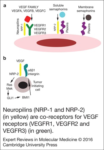
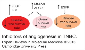
Pro-angiogenesis signatures may also identify cancers that are responsive to VEGF inhibition. A recent study showed that binding of VEGF to neuropilin 2 (NRP-2), which is primarily expressed in tumour-initiating cells, leads to activation of α6β1 integrin. This in turn facilitates induction of the Ras/MEK signalling pathway and overexpression of GLI1 (glioma-associated oncogene 1), a molecule involved in the Hedgehog signalling pathway, via autocrine pathways (Ref. Reference Basile, Afkhami and Gutkind76). Cleavage-resistant sema3C (FR-sema3C) is connected with the VEGF-C-induced phosphorylation of VEGFR-3, ERK1/2 and AKT and is related to the metastatic potential of TNBC (Ref. Reference Mumblat77). GLI1 upregulates the expression of BMI-1, a stem cell factor, which increases NRP-2 and α6β1 integrin expression, establishing an autocrine loop (Ref. Reference Goel78). Semaphorin mechanisms of action are shown in Figure 2. Thus, semaphorin signalling pathways could represent a potential therapeutic option for patients with TNBC. The benefit of exploring these pathways further is clear (Refs Reference Bender and Mac Gabhann35, Reference Ellis79). The relationships between various molecular drug targets involved in angiogenesis and survival rates in TNBC are shown in Figure 3.
(a) NRP-1 and NRP-2 (in yellow) are co-receptors for VEGF receptors (VEGFR1, VEGFR2 and VEGFR3) (in green). There are two types of semaphorins: soluble (light blue) and membrane (grey). Semaphorins use plexins (purple) as receptors. Many ligands from the VEGF family (red) and soluble semaphorins class (blue) use neuropilins as co-receptors. (b) When expressed on tumour initiating cells, NRP-2 stimulates the expression of α6β1 integrin, which induces the Ras/MEK signalling pathway and expression of GLI1. GLI1 upregulates the expression of BMI-1, which increases NRP2 and α6β1 expression, establishing an autocrine loop.

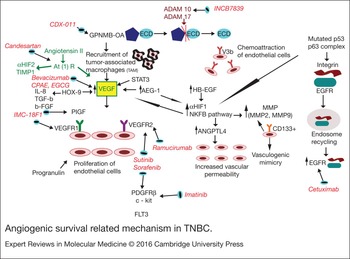
Angiogenic survival related mechanism in TNBC. An increased expression of the VEGF and IL-8 genes is associated with a shortened disease-free survival rate. Elevated levels of MMP-9 and AEG-1 correlate with shortened disease-free and overall survival rates. Higher EGFR levels are associated with short relapse-free survival rates

Semaphorin 3A (Sema 3A) represents a relevant candidate for inhibition of breast cancer progression in direct relationship with its co-receptor, neuropilin-1 (NRP-1). Sema 3A has also been observed to control NRP-1 via a PTEN-dependent mechanism, which ultimately leads to the activation of FOXO 3a (Ref. Reference Mishra80). Another study revealed a potential relationship between Sema 3A, osteoblastic differentiation, and nuclear β-catenin accumulation (Ref. Reference Shen81). Sema 3C has been shown to be related to breast cancer progression and angiogenesis (Ref. Reference Mumblat77). The highest expression level of Sema 3C has been identified in TNBC cases and, thus, represents a potential biomarker for TNBC disease progression and metastasis (Refs Reference Mumblat77, Reference Cole-Healy82). Sema 6D has been connected with breast cancer patients, particularly of the TNBC subtype (Ref. Reference Chen83). Semaphorins and their receptors have also been implicated in numerous mechanisms related to TNBC angiogenesis; therefore, they represent a new potential target for developing anti-angiogenic therapies in all cancers, not only in TNBC (Ref. Reference Gabrovska72).
Vasculogenic mimicry and vascular proliferation index (VPI)
The refractoriness of invasive cancers, such as TNBC, is closely connected to vasculogenic mimicry (VM) channels and the VPI, which display impressive drug resistance to standard chemotherapies because of the presence of vascular components (Ref. Reference Ju84). It is important to note that VM refers to the ability of tumour cells to mimic the patterns of embryonic vasculogenic networks (Ref. Reference Liu85) and this ability can be activated in pathological states similar to cancer and separately from the mechanism of angiogenesis.
Angiogenesis is one of the multiple hallmarks of cancer and several preclinical studies have focused on elucidating related mechanisms. To inhibit angiogenesis, several different nanocarriers have been developed that are able to target specific angiogenic markers (ICAM, MMP-9, VE-Cad, FAK, EphA2 and HIF-1α) (Ref. Reference Ju84). VM is associated with the expression of CD133 and very high levels of CD133 are seen in TNBC (Refs Reference Liu85, Reference Guo86, Reference Guo87). Lipocalin-2 (Lcn2) has been recognised as an angiogenic regulator. This protein can be detected in urine and is related to TNBC progression (Ref. Reference Guo87). ICAM-1 plays a significant role in angiogenesis and inflammation; overexpression of this protein has also been observed in TNBC tissues and in in vitro models for TNBC cancer. It is thus being presented as a molecular target for TNBC diagnosis and treatment (Refs Reference Guo86, Reference Guo87).
Another study presented a correlation between the expression of CD133 and VM and upregulation of MMP expression (Ref. Reference Chai88). VM and the CD133+ phenotype have been associated with upregulation of MMP2 and MMP9 in a holoclone of the MDA-MB-231 TNBC cell line and VM has shown a statistically significant correlation with CD133 expression and tumour relapse and progression (Ref. Reference Liu85). An understanding of the pathways that regulate VM is an important and significant advance because VM is a potential therapeutic target for the repression of angiogenesis and metastasis (Ref. Reference Liu85).
VPI is defined as the number of blood vessels showing evidence of endothelial cell proliferation. VPI calculations allow detection of the most active areas of tumour vasculature and appear to be a more sensitive method for quantifying angiogenesis than the traditional determination of microvascular density (Ref. Reference Nieto89). VPI can be used in breast cancer patient stratification and disease prognosis (Ref. Reference Kraby90). In addition, TNBC has numerous BL characteristics and VPI results have been shown to be high in this type of cancer. Evaluations of VPI thus have important clinical applications in patient stratification (Ref. Reference Rubovszky91).
The relationship between metabolic syndrome and angiogenesis in TNBC
The capacity to reprogram metabolism has recently been recognised as a new hallmark of cancer (Ref. Reference Serguienko92). Recent studies support a relationship between TNBC and metabolic syndrome, which consists of obesity, insulin resistance, decreased glucose tolerance, dyslipidaemia and arterial hypertension (Ref. Reference Davis and Kaklamani93). Moreover, the latest studies have revealed that these features are related to the activation of multiple metabolic pathways, including angiogenesis (VEGF and EGFR), along with cell proliferation and cell survival-related genes (cyclin D1, aromatase, STAT3, MAPK and AKT), cell-cycle regulation (p21) and apoptosis (caspase-9 and p53) (Refs Reference Davis and Kaklamani93, Reference Jarde94). Recently research has emphasised the reduction of metastatic potential in TNBC cases via caloric restriction, a process modulated by the miR-17–92 cluster (Ref. Reference Jin95).
It has been suggested that obesity decreases overall survival rates and increases the rate of recurrence in patients with TNBC. The insulin–leptin–adiponectin axis has been implicated in the pathogenesis of TNBC. It has been reported that insulin creates a link between obesity and breast cancer tumourigenesis by increasing oestrogen, testosterone and IGF-1 levels; this is accomplished by decreasing the level of sex hormone-binding protein (SHBG) and thereby stimulating the proliferation of breast cancer cells (Ref. Reference Maiti96). The mitogenic activity of IGF-1 has previously been demonstrated in seven TNBC cell lines (Ref. Reference Davison97). IGF-1 is also known to be involved in angiogenesis (Refs Reference Davis and Kaklamani93, Reference Bisso98, Reference Xu99).
Leptin is a hormone secreted by adipose tissue and its expression is connected to obesity rates (Ref. Reference Moschos, Chan and Mantzoros100). Leptin regulates metabolism and food intake, acting through the arcuate nucleus of the hypothalamus. Similarities between leptin resistance in obese individuals and insulin resistance in diabetic patients have been reported (Ref. Reference Bluher and Mantzoros101). Leptin resistance is presumed to occur as a result of reduced leptin receptor expression and impaired transport of leptin across the blood-brain barrier (Ref. Reference Munzberg102). Increased leptin levels have also been correlated with an increased risk of breast cancer (Ref. Reference Tessitore103). Leptin and IGF-1 interact through the metabolic mechanism. Whereas leptin directly increases IGF-1 receptor activity, IGF-1 upregulates leptin levels via phosphorylation. There is reciprocal crosstalk between upregulation of EGFR via the IGF-1 and leptin pathways (Ref. Reference Tessitore103).
Adiponectin, a protein secreted uniquely by adipose tissue, acts as an endogenous insulin sensitiser. An inverse correlation has been established between obesity and adiponectin levels and adiponectin may exert an antitumor effect in women with breast cancer (Refs Reference Korner104, Reference Miyoshi105, Reference Chen106). Low levels of adiponectin have also been associated with breast carcinogenesis. Although various mechanisms have been proposed, including interaction between leptin and insulin, inhibition of TNFα, promotion of angiogenesis and interactions among platelet-derived growth factor, bFGF and NFκB signalling (Refs Reference Kaaks107, Reference Ouchi, Shibata and Walsh108, Reference Wang109), further experiments are needed to elucidate the role of adiponectin in angiogenesis in different breast cancer subtypes, including TNBC (Ref. Reference Le Du110).
Role of ncRNA in targeting angiogenic pathways of TNBC
Until the beginning of the 21st century ncRNA was presented as ‘junk DNA’ because its functional elements are not transcribed in proteins, but are involved in transcription regulation (Ref. Reference Catana, Calin and Berindan-Neagoe111). NcRNAs are classified into microRNAs (miRNAs) and long noncoding RNAs (lncRNAs) (Refs Reference Berindan-Neagoe and Calin112, Reference Braicu113). miRNAs are small regulatory molecules (21–24 nucleotides in length) that control the expression of most genes at the posttranscriptional level (Refs Reference Berindan-Neagoe and Calin112, Reference Chang114).
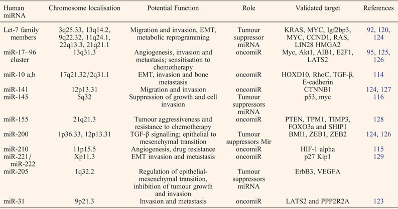
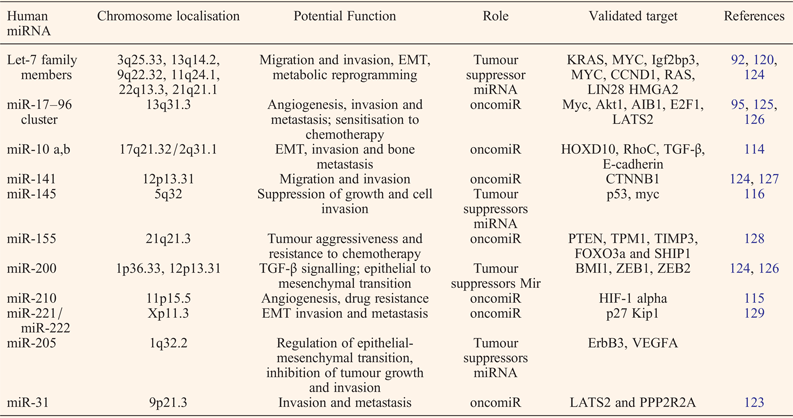
MiRNAs play a key role in TNBC pathogenesis through their ability to regulate fundamental pathways, such as cell growth and proliferation, angiogenesis and invasion (Refs Reference Catana, Calin and Berindan-Neagoe111, Reference Berindan-Neagoe and Calin112). MiRNA expression patterns contribute to the phenotypic diversity of TNBC (Refs Reference Danza64, Reference Braicu113). Recent miRNA profiling studies have identified miRNAs that are generally associated with carcinogenesis or activation of angiogenesis and metastasis (miR-21, miR-17–92 cluster and miR-10a/b). Some miRNAs are highly expressed in breast cancers (miR-210, let-7 and miR-143), whereas others are specific to TNBC (Table 1).
MiR-210 is a direct regulator of HIF-1α; it is also directly correlated with early angiogenic signals and overexpressed in TNBC. Moreover, miR-210 overexpression is linked with treatment recurrence and poor prognosis and it has been proposed as an independent prognostic factor for selecting the optimal adjuvant therapy (Ref. Reference Toyama115).
A recent study has demonstrated that miR-145 is correlated with cellular invasion; however, it does not affect cell proliferation and apoptosis. Instead, it influences invasion and migration mechanisms (Ref. Reference Eades116). MiRNAs related to angiogenic processes are presented in Table 2.
Examples of relevant miRNAs involved in TNBC, particularly those related to angiogenesis
lncRNAs are sequences of up to 200 nucleotides in length or even up to 15 kilobases (Ref. Reference Liu117). There have been an increased number of studies in recent years presenting the possible implications of lncRNA in TNBC (Refs Reference Liu117, Reference Jiang118, Reference Lv119). However, lncRNAs do not appear to be directly related to invasion and metastasis mechanisms. One of the first lcRNAs connected to cancer was the H19 gene, which has also been found to be overexpressed in TNBC (Ref. Reference Yan120). However, the most well-studied lncRNA is HOTAIR, which is overexpressed in BL and in certain HER-2-enriched subtypes of TNBC (Ref. Reference Lee121).
The LINK-A lncRNA has been found to activate HIF1-α signalling in the development of normoxic status with TNBC patients (Ref. Reference Lin122). LincRNA-RoR is significantly overexpressed in TNBC and has been demonstrated to regulate miR-145 via targeting of the small GTPase ADP-ribosylation factor 6 (Arf6). The lincRNA-RoR/miR-145/ARF6 pathway has also been connected with invasion and metastasis in TNBC (Ref. Reference Eades116). LOC554202 was observed to be downregulated in the basal TNBC subtype and overexpressed in the luminal subtype. LncRNA LOC554202 is associated with miR-31 expression and hypermethylation of its promoter-associated CpG island (Ref. Reference Augoff123).
Preclinical and clinical studies targeting angiogenesis in TNBC
Control of tumour angiogenesis is a widely agreed-upon therapeutic direction in cancer therapy. For example, one study used magnetic iron oxide nanoparticles (IONPs) conjugated with ICAM-1 antibodies (ICAM–IONPs) (Ref. Reference Guo86). Effective targeting of TNBC cells via the ICAM–IONPs leads to reduced cell proliferation and migration, which has been validated alongside antiangiogenic effects in animal models (Ref. Reference Guo86).
A study found that use of siRNA therapy targeting neutrophil gelatinase-associated lipocalin (NGAL, Lnc2) was related to decreased VEGF expression, leading to an inhibition of angiogenesis in both in vitro and in vivo models. Bortezomib, a proteasome inhibitor, is able to modulate multiple pathways in cancer cells. Bortezomib was able to induce apoptosis in a mechanism independent of proteasome inhibition in TNBC cells, but not in normal or double positive breast cancer cells (Ref. Reference Tseng130). Our studies have also demonstrated that p53 siRNA therapy diminishes cell proliferation and migration and activates apoptosis in TNBC cells (Ref. Reference Berindan-Neagoe, Braicu and Irimie131).
The central regulator for tumour angiogenesis is VEGFA, which binds to the tyrosine kinase (TK) receptors VEGFR1 and VEGFR2. Bevacizumab is a monoclonal antibody against VEGFA (Ref. Reference Bayraktar and Gluck132). The therapeutic use of bevacizumab in TNBC, in combination with taxanes, has been assessed in two phase III clinical trials (AVADO and ECOG2100). The combination of bevacizumab with docetaxel (in the AVADO trial) and with paclitaxel (in the ECOG trial) has been associated with enhanced progression-free survival (PFS) rates, compared to the use of single-taxane regimens (Ref. Reference Miller133). RIBBON-1 and -2 are phase III trials with the goal of assessing the efficacy of bevacizumab in combination with capecitabine, anthracyclines and taxanes. The combined use of capecitabine and bevacizumab has significantly increased PFS rate, whereas combinations of bevacizumab with anthracyclines/taxanes have been associated with less favourable outcomes (Ref. Reference Bayraktar and Gluck132).
The BEATRICE clinical trial was a multinational phase III study that led to unsatisfactory results because of a failure to augment adjuvant chemotherapy in early-stage TNBC (Ref. Reference Cameron134). Two additional clinical trials, GeparQuinto and NSABP-B40, are currently assessing the effect of bevacizumab in combination with neoadjuvant therapeutic agents (such as anthracyclines and taxanes) (Ref. Reference Miller133). TKs are cell-surface receptors that play a critical role in angiogenesis. The effects of TKs on the proliferation rate of endothelial cells have previously been assessed in TNBC. Sunitinib and sorafenib are TK inhibitors that target VEGFR2, PDGFRβ, FLT3 and c-kit. Randomised trials with sunitinib (alone or in combination with sorafenib) have shown no significant results (Refs Reference von Minckwitz135, Reference Spanheimer136). Although sorafenib has shown modest activity as a single agent, the combination of vinorelbine and sorafenib may successfully treat particular subtypes of TNBC (Ref. Reference Luu137). In combination with other chemotherapeutic agents, sorafenib treatment has been associated with improvement of overall survival rates in three phase IIb trials. In association with capecitabine, sorafenib showed a modest improvement in median PFS rate (Ref. Reference Baselga138). Icrucumab (IMC-18F1) targets VEGFR1, thereby preventing the binding of all other ligands to this target. It is a monoclonal antibody that also blocks placental growth factor (PLGF), which is its specific ligand (Ref. Reference LoRusso139).
The antimicrotubule agent noscapine, when used in combination with doxorubicin, inhibits angiogenesis in TNBC cell lines by acting on the NFκB pathway (Ref. Reference Chougule140). Propranolol, in combination with 5-FU and paclitaxel, was an effective adjuvant for antiangiogenic therapy in a mouse orthotopic model of TNBC (Ref. Reference Pasquier141). Ketorolac is a nonsteroidal anti-inflammatory drug that improved the post-operative survival rate in patients with breast cancer by interfering with the SDF1–CXCR4 pathway through sequential inhibition of COX-2, PGE and SDF-1. However, ketorolac acts by preventing recruitment of bone marrow-derived cells and the formation of new blood vessels. Further studies on TNBC subtypes are needed in order to validate this finding (Ref. Reference Chougule140).
Glycoprotein non-metastatic b (GPNMB) has also been identified as a novel gene with increased expression level in several cancer types, including TNBC (Ref. Reference Maric59). This metastatic mediator has been tested as a therapeutic target in TNBC. CDX-011 is an anti-GPNMB-OA antibody, and its efficacy is correlates with the levels of GPNMB-OA on the cell surface (Ref. Reference Witters142).
The protease ADAM10 represents another potential therapeutic target. Blockage of this enzyme inhibits cleavage of the GPNMB-OA ECD, thereby preventing migration of endothelial cells. INCB7839 is an inhibitor of ADAM10 and has been shown to act synergistically with TK receptor inhibitors targeting EGFR in breast carcinomas (Ref. Reference Witters142). EGFR inhibitors are a group of chemotherapeutic agents that have been successfully used to treat TNBC. Thus, the combination of INCB7839 and TK inhibitors may represent a promising option for TNBC therapy; however, further clinical trials are required (Ref. Reference Duffy, Crown and Mullooly63). Use of the leptin receptor antagonist peptide Allo-aca extended survival time by 80% in a mouse model for TNBC (Ref. Reference Otvos143). Preclinical trials proved the efficacy of this leptin agonist, but these results still need to be validated in humans (Ref. Reference Otvos143).
Another research direction includes the investigation of PI3K inhibitors, alone or in combination. One example is BEZ235, a dual PI3K/mTOR and MEK inhibitor that has been tested in a limited number of phase I/II clinical studies. Temsirolimus, another molecule that targets the mTOR pathway, is being explored in a phase I/II clinical trial. Importantly, mTOR inhibitors are also being investigated for metastatic TNBC and, in combination with anthracyclines and taxanes, in a phase III randomised neoadjuvant study (Ref. Reference Crown, O'Shaughnessy and Gullo144). VEGFC is overexpressed in the MSL subtype of TNBC, which indicates that this category of patients will respond to VEGFC inhibitors (Ref. Reference Hein145). Blocking endosome traffic, combined with EGFR inhibitors, may result in an increased therapeutic response for TNBC patients (Ref. Reference Williams146). The therapeutic targets in the angiogenesis pathway in TNBC are in Figure 4.
Inhibitors of angiogenesis in TNBC. The central regulator of tumour angiogenesis is VEGF, which binds to two TK receptors: VEGFR1 and VEGFR2. Bevacizumab is a monoclonal antibody that targets VEGF. CAPE and EGCG are two natural phytochemicals that target the VEGF pathway. Icrucumab (IMC-18F1) is a monoclonal antibody that targets VEGFR1, thereby preventing binding of all other ligands, including PLGF, to this target. Ramucirumab is a monoclonal antibody that targets VEGFR2. Sutinib and sorafenib are TK inhibitors that target VEGFR1 or VEGFR2, PDGFRβ and c-kit. Imatinib targets PDGFRβ and c-kit. GPNMB and ADAM10 are targeted by CDX-011 and INCB7839, respectively. Candesartan blocks angiotensin receptor AT1. Cetuximab inhibits the EFGR pathway, another important target interconnected with integrins and p63 pathways. The angiogenesis pathways are directly connected with the NFκB pathway or the chemoattraction capabilities of endothelial cells and VM.

Other studies are focused on peptide therapy as an alternative anti-angiogenic treatment for TNBC. One example, which has been validated in vitro and in vivo, is the SP2024 peptide that is derived from the noncollagenous domain of the α5 fibril of collagen IV. This compound exhibited a significant reduction with tumour growth in a xenograft model of TNBC, which makes it an attractive therapeutic agent either as a monotherapy or in combination with other agents (Ref. Reference Rosca147).
Alternative treatments targeting angiogenesis
Alternative remedies are widely used among patients with breast cancer. Among these patients, 63–83% use at least one alternative treatment and 25–63% use herbs and vitamins. A wide range of phytochemicals have been shown to reduce cell proliferation or to efficiently inhibit migration and invasion in TNBC in in vitro and in vivo models, with most of the beneficial effects being attributed to inhibition of MAPK-related pathways and antiangiogenic effects (Ref. Reference Ham148).
Propolis is one of the most widely used nutraceutical alternative therapies. One of its active components, caffeic acid phenethyl ester (CAPE), has been shown to inhibit angiogenesis in the MDA-MB-231 cell line by decreasing the secretion of VEGF and the formation of capillary-like tubes (Ref. Reference Wu149). Curcumin has also been shown to inhibit pro-angiogenic factors in a xenograft model of breast cancer (Ref. Reference Ferreira150). In addition, 1,2,3,4,6-O-penta-O-galloyl-β-D-glucose (PGG), a component of natural herbal products, has demonstrated inhibition of angiogenesis in TNBC by inhibiting the expression of STAT3, which is known to regulate VEGF levels in MDA-MB-231 cells (Ref. Reference Lee151). Although the molecular mechanisms have not yet been elucidated, earlier investigations have shown that green tea extracts may suppress cancer progression by blocking angiogenesis. Our recent studies show that epigallocatechin-gallate (EGCG) induces cell death and apoptosis, and inhibits proliferation and migratory behaviour in TNBC cells. It has also been suggested that EGCG regulates the angiogenic switch, tilting the balance in favour of anti-angiogenic factors and leading to modulation of cell migration (Ref. Reference Braicu152).
Conclusions
Recently, there has been a significant increase in interest regarding TNBC because of its extremely invasive behaviour and lack of response to current therapies. TNBC represents a heterogeneous group of diseases, divided into several distinct subtypes, with each displaying various molecular alterations. Angiogenic markers may have particular importance as diagnostic, prognostic and/or therapeutic targets for TNBC. Although multiple therapies with promising results have been tested, the future of TNBC treatment will be characterised by personalised therapy based on the development of novel array or sequencing methods, which will substantially contribute to a deeper understanding of this pathology. However, in spite of encouraging preclinical studies targeting angiogenesis, most of the current potential therapies are unable to meet the statistical criteria for efficacy in clinical trials. Consequently, it remains imperative to develop and validate novel and efficacious molecular targets for new TNBC therapies.
Acknowledgements
This study was supported by a POSCCE grant (709/2010, BREASTIMPACT).
Conflict of interest
None.