Congenital coronary artery anomalies are not uncommon, occurring in 1.3% – ranging from 0.3% to 5.6% – of the population.Reference Kayalar, Burkhart, Dearani, Cetta and Schaff1, Reference Angelini2 Most coronary artery anomalies do not cause myocardial ischaemia and are often found incidentally during angiographic evaluation for other cardiac diseases; however, approximately 20% of those anomalies may have a potential for life-threatening presentation.Reference Kayalar, Burkhart, Dearani, Cetta and Schaff1
Coronary artery fistula is an abnormal connection between a coronary artery and the pulmonary artery, coronary sinus, cardiac chamber, or systemic circulation. This defect can occur at any age and accounts for 0.27–0.40% of all congenital cardiac diseases,Reference Sunder, Balakrishnan and Tharakan3 with angiographic series revealing an incidence of 0.3–0.8%.Reference Kim, Song, Cho and Kim4 The single coronary artery is an even more rare anomaly and it is reported to occur in 0.04–0.19% of congenital cardiac anomaliesReference Ishii, Suzuki, Kobayashi and Arakawa5 and occurs when one coronary artery is absent and the others supply to the entire heart regardless of its distribution.
We report a case of single left coronary artery with an absence of the ostium for the right coronary artery. The course from the distal right coronary artery is in continuity with collateral vessels arising from the distal left circumflex artery and left anterior descending artery. This anomalous vessel is draining into the right ventricle.
Case report
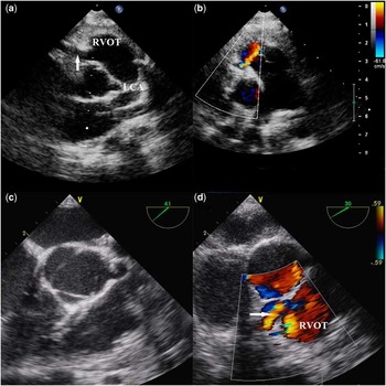
A clinically asymptomatic male child aged 4 years and 5 months with a continuous murmur was referred to our hospital. He had no significant illness before a heart murmur being identified in a routine medical examination. His body weight was 17.8 kilograms with a height of 102 centimetres. Auscultation of the chest disclosed a grade 3/6 continuous murmur along the left sternal border. The remainder of the physical examination was unremarkable. An electrocardiogram showed sinus rhythm, normal axis and intervals, and no ST-segment or T-wave changes. An echocardiogram showed prominent and dilated left coronary artery and did not identify the right coronary artery. Probable collateral vessels were seen in the ventricular septum and right ventricle free wall. Colour Doppler showed continuous flow in the right ventricular outflow tract below the pulmonary valve (Fig 1). There was no regional wall motion abnormality, and ventricular function was normal.
(a–b) Transthoracic echocardiography: prominent and dilated left coronary artery (LCA), and vessel (arrow) which ends in right ventricular outflow tract (RVOT). Colour flow imaging show an abnormal jet in the RVOT below pulmonary valve. (c–d) Transesophageal echocardiogram show the same.

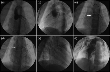
On cardiac catheterisation, the intracardiac and intravascular pressures were normal. Oxygen saturation did not show a step-up in the right ventricle, and the pulmonary/systemic flow ratio was 1:1. A selective left coronary angiogram showed a prominent left main coronary artery, a normal distribution of the left anterior descending artery, and circumflex artery. The left main coronary artery, with a 5-millimetre diameter, arose from the appropriate sinus without aneurysm or stenosis. After a slight delay, the posterior descending and right coronary artery opacified in a retrograde manner through collaterals of the left coronary system. The right coronary artery drained into the outflow portion of the right ventricle. A right coronary angiography was attempted, but the coronary ostium could not be found (Fig 2).
(a–b) Coronary arteriogram showed a coronary artery arised from the left coronary sinus. A dilated left coronary artery was saw. It gave off a normal appearing left anterior descending branch and normal distribution of the circumflex artery. No right coronary artery was visualized. (c–e) Selective left coronary artery injection showed retrograde filling of right coronary artery through collateral from left coronary system. This artery follows the course normally occupied by the right coronary artery. This anomalous vessel terminated in the outflow tract of the right ventricle (arrows). (f) The injection on the right ventricular outflow tract does not demonstrate the fistula origin.

A diagnosis of a coronary artery–right ventricle fistula was made and surgical correction was recommended. At operation, there was a single coronary artery originating from the aorta. This coronary artery was very dilated and tortuous between its origin from the aorta and insertion into the right ventricle. Under cardiopulmonary bypass, the site of entry of the fistula into the right ventricular cavity was confirmed before cross-clamping the aorta. Then, under cardioplegic arrest, the fistula was closed at the point where it drained into the right ventricle with continuous suture reinforced con Teflon (DuPont, Wilmington, Delaware, United States of America). The right ventricular cavity was checked and no residual leak was found. The post-operative and clinical courses were uneventful.
Discussion
Few cases of single coronary artery and coronary artery fistula have been reported so far.Reference Kayalar, Burkhart, Dearani, Cetta and Schaff1 Only 35 cases have been described in the literature. Of these, 19 patients provide sufficient information about the type of single coronary artery and coronary artery fistula, and the treatment received.Reference Kayalar, Burkhart, Dearani, Cetta and Schaff1
It is difficult to estimate the true incidence of coronary artery fistula because of its typically asymptomatic course in childhood, and the diagnosis is frequently made after the auscultation of a continuous murmur.Reference Sunder, Balakrishnan and Tharakan3 The chest radiography typically appears normal. The electrocardiogram is usually normal or may show non-specific abnormalities depending on the size and location of the coronary artery fistula and the degree of ischaemia due to coronary steal.
Cardiovascular ultrasonic diagnosis is an important method of determining coronary artery fistulas, although imaging of coronary arteries and the identification of the site of drainage may be difficult. Coronary angiography is the gold standard for determining coronary artery fistula, but it may fail to depict the drainage site of the coronary artery fistula or its relation to other structures. Magnetic resonance imaging helps in confirming the diagnosis, as the proximal coronary arteries and the entire length of the fistulous vessel can usually be well visualised. Recently, multidetector-row computed tomography cardiac imaging has provided excellent distal coronary artery and side branch imaging, and can be used for the assessment of coronary anomalies. The spatial resolution is superior to that of magnetic resonance imaging, but the exposure to radiation on multidetector computed tomography is not entirely risk free.Reference Zenooz, Habibi, Mammen, Finn and Gilkeson6 Perhaps in our case the realisation of one of these techniques have helped us to define a more precise location of the fistula drainage.
The natural history of coronary artery fistula is a progressive dilatation of the coronary artery to varying degrees depending on the shunt volume, accompanied by worsening of symptoms due to increased coronary steal. Spontaneous closure of small fistulae may occur in 23% of patients.Reference Sunder, Balakrishnan and Tharakan3 We know that the most common origin is the right coronary artery and a single termination is found in the majority of patients. The right ventricle is the most common site of drainage – in 45% of cases – followed by the right atrium – in 25% – and the pulmonary artery – in 15% of the cases.
Fistulae may cause arrhythmias, congestive cardiac failure, or aneurysm formation and are the sites of endocarditis in approximately 5% of cases. Myocardial ischaemia may develop in the portion of the myocardium supplied by the abnormally connecting artery owing to myocardial steal phenomenon or decreased myocardial blood flow distal to the fistulous connection.
With regard to surgical treatment, whether to operate on coronary artery fistula in childhood is controversial. Symptoms and complications are infrequent in children unless they have an additional cardiac anomaly and selective angiography that allows detection of a single coronary artery associated with coronary artery fistula in most patients during childhood. Adult patients often have symptoms, the incidence of congestive cardiac failure and angina increases with age, and they suffer from complications.
On the basis of these complications, there is a general agreement that closure of the fistula is desirable early in childhood regardless of the symptoms. This view is favoured because of the safety and efficacy of the operation and the low probability of spontaneous closure.Reference Ishii, Suzuki, Kobayashi and Arakawa5, Reference Hsieh, Huang and Lee7
Trans-catheter embolisation is an option for the treatment of coronary artery fistula. Embolisation coils, or recently developed cardiac devices, can be delivered directly into the fistula. After occlusion of the distal fistulous connection, complete thrombosis of the coronary artery fistula occurs.Reference Zaho, Jin and Wu8 In our case, we declined an interventional approach, and surgical repair was performed: first, because the anomalous vessel draining into the right ventricle was very difficult to cannulate due to the extreme tortuosity; second, and most importantly, because of the presence of a single coronary artery.
This type of pathology is important to describe because, in the revision of this kind of disease, this anomaly is very rare. The mortality and morbidity associated with surgical closure of coronary artery fistula is probably lower than the risk of complications developing after 20 years of age. Accordingly, we recommend elective surgical repair of the fistula during childhood in patients who have single coronary artery combined with coronary artery fistula.