Introduction
The family Steinernematidae includes more than 80 described species, the morphology of which has been established primarily by male and larval characters, such as the shape of spicules, position of excretory pore, and length of tail and hyaline layer portion in infective juveniles (Ijs). A lack of taxonomically important characters has been reported for steinernematid females (Hominick et al., Reference Hominick, Briscoe, del Pino, Jian, Hunt, Kozodoy, Mráček, Nguyen, Reid, Spiridonov, Stock, Sturhan, Waturu and Yoshida1997). Generally, nematode tails may be modified terminally by cuticular projections called mucrones (Maggenti, Reference Maggenti1981), sometimes also referred to as protuberances. Only such structures which are offset from the tail contour can be considered to be projections. Similarly, in steinernematids, several recently published species descriptions have reported minute tail projections on the tail tips of females of the first generation. Nguyen & Duncan (Reference Nguyen and Duncan2002) described five papillae-like structures (four visible in their Fig. 4D) on the tail tips of the first-generation females of Steinernema diaprepesi. Triggiani et al. (Reference Triggiani, Mráček and Reid2004) found similar projections on the tail tips of Steinernema apuliae, one large and two or three minute projections. All these observations have come from recently published papers, but the question remains whether such structures also occur in previously described species and whether they have a constant value. Therefore we focused our efforts on the study of steinernematid female tails and the clarification of any morphological differences in the tail projections among species.
Materials and methods
Seven steinernematid species were chosen for our study: Steinernema arenarium (Artyukhovsky) strain GELAN, S. carpocapsae (Weiser) strain Agriotos, S. ichnusae Tarasco, Mráček, Nguyen & Triggiani type strain, S. intermedium (Poinar) US strain, S. jollieti Spiridonov, Krasomil-Osterfeld & Moens type strain, S. longicaudum Shen & Wang China strain and S. kraussei SK89, topotype strain. The nematodes were reared on Galleria mellonella larvae. Cadavers containing the first-generation nematodes were dissected and females subsequently taken out and washed twice in Ringer's solution. The females of each species were divided into two batches, each of at least ten specimens. One batch was fixed in hot TAF (triethanolamine, formalin and water) (Southey, Reference Southey1970), transferred to glycerin by a rapid method (Seinhorst, Reference Seinhorst1959) and mounted in anhydrous glycerin on permanent mounts. For image capturing by light microscopy (LM) the Zeiss Axioplan 2 photomicroscope with DIC optics was used (Zeiss, Jena, Germany). The other batch was fixed in a 2.5% glutaraldehyde solution in a 0.2% cacodylate buffer at pH 7.2 for 24 h, kept at 4–6°C in a refrigerator, and post-fixed in 2% osmium tetroxide at room temperature for 4 h. After dehydration in graded alcohol, acetone/alcohol and an acetone series, desiccation at critical point and subsequent gold coating, the mounts were examined under a JEOL scanning electron microscope (Eching, Germany) (Mráček et al., Reference Mráček, Weiser and Gerdin1981) for scanning electron microscopy (SEM).
Results and discussion
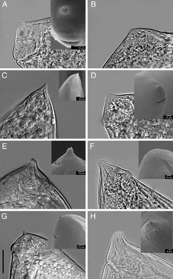
Steinernema jollieti has three equally protruding spine-like projections, either on the peg-like (younger specimen) or simply rounded (older specimen) tail tip (fig. 1A, B) whereas in S. ichnusae, spine-like projections (the middle of the three being more prominent) protrude slightly from a conical, pointed tail (fig. 1C). Steinernema longicaudum and S. arenarium possess characteristic peg-like tail tips with three spine-like projections, the more prominent of them being positioned dorsocentrally (fig. 1E, H). Steinernema kraussei possesses a conical tail with a longer filamentous projection on the tail tip and two minute papilla-like projections laterally (fig. 1D). In the present observations, S. intermedium is characterized by the peg on the tail bearing two minute dorsolateral papilla-like projections and one central, more prominent spine-like projection (fig. 1F). The tail of S. carpocapsae carries three projections. Of these, two minute papilla-like projections are positioned dorsolaterally with a short filament on the terminal papilla tip (under LM), whereas, in SEM, the terminal papilla-like projection is partly deformed (fig. 1G).
Tail projections in first-generation females of seven species of Steinernema; light microscopy and scanning electron microscopy (shown as insets in the top right-hand corners). (A, B) S. jollieti: (A) old and (B) young; (C) S. ichnusae; (D) S. kraussei; (E) S. longicaudum; (F) S. intermedium; (G) S. carpocapsae; (H) S. arenarium. Scale bar = 40 μm (A–D), 80 μm (E) or 55 μm (F–H).

All studied first-generation females of seven steinernematids have mammilate tail-bearing projections. These species represent four phylogenetic clades: kraussei (S. kraussei, S. jollieti and S. ichnusae), glaseri (S. longicaudum and S. arenarium), affine (S. intermedium) and carpocapsae (S. carpocapsae). Generally, these species can be divided into three basic types, all with a convex–conoid tail, often becoming broadly rounded in older specimens (fig. 1A, B). The first type has a conical tail (fig. 1C, D); the second, a blunt peg-like tail tip (fig. 1E, F, H); and the third has a simply rounded tail (fig. 1G). The projections do not vary in number. All species have three projections, but their shape varied from papilla-like to spine-like (fig. 1A–H). The number and position seem to be constant for each species. If the projections are not equally protruding, the most prominent projection is situated on the tail tip whereas the smaller two are located subventrally (fig. 1D, E). Under LM some sublateral projections may be overlooked if they do not protrude from the body contour, being overlapped by the nematode tail body. SEM preparations enable more precise determination of the number and position of the projections, but the mounting procedure may deform the shape of the projection (fig. 1G).
Though published descriptions of the morphology of tail cuticular structures have been mostly incomplete, or even completely lacking, the tail shapes and projections in the first-generation females of the family Steinernematidae have been mentioned in descriptions of several species. For example, Poinar (Reference Poinar1986) found wedge-like projections on the tail tips of the first-generation females of S. intermedium which, in fact, represent the peg. In Nguyen et al. (Reference Nguyen, Hunt, Mráček, Nguyen and Hunt2007), the authors reported that the tail of this species bears 2–4 minute papilla-like protuberances, whereas our light microphotography reveals three small spine-like projections, two dorsolaterally and one more prominent centrally (fig. 1F). Similarly, S. longicaudum was characterized by a rounded tail with one to several small projections at the terminus (Nguyen et al., Reference Nguyen, Hunt, Mráček, Nguyen and Hunt2007), while our observation identified three projections in all the slides (fig. 1D, E). The same authors have described the tail of S. carpocapsae as conical with or without a short spine on the tip, whereas in our LM and SEM observations three papilla-like projections with a short filament on the terminal papilla tip were found (fig. 1G). In the majority of steinernematid species, the tail projections in the first-generation females have not yet been described satisfactorily. For example, the tail of S. silvaticum Sturhan, Spiridonov & Mráček was characterized as broadly rounded, with only a slight terminal projection (Sturhan et al., Reference Sturhan, Spiridonov and Mráček2005), but observations of the real number and shape of the projections must be completed.
It seems that there are distinct differences among species of the family Steinernematidae in the tail morphology of the first-generation females. Projection(s) are probably always present. In our observations, they vary in their shape and protuberance, whereas their number is constant at three. The variety of projection shapes in our study supports the hypothesis that these structures may have taxonomic significance at the species level. Therefore, every description of a new species in the family Steinernematidae should contain a precise morphology of the female tail, focusing on these projections, using both light and scanning electron microscopy. Some older and recently described species should be revised for these structures, which may help significantly in easier identification of steinernematid species.
Financial support
The study was supported by the Czech Ministry of Education, Youth and Sport and Amvis in the programme KONTAKT II, project LH 12 105.
Conflict of interest
None.