INTRODUCTION
Marine tardigrades are found from various types of sediments from the deep sea to the coast. Adaptations to these sediments lead to their highly diversified morphology in contrast to their relatively simple freshwater and terrestrial siblings (Grimaldi de Zio et al., Reference Grimaldi de Zio, D'Addabbo Gallo and Morone De Lucia1987). The species described here is a member of Tanarctus Renaud-Debyser, 1959, a genus with very long leg appendages.
Tanarctus consists of eleven species and, from the literature, most are from the Atlantic where further species are expected (Hansen et al., Reference Hansen, Jørgensen and Kristensen2001; Jørgensen & Kristensen, Reference Jørgensen and Kristensen2001). In comparison, records of Tanarctus from the Pacific are scarce: T. velatus from the Galapagos Islands (McKirdy et al., Reference McKirdy, Schmidt and McGinty-Bayly1976), three undescribed species from Australia (Boesgaard & Kristensen, Reference Boesgaard and Kristensen2001), and a Tanarctus sp. from Okinawa, Japan (Noda, Reference Noda1994). Recently Suzuki (Reference Suzuki2010) reported T. cf. helleouetae and Tanarctus sp. from Shimabara Bay, Japan. One of the present authors (S.F.) collected additional specimens of Tanarctus during a survey of marine tardigrade fauna at Tanabe Bay, Japan. Detailed comparison of specimens from both localities revealed they belong to a single taxon, which we are describing here as a new species.
MATERIALS AND METHODS
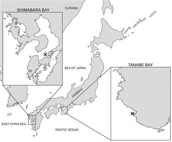
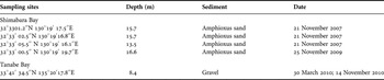
Sediment samples were collected from four sub-tidal sites at Shimabara Bay, Kumamoto, using a Smith–McIntyre grab, and from a sub-tidal site at Tanabe Bay, Wakayama, by diving (Figure 1; Table 1).
Map of the sampling localities.

List of sampling localities.

The collected samples were treated by freshwater shock technique (Kristensen, Reference Kristensen1983), and the supernatants were decanted through a 32 µm mesh net. Shimabara Bay samples were preserved in 4% buffered formaldehyde in seawater with rose Bengal, and tardigrades were sorted under a stereomicroscope and mounted on slides in glycerol. Tanabe Bay samples were sorted and observed live under a stereomicroscope prior to fixation with ethanol series or fixed in 3% formaldehyde in fresh water and then sorted under a stereomicroscope. They were mounted in Hoyer's medium or glycerol. Tardigrade specimens were observed with a differential interference contrast microscope (Nikon ECLIPSE E800 or Olympus BX51).
RESULTS
SYSTEMATICS
Order ARTHROTARDIGRADA Marcus, 1927
Family HALECHINISCIDAE Thulin, 1928
Subfamily TANARCTINAE Renaud-Mornant, 1980
Genus Tanarctus Renaud-Debyser, 1959
Tanarctus diplocerus sp. nov.
(Figures 2–6; Tables 2&3)
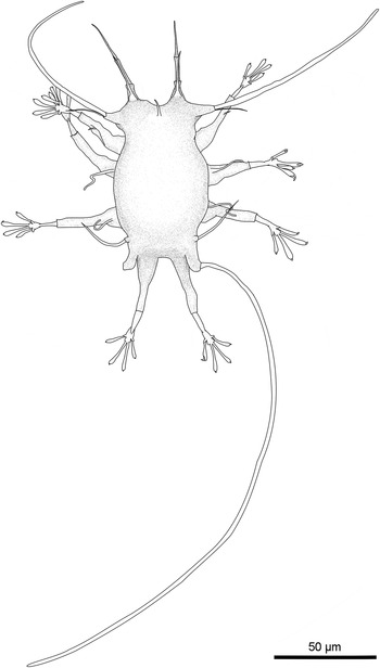
Tanarctus diplocerus sp. nov.: dorsal view of the holotypic female.

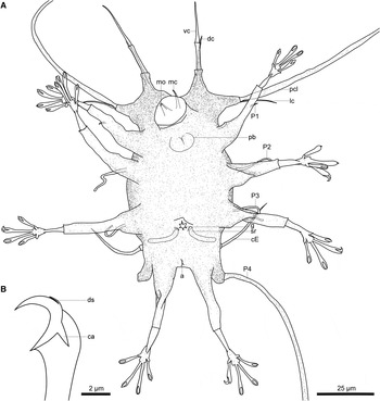
Tanarctus diplocerus sp. nov. holotypic female: (A) ventral view; (B) a claw of the internal digit. a, anus; ca, calcar; cE, cirrus E; dc, dorsal cirrus; ds, dorsal spur; g, gonopore; lc, lateral cirrus; mc, median cirrus; mo, mouth opening; pb, pharyngeal bulb; pcl, primary clava; P1-4, legs I–IV appendages; sr, seminal receptacles; vc, ventral cirrus.

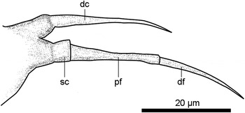
Tanarctus diplocerus sp. nov.: lateral view of an adult female's dorsal cirrus and ventral cirrus. The dorsal cirrus and the ventral cirrus are situated with no internal–external relation. dc, dorsal cirrus; df, distal flagellum of the ventral cirrus; pf, proximal flagellum of the ventral cirrus; sc, scapus of the ventral cirrus.

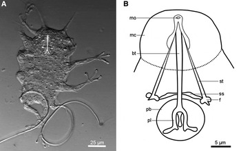
Tanarctus diplocerus sp. nov.: bucco-pharyngeal apparatus observed in a specimen mounted in water. (A) Photomicrograph; (B) drawing. f, furca; mc, mouth cone; mo, mouth opening; pb, pharyngeal bulb; pl, placoid; ss, stylet support; st, stylet.

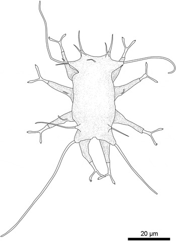
A probable two-digit juvenile of Tanarctus diplocerus sp. nov.: dorsal view. Some of the leg appendages were not observable. The flagellum of the ventral cirrus is not subdivided. The leg IV appendages are long unlike Tanarctus helleouetae.

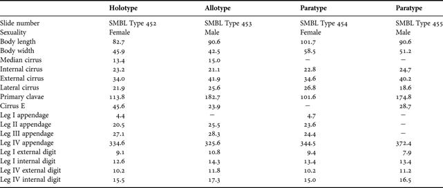
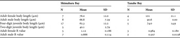
Morphometrics of the type series (in µm). A dash (−) indicates an unmeasured trait.
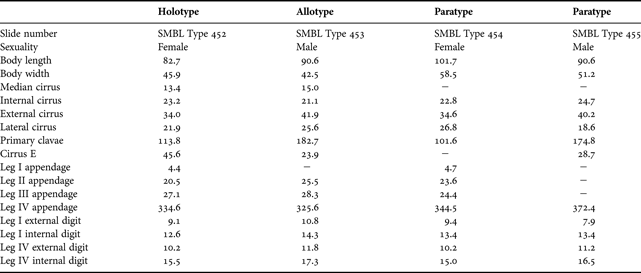
Summary of the specimens collected from the two localities.

N, number; SD, standard deviation.
TYPE MATERIAL
The following specimens collected from Tanabe Bay (33°41′34.5″N 135°20′17.8″E; water depth: 8.4 m) on 14 November 2010 were designated as the type series: the holotype [SMBL Type 452], an adult female; the allotype [SMBL Type 453], an adult male; the paratypes, an adult female [SMBL Type 454] and an adult male [SMBL Type 455]. These materials were mounted in Hoyer's medium. The type series is deposited in the Seto Marine Biological Laboratory, Kyoto University, Japan.
OTHER MATERIAL
The following additional specimens were examined: seven adult females, eight adult males, 26 four-digit juveniles and six probable two-digit juveniles from Shimabara Bay; two adult females and seven four-digit juveniles from Tanabe Bay.
DIAGNOSIS
Tanarctus with smooth cuticle, the cephalic internal and external cirri arranged in dorsal–ventral alignment, long flexible appendages on legs II and III, simple appendages on leg IV and no club-shaped secondary clavae.
ETYMOLOGY
The specific epithet, diplocerus, means ‘double horned’ in Greek referring to the cephalic morphology of the new species.
DESCRIPTION OF THE HOLOTYPE
Holotype: adult female (Figures 2 & 3A) 79.5 µm long with a maximum width of 45.9 µm. Cuticle smooth. Mouth cone protruded ventrally. Only the mouth opening and the ovoid pharyngeal bulb of the bucco-pharyngeal apparatus were observed due to the orientation of the mouth.
Primary clava (113.8 µm long) and the lateral cirrus (21.9 µm) share a common cirrophorus. The club-shaped secondary clavae was lacking from the cephalic appendages. Median cirrus (13.4 µm) inarticulate and positioned on the dorsal surface at a distance from the anterior margin. Cephalic internal and external cirri arranged in dorsal–ventral alignment with the inarticulate dorsal cirrus (23.2 µm) inserted near the base of the articulate ventral cirrus (34.0 µm). The robust ventral cirrus on a cirrophorus comprises a scapus (3.6 µm), a proximal flagellum (12.6 µm) and a distal flagellum (17.8 µm). The boundary between the cirrophorus and the scapus was indistinct. No eye spots present. Pigmented granules were observed in the cephalic region but disappeared after mounting in Hoyer's medium.
Leg I appendage (4.4 µm) an inarticulate short spine inserted distally. Leg II appendage (20.5 µm) and leg III appendage (27.1 µm) were inarticulate, long, flexible spines. Leg IV appendage (334.6 µm) arises from a cirrophorus (12.5 µm). Cirrus E (45.6 µm) consists of a cirrophorus (5.4 µm) and a flagellum (40.2 µm).
The legs were telescopic with lance-like tibia and conic tarsus. Internal digits were fused proximally forming a pretarsus; external digits with cuticular folds at the base. The external digits were always shorter than the internal digits. Claw of the internal digit with a calcar and a minute dorsal spur (Figure 3B). Claw of the external digit, simple with a calcar.
Female gonopore rosette-like and positioned 18.7 µm anterior to the anus. Two seminal receptacles open laterally to the gonopore.
OTHER REMARKS
Additional information about the bucco-pharyngeal apparatus, the pigmented granules in the cephalic region, the lateral view of the dorsal and ventral cirri (Figure 4), the sexual dimorphisms and the development stages, were obtained from the paratypes and other specimens from Shimabara Bay and Tanabe Bay.
The bucco-pharyngeal apparatus of the new species was observed in an adult female (body length: 102.7 µm) collected from Shimabara Bay (Figure 5). The smooth buccal tube was 21.6 µm long; outside diameter 2.0 µm wide near the mouth opening and 1.6 µm at the posterior end. The pharyngeal bulb was ovoid (major axis: 13.5 µm; minor axis: 11.8 µm) with three hooked placoids. Each placoid had a branch extending externally near the posterior end. The stylet was 18.5 µm long and the furca had a pointed apice adjacent to two rounded apices. The stylet support (5.4 µm) was inserted on the buccal tube 17.2 µm from the mouth opening.
Although the pigmented granules in the cephalic region disappeared in the holotypic female, about ten pigmented granules were visible in specimens mounted in either Hoyer's medium or glycerol.
The simple gonopore of the allotypic male was positioned 9.2 µm anterior to the anus.
The ratio ‘R’ of body length/primary clava length (as proposed by Grimaldi de Zio et al., Reference Grimaldi de Zio, Lamarca, D'Addabbo Gallo and Pietanza1999) was constant in the same sex and indicated secondary sexual dimorphism in the new species (Table 3). For Shimabara Bay specimens the primary clavae of adult males were longer than that of the adult female (P < 0.01, U-test). Tanabe Bay specimens also showed secondary sexual dimorphism, but the sample size was too small to perform the U-test.
The four-digit juveniles were distinguished from the adults by their lack of the gonopore. Two-digit juveniles found in the samples (Figure 6) were tentatively assigned to the new species. These juveniles had simple leg IV appendages, and the positions of the two pairs of cephalic cirri resembled that of the adults and four-digit juveniles of T. diplocerus sp. nov., though the ventral cirrus was inarticulate. Further study is required to confirm the relationship of the two-digit juveniles, e.g. molecular analysis.
Specimens were almost always found with detritus sticking to the periphery, and clear over the dorsal and ventral surfaces. This is similar to the alae of Florarctus Delamare Deboutteville & Renaud-Mornant, 1965, Wingstrandarctus Kristensen, 1984, Raiarctus Renaud-Mornant, 1981 and Actinarctus Schulz, 1935.
DISCUSSION
Differential diagnosis
Tanarctus and Zioella Renaud-Mornant, 1987 species with branched leg IV appendages form a monophylum within the subfamily Tanarctinae Renaud-Mornant, 1980 (Jørgensen & Kristensen, Reference Jørgensen and Kristensen2001). Tanarctus diplocerus sp. nov. forms a paraphyletic group together with the rest of Tanarctus that have simple leg IV appendages, i.e. T. gracilis Renaud-Mornant, 1980, T. heterodactylus Renaud-Mornant, 1980, T. minotauricus Renaud-Mornant, Reference Renaud-Mornant1984, T. ramazzottii Renaud-Mornant, 1975 and T. tauricus Renaud-Debyser, 1959. Compared with these five species, T. diplocerus sp. nov. resembled T. gracilis and T. heterodactylus in lacking the club-shaped secondary clavae. The new species differed from T. gracilis and T. heterodactylus by the presence of a smooth cuticle and from T. heterodactylus by the normally developed external digits. Furthermore, the dorsal–ventral alignment of the external and internal cirri in the new species is unique among the species of Tanarctus.
Topology of the ‘internal’ and ‘external’ cirri
The new species indicates a fluidity in the relationships of the internal and external cirri topology within Tanarctus. Halechiniscus Richters, 1908 is another genus with examples of this intra-generic variation of the cephalic cirri topology (e.g. Halechiniscus tuleari Renaud-Mornant, Reference Renaud-Mornant1979 and H. paratuleari Grimaldi de Zio, D'Addabbo Gallo & Morone De Lucia, Reference Grimaldi de Zio, D'Addabbo Gallo and Morone De Lucia1988 resemble the topology of the family Stygarctidae, which is opposite to the rest of Halechiniscus species with dorsal internal cirri and ventral external cirri). This variation is also apparent in species of the family Coronarctidae described in Renaud-Mornant (Reference Renaud-Mornant and Bertolani1987b). These variations among the taxa suggest there is high plasticity in the relationship of internal and external cirri. Since this plasticity might also exist in the dorsal–ventral and anterior–posterior organization of the cirri, the homology of the cirri must be dealt with prudently. Thus the relative positions of these cirri should be described attentively, as in some papers (Renaud-Mornant, Reference Renaud-Mornant1979, Reference Renaud-Mornant1987a, b; Grimaldi de Zio et al., Reference Grimaldi de Zio, D'Addabbo Gallo and Morone De Lucia1988, Reference Grimaldi de Zio, D'Addabbo Gallo, Morone De Lucia and Troccoli1990; Hansen et al., Reference Hansen, Gallo D'Addabbo and De Zio Grimaldi2003).
Difference between the two localities
While there is an apparent difference in body length between the two localities (Table 3), statistical analysis was complicated by the use of different mounting methods used in the two localities. Differences in body size could be due to the genetic variation between the two localities, but it might also be explained by the different environmental conditions (Kristensen, Reference Kristensen1978).
Secondary sexual dimorphism
Other species for which the secondary sexual dimorphism of the primary clava is reported include: Florarctus asper Renaud-Mornant, 1989, F. cinctus Renaud-Mornant, 1976, F. hulingsi Renaud-Mornant, 1976, F. pulcher Grimaldi de Zio, Lamarca, D'addabbo Gallo & Pietanza, 1999, Wingstrandarctus corallinus Kristensen, 1984, W. crypticus Renaud-Mornant, 1989, Tanarctus bubulubus Jørgensen & Kristensen, Reference Jørgensen and Kristensen2001, T. helleouetae Renaud-Mornant, Reference Renaud-Mornant1984 (the measurements of the holotypic female and the paratypic male in the original description were compared in this study) and T. heterodactylus Renaud-Mornant, 1980. These species have well-developed primary clavae which may be one reason the dimorphism is easily recognized. It is interesting to note that another well-developed trait of Tanarctus, the leg IV appendage, exhibits no dimorphism. Although the dimorphism of the primary clavae has not always held true in Florarctinae Renaud-Mornant, 1982 (Grimaldi de Zio et al., Reference Grimaldi de Zio, Lamarca, D'Addabbo Gallo and Pietanza1999) or in Tanarctinae, it could be considered that the function of the primary clavae is related to mating behaviour.
Leg IV appendage morphology in two-digit juveniles
The only previous report of the two-digit juvenile in Tanarctus is from the original description of T. helleouetae Renaud-Mornant, Reference Renaud-Mornant1984, a Tanarctus species with branched leg IV appendages. The immature leg IV appendage of this taxon was described as a short papilla (Renaud-Mornant, Reference Renaud-Mornant1984). It has been shown that many adult Halechiniscidae and Stygarctidae species have this trait and its distribution pattern on the molecular phylogenetic tree of Jørgensen et al. (Reference Jørgensen, Faurby, Hansen, Møbjerg and Kristensen2010) (withholding the aberrant position of Tanarctus) suggests that the long leg IV appendage in the two-digit juvenile in T. diplocerus could be viewed as an apomorphic trait that evolved within the paraphyletic group with simple leg IV appendages. The assessment of this trait's true value in phylogeny is an open question until the two-digit juveniles in many other species are discovered.
A TAXONOMIC KEY FOR THE SPECIES OF TANARCTUS
A taxonomic key for the twelve species of the genus Tanarctus is provided below:
1
a- Leg IV appendages simple ................ 2
b- Leg IV appendages branched ................ 7
2 (1)
a- Club-shaped secondary clavae present ................ 3
b- Club-shaped secondary clavae absent ................ 5
3 (2)
a- Club-shaped secondary clavae well developed and curved to the dorsal side ................ T. minotauricus
b- Club-shaped secondary clavae normally developed ................ 4
4 (3)
a- Primary clavae longer than body length ................ T. tauricus
b- Primary clavae shorter than body length ................ T. ramazzottii
5 (2)
a- Two pairs of cephalic cirri with no internal–external relation ................ T. diplocerus sp. nov.
b- Two pairs of cephalic cirri with internal–external relation ................ 6
6 (5)
a- Digits normally developed ................ T. gracilis
b- External digits reduced to cuticle outgrowths without claws ................ T. heterodactylus
7 (1)
a- Leg IV appendages terminate in spines ................ 8
b- Leg IV appendages terminate in balloon-like or leaf-like structures ................ 10
8 (7)
a- Leg IV appendages with secondary branches ................ T. longisetosus
b- Leg IV appendages with tertiary branches ................ 9
9 (8)
a- Tertiary branches short and simple ................ T. arborspinosus
b- Tertiary branches long and robust ................ T. dendriticus
10 (7)
a- Leg IV appendages terminate in balloon-like structures ................ T. bubulubus
b- Leg IV appendages terminate in leaf-like structures ................ 11
11 (10)
a- Cirrus E with accordion-like structure; leg III appendages present ................ T. helleouetae
b- Cirrus E simple; leg III appendages absent ................ T. velatus
ACKNOWLEDGEMENTS
We thank Dr Motohiro Shimanaga and Mr Hideyuki Shimazaki of Aitsu Marine Station, Kumamoto University, for their help in collecting Shimabara Bay samples. We also thank Dr Sandra McInnes for reading and refining our manuscript. This work was supported in part by Fujiwara Natural History Foundation (A.C.S.).