Introduction
Juvenile nasopharyngeal angiofibroma is a benign, highly vascular neoplasm.Reference Neel, Whicker, Devine and Weiland1 The lesion is described as primarily occurring in young males aged 10–24 years,Reference Glad and Vainer2 with initial unilateral nasal obstruction, recurrent recalcitrant epistaxis and nasal discharge symptoms. The reported incidence of juvenile nasopharyngeal angiofibroma is 0.4 per million inhabitants per year.Reference Glad and Vainer2
The tumour typically originates from the superior margin of the sphenopalatine foramen and spreads into the submucosal plane.Reference Neel, Whicker, Devine and Weiland1 Despite its benign nature, juvenile nasopharyngeal angiofibroma shows an aggressive growth pattern, especially among the youngest patients, causing the potentially life-threatening complications of haemorrhage and intracranial involvement.Reference Paris, Guelfucci, Moulin, Zanaret and Triglia3 Thus, it should be treated as radically as possible.
Here we present a unique case of bilateral juvenile nasopharyngeal angiofibroma. (Written informed consent was obtained from the patient for publication of this case report and any accompanying images.) It is the first reported case with clinical challenges and, to our knowledge, is the most extensive case of bilateral juvenile nasopharyngeal angiofibroma described in the English literature,Reference Rha, Byun, Kim and Kim4 and only the third case reported in the last 10 years.Reference Wu, Chark and Bhandarkar5
Case report
A 21-year-old male presented with a 6-month history of gradually increasing right-sided nasal stenosis and a 2-month history of increasing left-sided nasal stenosis that was not relieved by nasal decongestant spray. The patient experienced frequent bloody nasal secretions, but only one episode of epistaxis. He had no history of weight loss, fatigue or night sweats.
On physical examination, a closed rhinolalia was noted, and anterior rhinoscopy revealed complete obstruction on the left side by a mass. Nasal fibre-optic endoscopy revealed an obstructive reddish tumour on the right side occluding the nasal cavity from the mid-portion of the middle turbinate. The left-sided tumour was impassable.
The left-sided tumour was biopsied under local anaesthesia as juvenile nasopharyngeal angiofibroma was not initially clinically suspected. The biopsy showed polypoid tissue with central necrosis. Based on this unspecific histology, a freeze-drying biopsy was performed under local anaesthesia; the results indicated the diagnosis of juvenile nasopharyngeal angiofibroma. In both cases, temporary bleeding was managed by nasal packing. No biopsy was taken on the right side because of the suspicion of juvenile nasopharyngeal angiofibroma and risk of subsequent bleeding.
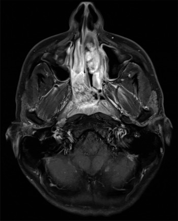
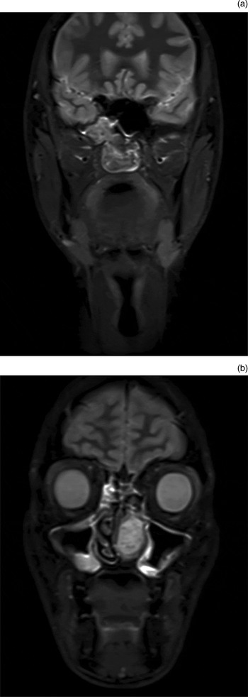
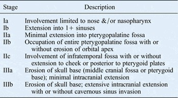
Computed tomography (CT) and magnetic resonance imaging (MRI) scans of the nose and sinuses (Figures 1 and 2) demonstrated a 5 × 3 × 2 cm tumour on the left side and a 4 × 4 × 3 cm tumour on the right side, corresponding to Radkowski et al. stages Ia and IIIa respectively (Table I).Reference Radkowski, McGill, Healy, Ohlms and Jones6 On CT and MRI, the right-sided tumour was in the proximity of the cavernous sinus, and enlargement of the sphenopalatine foramen was pronounced. There were no signs of nasal septum perforation. All laboratory test results were within normal levels.
Pre-operative axial, contrast-enhanced, T1-weighted magnetic resonance imaging scan showing the two independent tumours, sized 4 × 4 × 3 cm on the right side and 5 × 3 × 2 cm on the left side.

Pre-operative coronal, turbo inversion recovery magnitude magnetic resonance imaging scans showing: (a) angiofibroma in the right nasal cavity, extending through the right sphenopalatine foramen and fossa to the right infratemporal fossa; and (b) angiofibroma in the left nasal cavity.

Radkowski and colleagues' staging systemReference Radkowski, McGill, Healy, Ohlms and Jones6

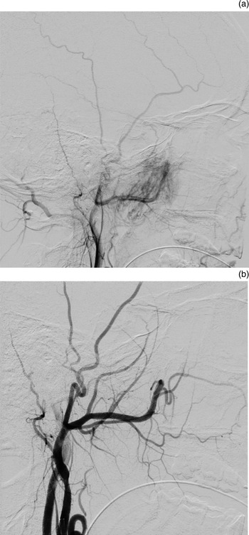
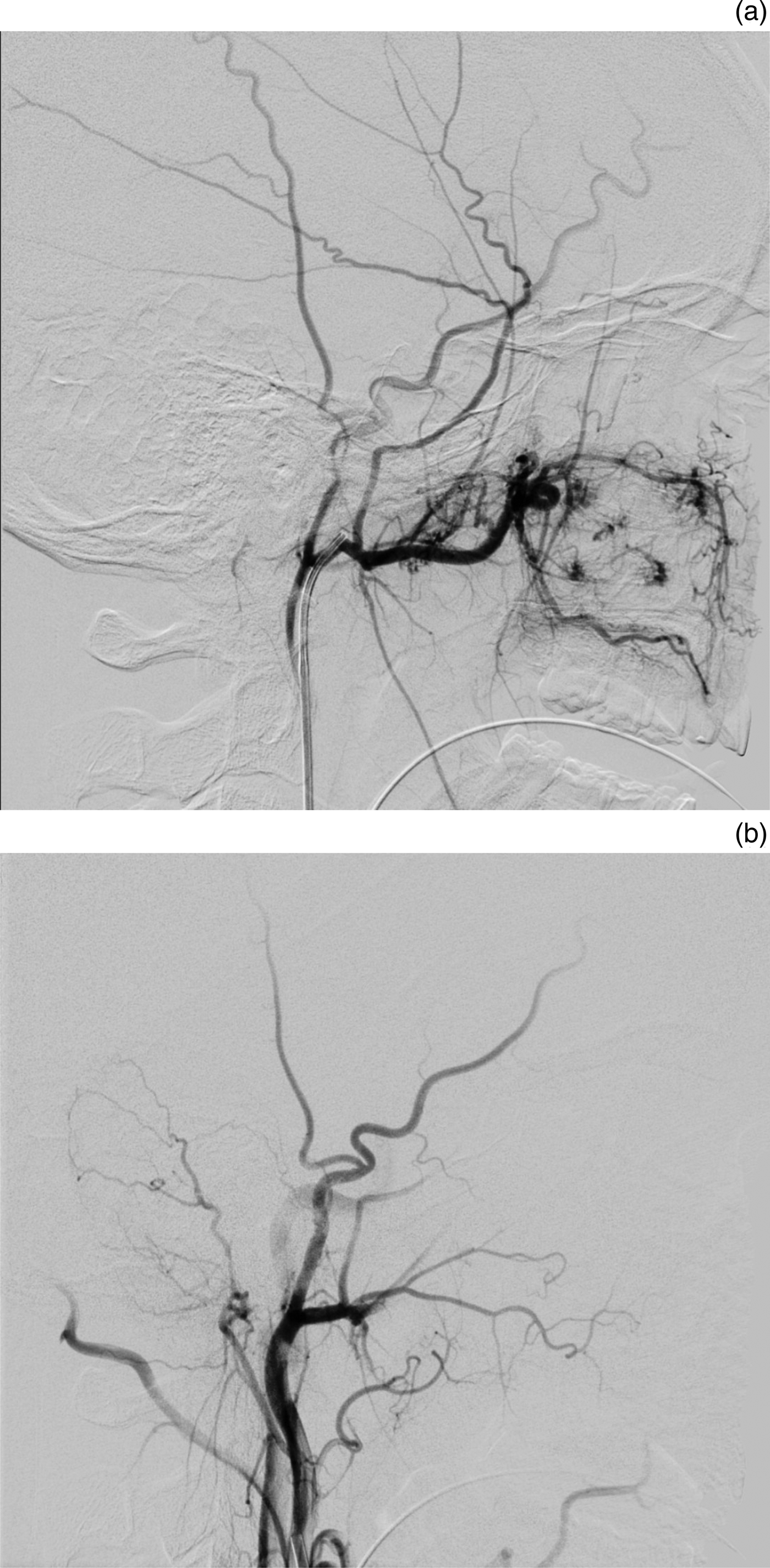
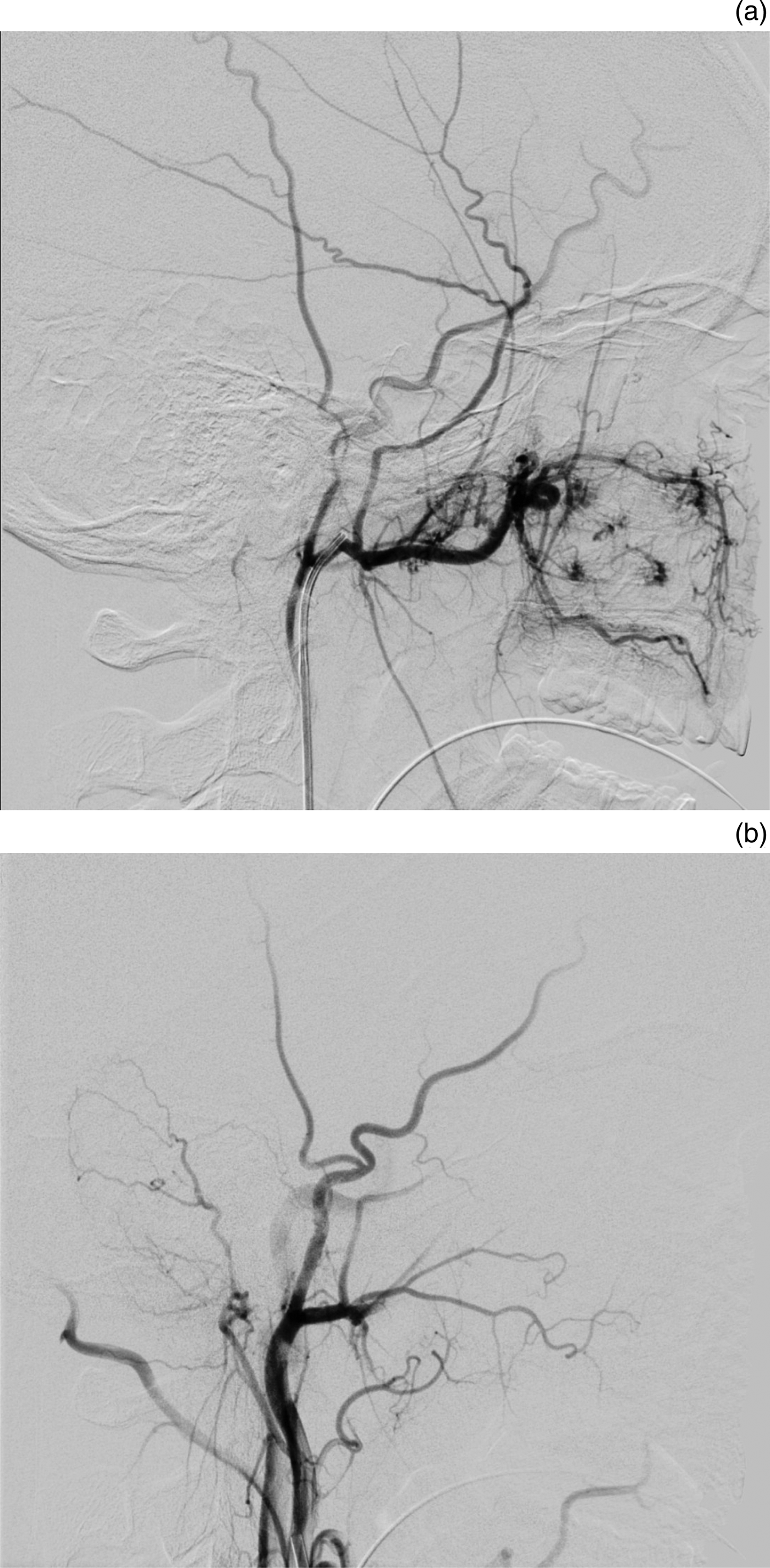
Pre-operative diagnostic digital subtraction angiography was performed bilaterally in the common carotid artery, internal carotid arteries and the external carotid artery to determine the vascular supply of the tumours and to embolise them. Both tumours were strictly ipsilaterally supplied by their respective sphenopalatine arteries (Figures 3 and 4). The right-sided tumour had a small additional supply from the ipsilateral Vidian artery, which branches from the internal carotid artery. Embolisation with 150–355 µ polyvinyl alcohol particles was performed bilaterally without complications.
Digital subtraction angiography pre-embolisation (a) and post-embolisation (b) images of the right external carotid artery. (a) Shows tumour blush from the sphenopalatine artery via the internal maxillary artery; (b) shows no visible tumour blush.

Digital subtraction angiography pre-embolisation (a) and post-embolisation (b) images of the left external carotid artery. (a) Shows nodular tumour blush from the sphenopalatine artery via the internal maxillary artery; (b) shows no visible tumour blush.
The following day, both tumours were successfully excised using CT and MRI guided functional endoscopic sinus surgery. The tumours from both sides were delivered transorally. The supplying arteries were electro-coagulated; the total peri-operative blood loss was 400 ml.
The post-operative course was uncomplicated. The patient was instructed to perform daily nasal irrigations and was discharged on the first post-operative day.
Upon follow-up examinations at zero, three, five, six and nine months post-operatively, the patient had no complaints, and there were no signs of non-radical surgery or recurrence upon nasal fibre-optic endoscopy. The absence of recurrence was confirmed by MRI nine months post-operatively.
Discussion
Juvenile nasopharyngeal angiofibroma is a rare tumour, with a reported incidence of only four to six new cases in Denmark per year. Juvenile nasopharyngeal angiofibroma may be misdiagnosed initially, especially in cases of bilateral juvenile nasopharyngeal angiofibroma; the current case is only the third reported case of bilateral juvenile nasopharyngeal angiofibroma in the literature.Reference Rha, Byun, Kim and Kim4, Reference Wu, Chark and Bhandarkar5
Juvenile nasopharyngeal angiofibromas are composed of a rich, irregular, vascular network, completely devoid of any muscular layer.Reference Girgis and Fahmy7 Early suspicion of juvenile nasopharyngeal angiofibroma is important, as biopsying in juvenile nasopharyngeal angiofibroma cases can cause massive bleeding.
The typical clinical presentation of juvenile nasopharyngeal angiofibroma is the triad of unilateral nasal obstruction, recurrent epistaxis and nasal discharge, primarily occurring in adolescent males,Reference Neel, Whicker, Devine and Weiland1 but recognition of the extremely rare case of bilateral juvenile nasopharyngeal angiofibroma based on symptoms is difficult. The bilateral obstruction, as observed in the current case, can be mimicked by a unilateral tumour extending to the contralateral side of the nasopharynx. The possibility of bilateral juvenile nasopharyngeal angiofibroma must, however, be considered. Thorough clinical and radiological investigation is necessary to optimise pre-surgical planning, minimising the risk of massive peri-operative bleeding and ensuring radical removal of the tumours.
In the present case, the left-sided tumour was biopsied, as juvenile nasopharyngeal angiofibroma was not suspected initially, and this caused some bleeding. The biopsy did not reveal the typical pattern of juvenile nasopharyngeal angiofibroma. Bremer et al. reported that a superficial biopsy of a juvenile nasopharyngeal angiofibroma tumour mass might be misleading and that the classical histological appearance of juvenile nasopharyngeal angiofibroma is generally found in the deeper aspects of the tumour.Reference Bremer, Neel, DeSanto and Jones8 In the present case, the second biopsy and the arteriography revealed the diagnosis of bilateral juvenile nasopharyngeal angiofibroma.
Magnetic resonance imaging was found to be superior to CT in detecting the soft tissue extensions of the juvenile nasopharyngeal angiofibroma.Reference Gomaa, Hammad, Abdelmoghny, Elsherif and Tawfik9 Computed tomography was used to display potential bone changes, including the typical enlargement of the sphenopalatine foramen in juvenile nasopharyngeal angiofibroma cases.Reference Lloyd and Howard10 In this particular case, it was not possible to compare an affected foramen to an unaffected foramen.
Pre-operative identification of the total blood supply is essential in planning the surgical approach of these highly vascular tumours. This is achieved by angiography, which also enables pre-operative embolisation. The blood supply typically arises from the internal maxillary artery. With the continuing growth of a juvenile nasopharyngeal angiofibroma, an additional blood supply is also induced from the internal carotid system,Reference Glad and Vainer2 as was the case for the right-sided juvenile nasopharyngeal angiofibroma in our patient.
Pre-operative embolisation has been shown to reduce peri-operative blood loss and the need for blood transfusions,Reference Ballah, Rabinowitz, Vossough, Rickert, Dunham and Kazahaya11 and is now considered standard treatment. This rare case of bilateral juvenile nasopharyngeal angiofibroma emphasises the importance of bilateral embolisation, which reduces the risk of heavy bleeding from two possible sources upon excision. Our pre-operative intervention, with embolisation on both sides conducted in one procedure, was successful. It allowed for uncomplicated, one-step radical removal of both juvenile nasopharyngeal angiofibromas, with minimal blood loss and no subsequent necrosis of the nasal mucosa.
Surgical excision is the ‘gold standard’ treatment. During the last two decades, endoscopic excision has been shown to be less disfiguring and to have lower recurrence rates than open surgical approaches.Reference Khoueir, Nicolas, Rohayem, Haddad and Abou Hamad12
• This is the first case of clinically challenging bilateral juvenile nasopharyngeal angiofibroma
• The complications are potentially life-threatening
• Both tumours were pre-operatively embolised
• One-step bilateral resection was performed; blood loss was minimal and recovery was rapid
The overall recurrence rate of juvenile nasopharyngeal angiofibroma is reported to be about 23 per cent.Reference Glad and Vainer2 However, endoscopic excision might reduce the recurrence rate to about 10 per cent.Reference Khoueir, Nicolas, Rohayem, Haddad and Abou Hamad12 This might be a result of technological development within embolisation, imaging and surgery. There are no records of malignant transformation of juvenile nasopharyngeal angiofibromas.
Conclusion
In this report, we describe the first case of clinically challenging bilateral juvenile nasopharyngeal angiofibroma, which warranted thorough pre-operative planning, successful bilateral embolisation and total resection of both juvenile nasopharyngeal angiofibromas. The patient was discharged the day after surgery.