Background
Pericardial effusion is commonly caused by viruses in pediatric population, and the clinical course of the majority of these effusions is mild, with spontaneous resolution. Agents identified as causing pericarditis include the enteroviruses, influenza, adenovirus, respiratory syncytial virus, and parvovirus. Non-infectious causes include autoimmune, rheumatologic, and connective tissue disorders. Most uncomplicated cases of pericarditis are treated with NSAIDs (nonsteroidal anti-inflammatory drugs), steroids, or a combination of both with expectation of recovery within few days.Reference Carvalho, Kenney, Carrington and Hall 3 – Reference Adler, Charron and Imazio 5
Kwashiorkor is an unusual cause of pericardial effusion. We describe a complicated case of a child presenting with pericardial effusion and pneumonia. Although less common in developed countries, the diagnosis of kwashiorkor in our case was considered and reached by supporting history, examination, and laboratory findings.
Case presentation
A 13-year-old fully-immunised male with trisomy 21 and complex past medical history was transferred from a peripheral hospital for lethargy, pneumonia, and worsening respiratory distress. Due to significant work of breathing, he was admitted to the regional pediatric ICU, where a large pericardial effusion was confirmed on echocardiography.
The past medical history was significant for trisomy 21, seizure disorder, global developmental delay, small atrial septal defect, obstructive sleep apnea, gastro-oesophagealreflux disease, and hypothyroidism. His medication regimen on admission included valproic acid, thyroxine, lansoprazole, and domperidone. The family and social histories were unremarkable. The patient was born in Canada. The family is of East Indian descent (unknown exact geographic region). Beside the trisomy 21 features, the physical examination revealed a pale, lethargic boy with facial oedema, hepatomegaly, ascites, pedal oedema, and non-specific dermatitis. His hemodynamic exam revealed tachycardia but no other signs of pericardial effusion, no rub, and no muffled heart sounds. However, it was noted that he would prefer to stay in sitting position most of the time. His weight on admission was well below the 5th percentile on both the traditional Center for Disease Control Growth Charts for Children with trisomy 21, as well as the newer growth charts based on the Down Syndrome Growing Up Study.Reference Zemel, Pipan and Stallings 4 His mid-upper-arm circumference was <11.5 cm; weight-for-height z score < 3.
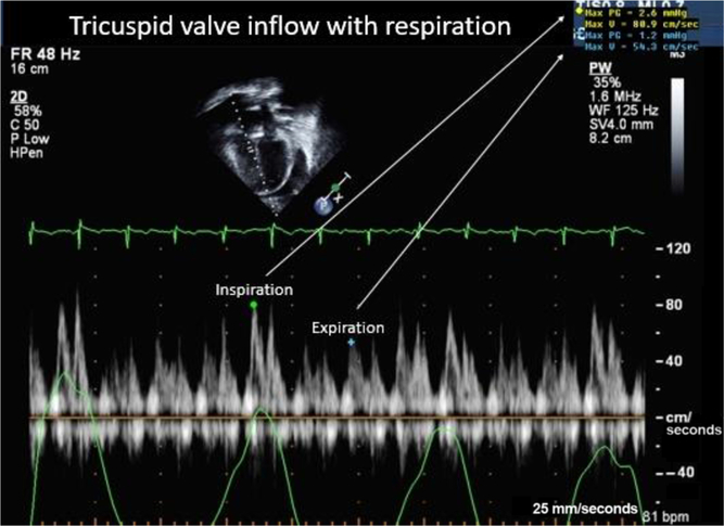
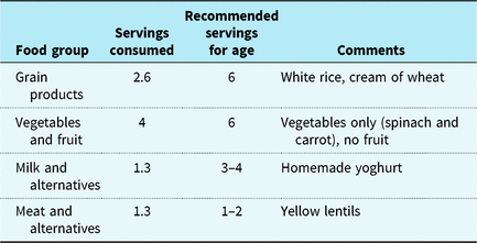
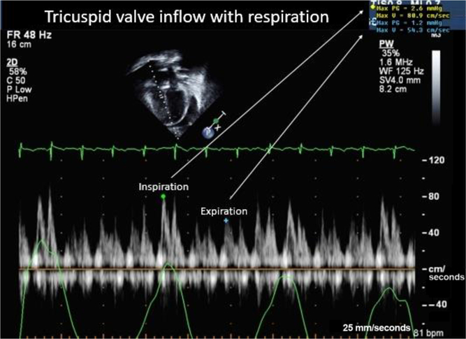
Initial echocardiography showed a significant circumferential pericardial effusion with no echocardiographic evidence of tamponade, normal biventricular systolic function, as well as the known small atrial septal defect (Fig 1). This was managed with high-dose ibuprofen for 10 days followed by a 7-day course of prednisone, with no significant improvement in the effusion. Because of the lack of response to the anti-inflammatory drugs, other causes of pericardial effusion were considered. Dietary history revealed a prolonged, inadvertent administration of a restricted diet, deficient in protein and several other nutrients. Parents believed their child had a milk allergy and consequently restricted his daily intake (Table 1).
Pericardial effusion in a patient with Kwashiorkor: tricuspid valve inflow during respiration with no evidence of pericardial tamponade.
Number of servings of the various food groups and how it compares to the Canadian Food Guide.
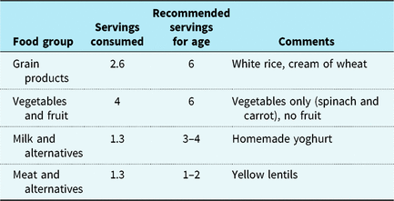
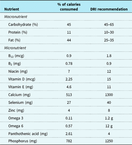
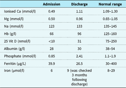
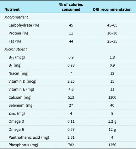
Taking into consideration the history of poor weight gain, dietary restrictions, and electrolyte abnormalities, the diagnosis of kwashiorkor was considered as a potential cause for the pericardial effusion. Our Registered Dietitian conducted a 24-hour dietary recall with the patient’s mother and used the aid of measuring cups to help estimate usual volume. The patient’s mother reported that the child’s diet consisted exclusively of pureed or well-mashed foods of cream of wheat, white rice, small amounts of lentils, spinach, carrots, and unfortified (homemade) goat-based yoghurt and ghee. When comparing his best usual intake to Canada’s Food Guide to Healthy Eating, on average, his intake was well below the recommendations for age for the majority of Food Groups (Table 1). The 24-hour dietary recall was than analysed using ESHA (Elizabeth Stewart Hands and Associates), a computer diet analysis program to help assess the intake of micro- and macronutrients. The patient’s intake was found to be well below recommendations for a variety of macro- and micronutrients (Table 2). The child was found to have multiple severe nutritional deficiencies, including hypoalbuminaemia, calcium, magnesium, 25-OH Vitamin D, phosphate, and iron (Table 3). His serum potassium level was adequate. Celiac screening was negative. He had low ionised calcium levels with no obvious clinical signs or increasing seizure activity. He required supplementation with intravenous calcium gluconate as well as calcium carbonate via NG (nasogastric tube) for his long-term supplementation. The patient, on his best intake, consumed 1.47 g/kg of protein (but more recently consuming 50% of this or 0.74 g/kg) which compares to 0.95 g/kg recommended for age. Once the patient became stable medically, the Registered Dietitian met with mom and dad on a number of occasions to provide nutrition education and a Pureed Diet Plan for home. This plan included 1800–1900 calories per day and provided recommended number of servings, serving sizes, and additional recommendations for the parents to follow to help ensure that the patient had a well-balanced variety of macro- and micronutrients. A written copy of the plan was given to parents for their use at home as the plan for a gradual increase in his caloric intake to avoid refeeding syndrome which was monitored.
Distribution of nutrients.
Nutritional and electrolyte deficiencies in a patient with kwashiorkor malnutrition.
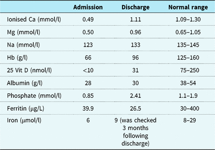
These values are the actual laboratory results of this patient’s blood work. All admission values are within 2 days of the patient’s initial admission to hospital, with the exception of his sodium, phosphate, and haemoglobin values, which denote the nadir of his anaemia, hypophosphataemia, and hyponatraemia rather than his admission blood work. The discharge results are the most recent values prior to discharge and may not have been drawn on the exact discharge date. The findings show a significant improvement in all categories following refeeding, nutritional supplementation, and medication administration.
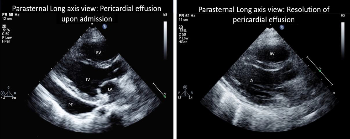
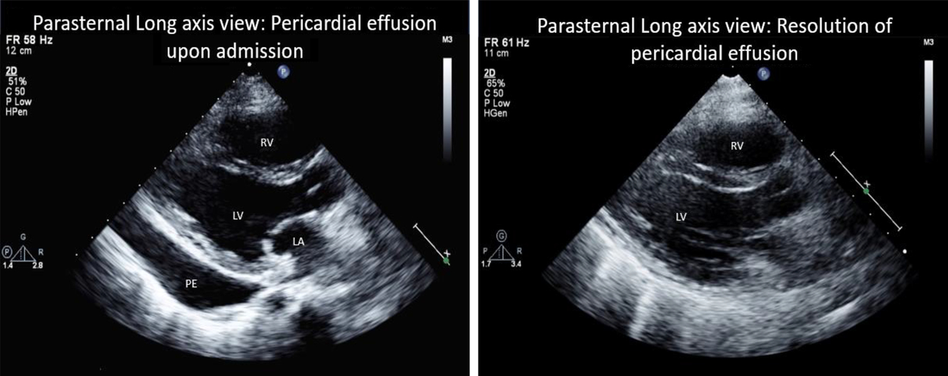
This patient’s history, clinical picture, various electrolyte disturbances, and complications were consistent with Kwashiorkor malnutrition. With appropriate nutritional and caloric requirements, he became increasingly active. His ascites and hepatomegaly also resolved. The pericardial effusion, which had been unresponsive to treatment, also improved with appropriate targeted nutritional therapy (Fig 2). After more than a month-long hospital stay, all biochemical parameters had returned to normal; the patient had resumed oral feeding and was discharged in a stable condition on his home medications as well as several nutritional supplements.
Pericardial effusion in a patient with Kwashiorkor: pericardial effusion on admission (left) with subsequent resolution (right) following appropriate therapy. The effusion was initially treated with a course of ibuprofen and prednisone, with minimal improvement. It was not until the Kwashiorkor diagnosis and ensuing management targeted at malnutrition that the effusion resolved.
Limitations
It is important to note that dietary analysis is limited to a single 24-hour recall provided by the patient’s mother and that the recall estimated the patient’s “best” intake. DRI’s (Dietary References Intakes) are good population references; however, it is difficult to apply them for patients with trisomy 21.
Discussion
Kwashiorkor is a type of protein–calorie malnutrition, most often seen in children of impoverished countries or areas affected by famine. It was first described by Cicely Williams in 1935 and is thought to be secondary to inadequate dietary protein intake in the presence of sufficient caloric intake. Reference Krebs, Primak, Hambridge, Hay, Levin, Sondheimer and Deterding1,Reference Williams2 Associated physical findings may include dermatitis, protuberant abdomen, thinning hair, and “bull-dog” face. Clinical findings may also include weight loss/failure to thrive, delayed linear growth, irritability, and lethargy. Kwashiorkor is a rare cause of pericardial effusion and can be severe enough, as in our case, causing echocardiographic evidence of tamponade. There are many cases of Kwashiorkor malnutrition in the United States literature that have described infants who have suffered significant dietary restriction by ‘‘well-intentioned”, but ultimately misguided, parents, as was the case in our patient. This child had previously been treated for inflammatory causes of pericardial effusion with no response. Additionally, his primary health care provider had never addressed his poor weight gain and nutritional status. The diagnosis of Kwashiorkor was made subsequently upon discovering multiple nutritional deficiencies and clinical nutritional assessment.
Kwashiorkor is often underdiagnosed or misdiagnosed in high-income countries. The results, if unrecognised, may be devastating. This makes it imperative that physicians consider this diagnosis, recognise potential risk factors, and be prepared to accurately assess overall nutritional status of patients, especially vulnerable populations such as those with trisomy 21. Additionally, protein malnutrition should be considered in any child presenting with pericardial effusion unresponsive to conventional treatment.
Conclusion
Although less commonly encountered in developed countries, malnutrition should be considered as a potential contributor to diseases, especially in patients with multiple comorbidities. Not addressing nutritional aspects in these cases may have significant impact on health.
Acknowledgements
None.
Supplementary material
To view supplementary material for this article, please visit https://doi.org/10.1017/S1047951119002129
Financial Support
No funding was secured for this study.
Conflict of Interest
All authors have no conflicts of interest to disclose.
Ethical Standards
None.
Authorship
Dr Aimann Surak conceptualised and designed the case report, drafted the initial manuscript, and reviewed and revised the manuscript. Dr Grace Bravo helped in conceptualising the study and drafting the manuscript. Amanda McMurray (registered dietitian) helped in providing the dietary history as well as the management plan provided in the tables. Dr Luis Altamirano-Diaz is the cardiologist who revised the manuscript and provided the echocardiographic images. Dr Sepideh Taheri initiated the case report and supervised the writing of the manuscript. All authors reviewed and participated in the writing of the manuscript.