Introduction
Congenital midline nasal dermoid sinus cysts account for 10 per cent of head and neck dermoids and 1 per cent of all dermoids.Reference Crawford and Webster1
Development of nasal dermoids may be related to the dural process in the prenasal space. Initially, the dura and skin are in direct contact, but if there is failure of the skin to separate from the dura then a dermoid cyst may be formed.Reference Pratt2 If the dermoid and tract communicate with the skin via a punctum, then a dermoid sinus is formed.Reference Nydell and Masson3 It has also been suggested that the variable location of the nasal dermoid sinus cyst represents the point where ectodermal inclusion occurred during embryological development. This point occurs at variable positions along an anatomical line extending from the midface to the anterior cranial fossa.Reference Charrier, Rouillon, Roger and Denoyelle4
Nasal dermoids most commonly present as a midline nasal mass along the dorsum of the nose between the columella and the glabella.Reference Blake, Cow, Holmes and Meara5 Given the potential communication with the anterior skull base, there is a risk of meningitis if they become infected. There is also a potential risk of cerebrospinal fluid leakage if they are biopsied. Given these risks, early imaging and subsequent complete surgical excision is the management of choice. For this reason, early diagnosis and prompt treatment are essential.Reference Zapata and Kearns6
Nasal dermoids are unusual. From our experience, we discuss three variants of this condition which are more unusual still, and which presented unique challenges to the surgeon.
Method
Three unusual cases of nasal dermoid cysts are presented here. This was a retrospective analysis of three cases referred to the Royal Hospital for Sick Children, Glasgow, over the past three years. Once the cases were identified, the details of the initial presentation were obtained from the case notes, along with the management and outcome.
Results
Case one
The first case was a two-year-old boy referred to ENT from the emergency department. He had initially presented with a nasal injury resulting from a fall. However, the attending doctor had noted a discharging sinus on his upper lip. He had had only one episode of discharge from the sinus, and was otherwise healthy.
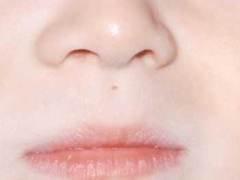
Examination revealed a small sinus in the philtrum of the upper lip (Figure 1), but no nasal swelling or other abnormality. Intraoral examination was normal.
Clinical photograph of case one showing the sinus on the philtrum of the upper lip.

A magnetic resonance imaging (MRI) scan revealed a tract passing from the philtrum superiorly within the nasal septum to the crista galli. There was associated widening of the nasal septum and crista galli.
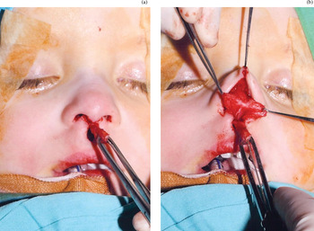
The lesion was excised via an external rhinoplasty approach. A small incision around the sinus opening on the upper lip permitted excision of the sinus. The external rhinoplasty incision allowed the tract to be followed superiorly and posteriorly through the septum into the crista galli. Figure 2 shows the punctum and tract excised from the philtrum, and also the tract passing through the nasal spine into the nasal septum.
Intra-operative photographs of case one, showing (a) the sinus (excised from the philtrum) and (b) the sinus tract (followed to the nasal spine).

There were no intra-operative or post-operative complications. Both wounds healed very well, and the nasal shape was not affected. There was no evidence of recurrence during the six months of follow up.
Case two
The second case was a 15-month-old boy who was referred from another ENT department with two separate puncta on the dorsum of his nose. There was no history of discharge or swelling. He was otherwise fit and well.
Examination revealed two small sinuses on the nasal dorsum, 10 mm apart, with no palpable masses. Figure 3 shows the two puncta on the dorsum of the nose. An MRI scan revealed a cyst lying deep to the nasal bones, with no obvious communication with the anterior cranial fossa.
Clinical photographs of case two, showing the two puncta visible on the (a) upper and (b) lower dorsum of the nose.

This patient underwent excision of his nasal dermoid via an external rhinoplasty approach. A small ellipse of skin was excised with each of the sinus openings. The inferior sinus was traced deep to the nasal bones and into the inferior aspect of a dermoid cyst lying deep to the nasal bones. The upper sinus arose from the superior aspect of this dermoid cyst. These lesions were all removed in continuity. There was no extension into the anterior cranial fossa.
There were no operative complications. All the wounds healed well, and the patient's parents were happy with the cosmetic outcome. Over three years of follow up, there was no evidence of recurrence, and nasal growth was not affected.
Case three
The third case was a 17-year-old male adolescent with neurological disability, who was referred to the salivary control clinic for management of drooling. As an incidental finding, he was noted to have a sinus opening on the dorsum of his nose, with protruding hairs. There was no history of nasal swelling or sinus discharge.
Examination confirmed a single sinus with no palpable cyst. The patient had quadriplegic cerebral palsy with seizures. His seizures were poorly controlled, and he had previously been prescribed many different treatments, including sodium valproate, clobazam, oxcarbazepine, zonisamide, topiramate, phenytoin, piracetam, leveticacetan bromide, nitrazepam and lamotrigine, as well as a ketogenic diet.
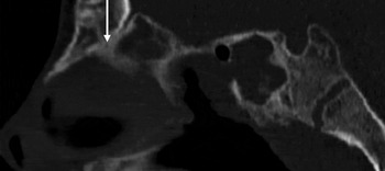
Computed tomography (CT) scanning revealed a sinus tract extending from the crista galli, which was bifurcated, into the anterior skull base, but not connected to the superficial tissues of the nose. The bone of the anterior skull base was generally thickened with bony overgrowth (Figure 4).
Sagittal computed tomography scan of the anterior skull base in case three, showing the partly obliterated tract (arrow).

Due to the low risk of infection and the patient's intracranial complication, the decision was made not to excise the lesion. There were no concerns about the current cosmetic appearance, and the parents were very reluctant for any surgical intervention.
During the one year of follow up, there were no problems associated with the sinus tract.
Discussion
It is well accepted that the standard treatment for nasal dermoid sinus cysts is primary complete excision. This is because of the potential for infection and abscess formation. There is also a risk of meningitis if the tract communicates with the anterior cranial fossa.Reference Blake, Cow, Holmes and Meara5
To avoid confusion between sinuses and fistulae, we define a sinus as a blind-ending epithelial tract connected to the surface epithelium. We define a fistula as an epithelialised communication between two epithelial surfaces.
Given the proposed hypotheses for nasal dermoid development, it is clear that there will inevitably be variability in nasal dermoid position, size and association with surrounding structures. There are several theories about the development of nasal dermoid sinus cysts; one suggests that if there is failure of separation of the ectoderm from the dura then the ectoderm will be pulled into the prenasal space.Reference Brunner and Donnelly7 Charrier et al. propose that the variability of the position of anterior nasal dermoid sinus cysts reflects the variable point of ectodermal inclusion during embryonic development.Reference Charrier, Rouillon, Roger and Denoyelle4
These theories explain the occurrence of an anteriorly placed cyst with a potential sinus positioned somewhere along a line from the base of the columella to the nasofrontal area.
However, our second case raises questions about the formation of nasal dermoids. This case had a cyst deep to the nasal bones and two separate sinus tracts to the skin, one superior and one inferior to the nasal bones. This differs from the accepted idea of a sinus tract, containing a cyst, which runs from a solitary punctum on the nasal dorsum and passes superiorly anterior to the nasal capsule to terminate at some point towards the anterior cranial fossa. There is previously reported evidence of bifurcation of sinus tracts. Tiwari has described a nasal dermoid with a sinus on the nasal dorsum and a tract bifurcating deep to the nasal bones, with one branch going to the anterior cranial fossa and the other running anterior to the frontal bone.Reference Tiwari8
Median upper lip sinuses are said to be extremely rare, and may represent ectodermal inclusion during fusion of the intermaxillary process.Reference Charrier, Rouillon, Roger, Denoyelle, Collon and Garabedian9 Median upper lip sinuses have not been described before in association with nasal dermoid sinus cysts. Sancho et al. have added three new cases to the existing literature of 25 reported cases of upper lip sinuses.Reference Sancho, Albert, Cusi, Grande, Aguilar and Morales10 All these three cases had a punctum in the philtrum and were blind-ending. Intra-operatively, one was noted to extend to the nasal spine. Charrier et al. have reported a case with a sinus on the philtrum extending 1.5 cm and ending at the nasal spine.Reference Charrier, Rouillon, Roger, Denoyelle, Collon and Garabedian9 Asahina et al. have described a further case of an upper lip sinus with a blind-ending tract, which they excised via an intraoral approach.Reference Asahina, Sakakibara, Miyashin, Tachikawa and Enomoto11
In our first case, the tract passed from the upper lip to the nasal spine and through the middle of the nasal septum to the anterior skull base. The presence of a nasal dermoid sinus extending from the philtrum to the skull base would suggest that it is formed by ectodermal inclusion before development of the nasal capsule, as the lesion passes through the middle of the septum. In previous studies assessing median upper lip sinuses, the tract was found to extend towards the nasal spine. Other studies refer to these ‘fistulae’ or tracts as sinuses, with no intraoral component.Reference Illing, Field, McNamara and Sandy12 We suggest that not only do nasal dermoid sinus cysts and median upper lip fistulae have a common developmental relationship, but they could represent the same condition. We propose that a nasal dermoid can have a cyst or sinus anywhere from the philtrum of the upper lip or columella, via the nasal dorsum, to the anterior skull base.
Surgical excision by external rhinoplasty has been well described as a method of removing nasal dermoids, with good results.Reference Bilkay, Gundogan, Ozek, Tokat, Gurler and Songur13 In our first two cases, an external rhinoplasty approach allowed complete identification of the dermoid sinus and tract, and enabled complete excision without restriction of access. This approach provided excellent views of the nasal septum in case two. It also resulted in excellent post-operative cosmesis. In our second case, the nasal bones had already been splayed by the dermoid cyst; however, during follow up they showed evidence of remodelling.
During the management of nasal dermoids, it is crucial to perform pre-operative anterior skull base imaging. This should involve MRI if possible, due to this modality's superior soft tissue detail, in order to better assess the sinus tract and to examine for intracranial extension or an intracranial dermoid cyst. Pre-operative imaging also allows identification of any unusual arrangements of the nasal dermoid cyst which may prompt the surgeon to use a particular technique to excise the cyst and sinus. Studies have shown that MRI has few false positive and false negative results for nasal dermoids.Reference Blake, Cow, Holmes and Meara5, Reference Bloom, Carvalho, Dory, Brewster, Wickersham and Kearns14 In our institution, MRI with a facial coil is used to provide very high resolution images of the anterior skull base.
• Nasal dermoids can present as a pit or sinus anywhere from the columella, via the nasal dorsum, to the anterior skull base
• Standard treatment for nasal dermoid sinus cysts is primary complete excision
• The presented three cases show how dermoids may arise from the philtrum or develop in an unusual manner
• Nasal dermoids may vary from conventional descriptions; management requires great care
Antiepileptics have been reported to have possible effects on bone mineralisation and growth. Phenytoin has been linked to skull thickening and hyperostosis frontalis interna.Reference Chow and Szeto15 Both lead to thickening of bone in the frontal region. In our third case, CT imaging confirmed that any communication between the intracranial dermoid tract and the nasal sinus had been obliterated by bony overgrowth. This case was complicated by the fact that the patient had significant and life-threatening co-morbidities, which had led to delays in the patient's referral to ENT for diagnosis and management. These factors also limited surgery to immediately life-threatening conditions only. In this case, imaging enabled the standard management of nasal dermoids to be changed to suit the patient.
Conclusion
These three cases show that nasal dermoid sinus cysts can have atypical arrangements, and that great care must be taken, during pre-operative planning and in the operating theatre, to ensure that all of the cyst and tract are identified and completely excised, in order to obtain the best outcome for the patient. The third case proves the exception to the rule, showing that not all nasal dermoid cysts need to be excised. These cases also add to the debate over the developmental causes of nasal dermoid and their association with median upper lip fistulae.