Highlights
-
The association of time-to-treatment intervals and baseline imaging characteristics with outcomes was analyzed in late-window (>6 hours) EVT patients of the pre-DAWN and DEFUSE-3 era.
-
Outcome was not associated with workflow time intervals.
-
Favorable imaging profiles (slow infarct progressors) may have driven patient selection for EVT in the late window.
Background
Acute ischemic stroke (AIS) treatment is exquisitely time-sensitive, particularly when caused by a large vessel occlusion (LVO). Endovascular treatment (EVT) is a highly effective treatment for AIS due to LVO. Initially, the effect of EVT was only shown in a narrow time interval after symptom onset. Reference Goyal, Menon and van Zwam1 Shortly thereafter, prospective randomized trials, namely, DAWN, Reference Nogueira, Jadhav and Haussen2 DEFUSE-3 Reference Albers, Marks and Kemp3 and MR CLEAN LATE, Reference Olthuis, Pirson and Pinckaers4 showed that patients with AIS due to LVO also benefit when EVT is performed > 6 hours after the time of last known well. Recently, large core trials like SELECT-2 and ANGEL-ASPECT were published, which all showed the benefit of EVT in strokes up to 24 hours after last known well with established large infarct. Reference Sarraj, Hassan and Abraham5,Reference Huo, Ma and Tong6 In DAWN and DEFUSE-3, eligible patients were selected based on perfusion imaging and clinical criteria showing potentially salvageable tissue. In MR CLEAN LATE, patients were selected for EVT primarily based on the collateral status of the affected brain hemisphere on CT angiography. In the large core trials, patients were selected based on non-contrast CT alone or in combination with CT perfusion or diffusion-weighted MRI.
For AIS patients with anterior circulation LVO who present in the early time window (within 6 hours from last known well), it has been shown that a short time from last known well to reperfusion is associated with better outcome. Reference Saver, Goyal and van der Lugt7 Interestingly, studies investigating the time interval from last known well to qualifying imaging or arterial access for EVT have failed to show a significant association with clinical outcome, while the time from qualifying imaging or arterial access to reperfusion shows clear evidence of treatment effect modification. Reference Menon, Sajobi and Zhang8 The principal reason for this effect is thought to be that by selecting patients with favorable baseline imaging profiles for treatment, those patients who are ultimately selected for treatment are more likely to have slower infarct progression. While this phenomenon persists in randomized trials as well as in real-world settings, Reference Kaesmacher, Maamari and Meinel9 it has only been shown when EVT has been performed within 6 hours after last known well.
We aimed to investigate associations of clinical outcomes in AIS patients with anterior circulation LVO that were treated with EVT > 6 hours after last known well and both the time from last known well to arrival at the EVT hospital and the time from EVT hospital arrival to arterial access.
Methods
This is a secondary analysis of the ESCAPE-LATE study, which was an international, multicenter, prospective cohort study of AIS patients ≥ 18 years with LVO in the anterior circulation who were treated with EVT > 6 hours after last known well in routine clinical practice. There were no imaging inclusion criteria. The data were acquired at 10 high-volume ESCAPE trial sites from February 2015 to December 2017. This time frame is confined by the conclusion of patient enrollment of the ESCAPE trial (November 2014) and the publication of DAWN and DEFUSE-3 (January 2018). All variables collected were captured as part of clinical routine care; therefore, the local ethics boards approved the study with waiver of consent.
The current study only included patients with anterior circulation LVO in order to make the analysis more comparable with the existing literature.
Outcome
The primary outcome was the functional outcome measured on the modified Rankin Scale (mRS; range 0 [no symptoms] to 6 [death]) 90 days after stroke. Secondary outcomes were dichotomized mRS (0–2 [good functional outcome] and 5–6 [poor functional outcome]), ordinal National Institutes of Health Stroke Scale (NIHSS) score at 24 hours after EVT and dichotomized 24-hour NIHSS score 0–2 (good short-term functional outcome). NIHSS at 24 hours was chosen as a secondary outcome since it was expected that in clinical routine, not all patients may have returned to the hospital for a 90-day mRS assessment, and the 24-hour NIHSS has been shown to be closely associated with the 90-day mRS. Reference Rangaraju, Frankel and Jovin10
Statistical analysis
Patient clinical and imaging baseline characteristics and workflow time intervals are reported using descriptive statistics, namely, counts and frequencies for categorical data and medians and interquartile ranges (IQR) for continuous data. Although ESCAPE-LATE did not have any imaging-based eligibility criteria based on tissue viability, there was still a chance that physicians may have intuitively selected patients with favorable imaging profiles. Therefore, we were particularly interested in baseline collateral status and Alberta Stroke Program Early CT Score (ASPECTS, range 0–10), which were reported as part of the imaging baseline characteristics.
Unadjusted associations of time intervals and outcomes were performed using Wilcoxon rank sum tests (dichotomous outcomes) and Spearman rank correlation (ordinal outcomes). Adjusted ordinal and binary logistic regression models were used to investigate the association of time from last known well to EVT hospital arrival and time from EVT hospital arrival to arterial access with clinical outcomes after reperfusion. Ordinal logistic regression models were used to investigate the association of ASPECTS and collateral status on initial neurovascular imaging with the time from last seen well to hospital arrival. If not further mentioned, all adjusted analyses were adjusted for age, sex, baseline NIHSS, intravenous alteplase treatment, occlusion site (intracranial internal carotid artery [ICA] vs. extracranial ICA vs. M1 vs. M2), collateral status (poor [<50% filling of leptomeningeal collaterals compared to the contralateral side] vs. moderate [>50%–75% filling of leptomeningeal arteries compared to the contralateral side] vs. good collateral status [>75% filling of the leptomeningeal arteries compared to the contralateral side]) and baseline ASPECTS. Furthermore, we also assessed the association of baseline ASPECTS and collateral status with workflow interval times using univariable and multivariable logistic regression with adjustment variables as described above. Adjusted effect size estimates for time intervals were reported for 10-minute increments. We performed a complete case analysis without imputation for missing data. We deliberately did not attempt to impute missing mRS data in the main analysis because we cannot assume that data were missing at random. However, we performed a sensitivity analysis in which imputed binary mRS was used as a primary outcome: patients discharged home were imputed as having achieved a 90-day mRS 0–2 while those discharged to another location than home (e.g., in-patient rehabilitation facility or long-term care) were imputed as 90-day mRS > 2.
Statistical significance was assumed at the alpha = 0.05 significance level, and all tests were two-sided. Statistical analyses were performed using Stata MP 17.0.
Results
A total of 464 patients were included in ESCAPE-LATE, of which 211 patients were excluded for this analysis because the time from last known well to treatment was less than 6 hours. These cases were identified as false inclusions during data review and were excluded to ensure the analysis adhered to the current study’s inclusion criterion of treatment beyond 6 hours from last known well. Another 53 patients with vessel occlusions in the posterior circulation were excluded, leaving a total of 200 patients for the analysis, 141 (70.5%) of whom had an available 90-day mRS. The 24-hour NIHSS was available for 125 (62.5%) patients (Figure 1). Of the 200 included patients, 85 (42.5%) patients were women, and 115 (57.5%) were men. The median age was 72 years (IQR 62–81 years). Thirty-nine patients (20%) had occlusions in the intracranial ICA, 140 (70%) in the M1 segment and 35 (18%) in the M2 segment, with some patients having multiple occlusions. Thirteen of 200 (6.5%) patients had an additional extracranial ICA occlusion. Hundred-thirty-five of 150 (90%) patients had moderate-to-good collateral status. Median ASPECTS was 8 (IQR 7–10). Sixty-six out of 141 (46.8 %) patients had mRS 0–2, and the median 24-hour NIHSS was 6 (IQR 3–15). Supplementary Figure 1 shows the distribution of the 24-hour NIHSS and the 90-day mRS of the total population.
Flowchart with inclusion and exclusion of patients.Note: EVT = endovascular thrombectomy; LVO = large vessel occlusion.

Baseline characteristics and workflow times for all patients included in ESCAPE-LATE with anterior circulation EVT > 6 hours from last known well are shown in Table 1.
Patient characteristics for the total population and for patients with available 90-day mRS only

Note: IQR = interquartile range; NIHSS = National Institutes of Health Stroke Scale; mRS = modified Rankin Scale; TIA = transient ischemic attack; ASPECTS = Alberta Stroke Program Early CT Score; ICA = internal carotid artery; occlusion site * = some patients had multiple occlusions; min = minutes.
Association of time to EVT hospital arrival with outcome measures
Unadjusted and adjusted analyses were performed for ordinal mRS and dichotomized mRS for patients with available 90-day mRS (n = 141) and for 24-hour NIHSS (n = 125) as an alternative outcome including 49 patients with missing 90-day mRS. The complete adjusted regression models testing the association between time to EVT hospital arrival and the clinical outcome measures (i.e., ordinal and dichotomized (0–2) 90-day mRS) are shown in Supplemental Tables 2–5.
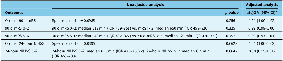
The time interval from last known well to arrival at the EVT hospital showed no association with ordinal mRS at 90 days after EVT (Spearman correlation coefficient = 0.10, p = 0.256; adjusted odds ratio [OR] 1.01 [95% CI 1.00–1.02]) and no association with good clinical outcome (90-day mRS 0–2: median 617 min [IQR 469–751] vs. 90-day mRS > 2: median 650 min [IQR 458–826], p = 0.325; adjusted OR = 0.99, 95% CI 0.98–1.00) or with poor clinical outcome (90-day mRS 5–6: median 643 min [IQR 458–826] vs. mRS < 5: median 620 min [IQR 476–771], p = 0.957; adjusted OR 0.99 (95% CI 0.97–1.01). A sensitivity analysis of the association of 90-day mRS 0–2 and the time from onset to EVT hospital arrival with imputed values for the dichotomized 90-day mRS 0–2 showed similar results (Supplemental Table 1). There was no association of time from last known well to arrival at the EVT hospital with ordinal 24-hour NIHSS (Spearman correlation coefficient = 0.663, p = 0.663; adjusted OR 1.01 (95% CI 1.00–1.02) nor with good clinical outcome in form of the dichotomized 24-hour NIHSS 0–2 (24-hour NIHSS 0–2: median 613 min [IQR 475–730] vs. 24-hour NIHSS > 2: median 615 min [IQR 458–789], p = 0.664; adjusted OR = 0.98, 95% CI 0.95–1.01). Outcome measures and their association with the time from last known well to arrival at the EVT hospital are summarized in Table 2.
Associations between time from last known well to EVT hospital arrival and stroke outcomes

Note: 90 d mRS = modified Rankin Scale 90 days after stroke; a(c)OR = adjusted (common) odds ratio; IQR = interquartile range; CI = confidence interval; ICA = internal carotid artery; ASPECTS = Alberta Stroke Program Early CT Score; NIHSS = National Institutes of Health Stroke Scale.
* Adjusted analyses were conducted with multiple logistic regression models and ordered logistic regression models. Adjusted common odds ratios are reported for ordinal 90 d mRS and 24-hour NIHSS to describe the odds of a 1-point increase of the 90 d mRS or the 24-hour NIHSS score, respectively, per 10 min increase in time from last known well to arrival at the EVT hospital. Adjustments were made for age, sex, baseline NIHSS, intravenous alteplase treatment, occlusion site (extracranial ICA vs. intracranial ICA vs. M1 vs. M2), collateral status (poor vs. moderate vs. good collateral status) and baseline ASPECTS. The proportional odds assumption for the 90 d mRS 0–6 was met with a p-value (Brant test) = 0.4138, as well as for the 24-hour NIHSS with a p-value (Brant test) = 0.5636. Adjusted odds ratios were reported for the dichotomized 90 d mRS 0–2, the dichotomized 90 d mRS 5–6 and the dichotomized 24-hour NIHSS 0–2, to describe the odds of achieving a 90 d mRS of 0–2, a 90 d mRS of 5 or 6 and the odds of achieving a 24-hour NIHSS of 0–2, per 10 min increase in time from last known well to arrival at the EVT hospital.
Association of outcome measures with the time from EVT hospital arrival to arterial access
For the time interval from arrival at the EVT hospital to arterial access, there was no association with ordinal mRS at 90 days (Spearman correlation = 0.558; adjusted OR = −0.01, 95% CI −0.02–0.00), with good clinical outcome (90-day mRS 0–2: median 76 min [IQR 56–106] vs. 90-day mRS > 2: median 83 min [IQR 35–125], p = 0.596; adjusted OR = 1.02, 95% CI 0.99–1.05) or with poor clinical outcome (90-day mRS 5–6: 84 min [IQR 43–146] vs. 90-day mRS < 5: median 80 min [IQR 60–106], p = 0.567; adjusted OR = 0.99, 95% CI 0.97–1.02). Figure 2 shows the distribution of good and poor functional outcomes depending on the time from symptom onset to EVT hospital arrival (Panels A and B) and the time from EVT hospital arrival to arterial access (Panels C and D). A sensitivity analysis of the association of 90-day mRS 0–2 and the time from EVT hospital arrival to arterial access with imputed values for the dichotomized 90-day mRS 0–2 showed similar results (Supplemental Table 1).
Violin plots displaying the time from (a) last known well to hospital arrival and (b) hospital arrival to arterial access stratified by 90-day mRS scores. (A) Time from last known well to hospital arrival of patients who were functionally independent (mRS 0–2) versus patients who were functionally dependent (mRS > 2) after 90 days. (B) Time from last known well to hospital arrival of patients with poor (mRS 5–6) versus good-to-moderate (mRS < 5) functional outcome. (C) Time from arrival at the EVT hospital to arterial access for EVT for patients with functionally independent (mRS 0–2) versus dependent (mRS > 2) at 90 days. (D) Distribution of the time from arrival at the EVT hospital to arterial access for EVT of patients with poor (mRS 5–6) versus good-to-moderate (mRS < 5) functional outcome. Note: mRS = modified Rankin Scale.

There was no association of the time from EVT hospital arrival to arterial access with ordinal 24-hour NIHSS (Spearman correlation coefficient = 0.141, p = 0.141; adjusted OR = 1.00, 95% CI 0.99–1.01), nor with good short term clinical outcome, that is, 24-hour NIHSS 0–2 (24-hour NIHSS 0–2: median 63 min [IQR 36–93] vs. 24-hour NIHSS > 2: median 80 min [IQR 46–113], p = 0.155; adjusted OR = 0.93, 95% CI 0.83–1.04). Outcome measures and their association with the time from hospital arrival to arterial access for EVT are summarized in Table 3. The complete adjusted regression models testing the association between time from EVT hospital arrival to arterial access and the clinical outcome measures (i.e., 90-day ordinal and dichotomized (0–2) mRS) are shown in Supplemental Tables 2–5.
Associations of outcomes and time from hospital arrival to arterial access at the EVT hospital

Note: 90 d mRS = modified Rankin Scale 90 days after stroke; a(c)OR = adjusted (common) odds ratio; CI = confidence interval; IQR = interquartile range; min = minutes.
* Adjusted analyses were conducted with multiple logistic regression models and ordered logistic regression models. Adjusted common odds ratios are reported for ordinal 90 d mRS and 24-hour NIHSS to describe the odds of a 1-point increase of the 90 d mRS or the 24-hour NIHSS score, respectively, per 10 min increase in time from arrival at the EVT hospital to arterial access. Adjustments were made for age, sex, baseline NIHSS, intravenous alteplase treatment, intracranial occlusion location (intracranial ICA vs. M1 vs. M2), extracranial ICA occlusion, collateral status (poor vs. moderate vs. good collateral status) and baseline ASPECTS. The proportional odds assumption for the 90 d mRS 0–6 was met with p-value = 0.4138, as well as for the 24-hour NIHSS with p-value = 0.5636. Adjusted odds ratios were reported for the dichotomized 90 d mRS 0–2, the dichotomized mRS 5–6 and 24-hour NIHSS 0–2 to describe the odds of achieving a 90 d mRS of 0–2 or a 90 d mRS of 5 or 6, respectively, as well as the odds of achieving a 24-hour NIHSS of 0–2 per 10 min increase in time from arrival at the EVT hospital to arterial access.
A 10 min increase in time from last known well to arrival at the EVT hospital did not change the odds of ASPECTS 7–10 (vs. ASPECTS < 7) on the baseline CT scan (adjusted OR = 1.01, IQR 0.99–1.03). Similarly, the odds of having a moderate or good collateral status on the first CT scan after hospital arrival did not significantly change per 10 min increase in time from last known well to arrival at the EVT hospital (adjusted OR = 1.00, 95% CI 0.99–10.2). However, Figure 3B shows that patients presenting very late mostly had a moderate or good collateral score.
Violin plots showing the time from last known well to qualifying CTA for each ASPECTS (A) and each collateral score (B). Note: CTA = CT angiography.

Discussion
We found no association of clinical outcomes with either time from last known well to hospital arrival or time from EVT hospital arrival to arterial access. The most likely explanation for this real-world data finding is that patients were more likely to have been selected for treatment using imaging, and this resulted in a population of patients with slow-progressing infarcts.
In workflow analyses of EVT patients < 6 hours after last known well, shorter times to reperfusion have been clearly associated with better outcomes. Reference Menon, Sajobi and Zhang8,Reference Goyal, Jadhav and Bonafe11 The time from arrival at the EVT hospital to arterial access was constantly shown to have a stronger impact on outcome than the time from last known well to hospital arrival. Reference Menon, Sajobi and Zhang8,Reference Kaesmacher, Maamari and Meinel9,Reference Goyal, Jadhav and Bonafe11 This may be an artifact related to the “equalizing effect” of selecting patients with favorable imaging characteristics upon hospital arrival and the exclusion of patients with extensive infarcts and/or poor collateral status. Interestingly, in this cohort of late-window EVT patients, even the time interval from EVT hospital arrival to arterial access did not show an association with outcome.
Imaging profiles of the included patients were generally very favorable, with a median ASPECTS of 8 and 90% moderate-to-good collaterals. Collaterals play a crucial role in determining the rate of infarct progression in AIS. Reference Liebeskind12 Patients with good collateral status have a higher probability for slow progression of the infarct core. Reference Liebeskind, Jahan, Nogueira, Zaidat and Saver13,Reference Menon, Qazi and Nambiar14 The fact that 90% of the patients in ESCAPE-LATE had a moderate or good collateral score (Figure 3B) suggests that the study may have included a high percentage of “slow progressors.” This hypothesis is supported by the overall high ASPECTS despite the late time window (Figure 3A). Selective inclusion of slow progressors would result in delays in the time interval from hospital arrival to arterial access being less detrimental than in a general stroke population that includes all patients along the spectrum of fast and slow infarct progression. Therefore, the lack of a significant association between time from hospital arrival and arterial access may indeed be explained by preferential inclusion of slow progressors in ESCAPE-LATE.
In workflow analyses of the randomized ESCAPE Reference Menon, Sajobi and Zhang8 and SWIFT-PRIME Reference Goyal, Jadhav and Bonafe11 trials, patients were selected by clinical and imaging criteria suggesting salvageable tissue, namely, ASPECTS and collateral status in ESCAPE Reference Goyal, Demchuk and Menon15 and perfusion profiles in SWIFT-PRIME. Reference Saver, Goyal and Bonafe16 The ESCAPE-LATE study, Reference Ospel, Zerna and Harrison17 on the other hand, had no specific imaging requirements or imaging-based enrollment criteria other than the presence of an LVO. Since the ESCAPE-LATE study has been completed, the DAWN and DEFUSE-3 trials have been published and have provided substantial evidence that late-window stroke patients with a mismatch between clinical deficit and infarct volume, which are likely slow progressors, benefit from EVT. These two trials mandated MRI or perfusion imaging for patient selection. Patients in the current study, on the other hand, were enrolled based on pragmatic imaging protocols that reflected local institutional protocols and “real-world availability” and accessibility of imaging. Interestingly, the results from our study suggest that even in a real-world setting in the pre-DAWN and pre-DEFUSE-3 era, in the extended time window, physicians intuitively selected patients with favorable baseline imaging profiles, even if this was not required for enrollment in ESCAPE-LATE. This may have led to preferential enrollment of slow progressors in the extended time window.
Strengths and limitations
As opposed to clinical trials, which commonly apply rather stringent eligibility criteria for patient selection, the ESCAPE-LATE prospective cohort study presents a real-world documentation of how EVT performs outside of study settings. However, the pragmatic study design yields several limitations. First, the 90-day mRS, which was the primary outcome in our study, was assessed in an unblinded fashion, which, in theory, could have led to some degree of bias. Second, the 90-day mRS was only available for 70.5% of the population, reflecting the real-world proportion of patients returning for a post-stroke follow-up visit. This presents a major limitation. Third, we had a relatively large number of patients with missing 90-day mRS (59 out of 200 [29.5%]). Therefore, we also performed an analysis in which we used the 24-hour NIHSS as an outcome, which was however also only available for 125 out of 200 (62.5%) patients. The results were similar compared to the primary analysis. Fourth, since EVT tools and techniques have evolved over time and operators have gained more experience performing EVT, there is a chance that the results may look different if the study were to be repeated today. Lastly, since the pragmatic design of ESCAPE-LATE did not include screening logs and captured no information on screen failures (i.e., late time window patients that were not treated with EVT), we can only hypothesize what the reasons for the observed lack of associations between time intervals and outcomes were, but we are unable to prove these assumptions.
Conclusion
Workflow time intervals showed no significant association with functional outcome in patients undergoing EVT > 6 hours after last known well. The reason for this may be intuitive imaging-based selection of patients for late time window EVT who are likely to be slow progressors.
Supplementary material
The supplementary material for this article can be found at https://doi.org/10.1017/cjn.2025.44
Author contributions
SLB, AS and JMO did the statistical analysis and wrote the manuscript. EH, TK, VP, DPOK, BG, AYXY, BVA, JS, RM, VP, DFF, MG and MDH did the data collection, and CZ did the data curation.
Funding statement
The authors declare that no funding was received for the preparation or submission of this manuscript.
Competing interests
The other authors have no competing interests.
Disclosures
AY holds a Canada Research Chair (Tier 2) in data-driven design of stroke systems. MG is a consultant for Medtronic, Mentice, Microvention, Philips and Circle. MG received grants from Medtronic and Cerenovus. AS received a stipend for his research fellowship from the Swiss Society of Radiology.