Introduction
Median sternotomy has been the standard approach for surgical correction of most CHDs, providing optimal exposure and access to all cardiac structures. However, it has several inherent drawbacks, including postoperative pain, long time need for recovery, and a visible sternotomy scar. Cosmetic disadvantages and the sternotomy scar may have a long-term psychological impact, especially on females. Minimally invasive techniques have been evaluated and widely used since several years in adult cardiac surgery. Requests by parents, competition with catheter-based percutaneous interventional techniques, and good results of minimally invasive cardiac surgery in adults have played a role towards developing and expanding minimally invasive surgical approaches in paediatric cardiac surgery as well.
The vertical right axillary thoracotomy, which was proposed about 2 decades ago,Reference Prêtre, Kadner, Dave, Dodge-Khatami, Bettex and Berger1 is nowadays a good minimally invasive alternative to the median sternotomy. The mini right axillary approach is a muscle-sparing midline mini-thoracotomy incision, providing the necessary exposure needed to perform the repair of a wide spectrum of CHDs. This approach offers a smaller, almost invisible cosmetical scar, avoids the highly visible sternotomy scar, and has demonstrated comparable clinical results for various CHD repairs.Reference Prêtre, Kadner, Dave, Dodge-Khatami, Bettex and Berger1–Reference Dodge-Khatami, Gil-Jaurena and Hörer5 Multiple studies have reported the various benefits of mini right axillary approach, which have positioned it as a valuable alternative to traditional median sternotomy for the repair of many defects.Reference Said, Mashadi and Essa2 Despite this data, the approach is not widely used, and only increasingly explored and implemented only by experienced centres. The objective of this study is to report our experience in introducing a new minimal invasive protocol in an emerging programme, with its inherent challenges, and the outcomes in repairing different selected CHDs.
Material and methods
This is a retrospective single-centre study and approved by our Institutional Ethics Committee. The data were collected from clinical records, surgery and follow-up reports of patients operated on between April 2022 and December 2025 at Merkezi Klinika in Baku, Azerbaijan. All paediatric patients (aged <13 years) who underwent minimally invasive right axillary thoracotomies for correction of CHDs with central cannulation were included in this study. The total number of patients is 152. The median age was 4.9 years (minimum 4 months, maximum 13 years), and median weight was 16 kg (minimum 6 kg, maximum 41 kg). Contemporary patients weighing more were candidates for femoral cannulation and were excluded from this study (n = 20, underwent minimal invasive mini axillary repair but without central cannulation). The exclusion criteria were another minimally invasive technique used (ie. partial sternotomy, sub-mammary incision) and use of peripheral cannulation. Results are given as median ± standard deviation and/or median and range when relevant. The median follow-up was 16 months (range 5 months–4 years).
New pathway implementation
Our cardiac centre has been performing surgery for acquired lesions for decades, including CHDs, and is the biggest centre in Azerbaijan for a population of 10 million. Starting in 2018, a formal dedicated congenital heart programme was initiated including all subspecialties evolving around the care for our paediatric population. As such, and only 4 years into its inception, we commenced the minimal invasive surgical approach using mini right axillary incisions in 2022. Prior to starting, diagnostic, surgical, anaesthesia, intensive care and nursing protocols, and teaching were introduced to ensure a smoother workflow and optimise comfort when initiating the new care pathway.
Prior to starting minimally invasive paediatric cardiac surgery, our team gained experience with minimally invasive cardiac surgery in adult-sized patients (including CHDs such as atrial septal defects, and partial atrioventricular canal defects). In addition, we gained knowledge through exchange with international centres experienced in minimally invasive paediatric cardiac surgery, particularly in Switzerland and in Germany, including on-site surgical visits. Surgical techniques and perioperative management protocols were further refined through educational resources, including established platforms such as the Multimedia Manual of Cardiothoracic Surgery including valuable educational surgical videos.
Based on these cumulative experiences, we developed our own protocol to ensure a smoother and informed workflow, optimize comfort and procedural safety, when initiating the new care pathway. Selective educational sessions were organized for all multi-disciplinary teams starting 2 months prior to the initial paediatric case.
The programme was introduced in a selective and stepwise manner. Initially, only patients with a relatively “simple” diagnoses (atrial septal defect) with the weight between 10 and 15 kg were selected. Following the successful completion of initial cases and accumulation of the institutional experience, the indications were gradually expanded to simple ventricular septal defects. Afterwards, as the team’s surgical and anaesthesiologic experience and confidence increased, more complex pathologies were included. In the beginning, the numbers were 1 patient per month due to a heightened selection process, gradually increasing to 1–2 per week. By implementing the protocol, the whole team was familiarised with minimal invasive surgery, and the approach gradually transformed itself into almost a standard approach for the currently selected diverse cases.
Along with the programme’s expansion, a structured local training model was implemented. Less complex cases were gradually mentored on, and then transferred to junior surgeons, while more complex procedures remained with the most experienced hands. This approach allowed both the safe transfer of technical skills and sustainable growth of the programme. The anaesthesiology team also implemented their teaching protocol with regards to intra-operative airway management, postoperative pain control pathways introducing regional blocks, and strategies towards rapid postprocedural extubation in the first hours, which will hopefully advance towards in-theatre extubation with more experience moving forward. Protocols were implemented accordingly regarding intensive care stay and nursing, to increase awareness regarding approach-specific handling.
Surgical technique
The vertical right axillary thoracotomy was used as the surgical approach for all patients included in the study. After general anaesthesia with a single-lumen tube, the patient is placed in a left lateral decubitus position. The right arm was placed cranially for small patients <10 kg (Figure 1), or suspended on a head brace (for larger patients). This approach involves a muscle-sparing vertical incision made in the mid-axillary line. The incision corresponds exactly to the anterior border of the latissimus dorsi muscle.Reference Dodge-Khatami, Dodge-Khatami, Nguyen and Rüffer3,Reference Dodge-Khatami and Dodge-Khatami4 The right groin and sternum are prepped in a sterile fashion in case peripheral cannulation or conversion to sternotomy becomes necessary. A small vertical incision of about 3–5 cm was made and the chest was entered through the 3rd or 4th intercostal space, depending on planned surgery. A soft tissue retractor together with a rigid tissue retractor was placed to better exposure. For smaller patients (<7–8 kg) only rigid retractors were used. After the retracting the right lung, the right half of the thymus is generally resected. Then, the pericardium is incised 1–2 cm anterior to phrenic nerve and several retraction stiches are placed to help exposure of the heart.Reference Dodge-Khatami and Dodge-Khatami4 After heparinization, central aortic and bicaval venous cannulation is performed (Figure 2). After initiating cardiopulmonary bypass, the aorta and the pulmonary artery were mobilised to achieve a better surgical approach for more complex cases. After the defect is repaired, weaning from cardiopulmonary bypass, decannulation and haemostasis is performed similar as with the sternotomy approach. Then, a single chest tube is placed in the right pleural cavity, and the thoracotomy is closed with gentle re-approximation of the inferior rib and periosteal cuff of tissue from the intercostal space above. This avoids rib fusion and contributes to more easy postoperative pain management.Reference Dodge-Khatami, Dodge-Khatami, Nguyen and Rüffer3 A fast-track protocol with a regional block was used in most of our patients. Our fast-track protocol is comprised of early extubation in ICU (within first 3 hours of surgery), allowing for more rapid discharge from the ICU and hospital.
Positioning and aspect of the mini right vertical axillary thoracotomy in a small child.

Operative situs after central aorto-bicaval cannulation for cardio-pulmonary bypass through the same mini thoracotomy incision.

Results
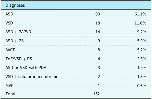
The study included 152 paediatric patients operated at a single centre. The operative details and postoperative outcomes are summarised in Tables 1 and 2. The median age and weight of patients at the time of surgery were 4.9 years (4 months–13 years) and 16 kg (6 kg–41 kg). Diagnostic modalities included routine transthoracic echocardiography confirmed with CT when there was doubt about the diagnosis. The most frequent primary diagnosis was atrial septal defect (n = 93), followed by ventricular septal defect (n = 18). However, there is a wide range of different pathologies in our cohort, including sinus venosus type atrial septal defect with partially anomalous pulmonary venous drainage, associated pulmonary valvar stenosis, ventricular septal defect with pulmonary stenosis, tetralogy of Fallot, partial and complete atrioventricular canal defects, subaortic membrane stenosis resection, and mitral valve surgery.
CHDs operated on through a mini right vertical axillary thoracotomy

Table 1. Long description
A table summarizing diagnoses of pediatric patients operated on through a mini right vertical axillary thoracotomy. The table has 10 rows and 3 columns. The columns are labeled Diagnoses, number of cases, and percentage. The rows list different types of congenital heart defects (CHDs) and their corresponding number of cases and percentages. Row 1: ASD, 93, 61.1 percent. Row 2: VSD, 18, 11.8 percent. Row 3: ASD + PAPVD, 14, 9.2 percent. Row 4: ASD + PS, 9, 5.9 percent. Row 5: AVCD, 8, 5.2 percent. Row 6: ToF/VSD + PS, 4, 2.6 percent. Row 7: ASD or VSD with PDA, 3, 1.9 percent. Row 8: VSD + subaortic membrane, 2, 1.3 percent. Row 9: MVP, 1, 0.6 percent. Row 10: Total, 152.
ASD = atrial septal defect; VSD = ventricular septal defect; PAPVD = partial anomalous pulmonary venous drainage; PS = pulmonary stenosis; AVCD = atrio-ventricular canal defect; Tof = Tetralogy of Fallot; PDA = patent ductus arteriosus; MVP = mitral valve plasty.
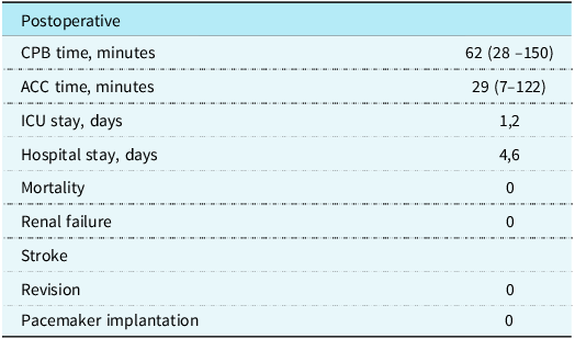
Intra-operative and in-hospital outcomes

Table 2. Long description
The table presents postoperative outcomes for pediatric patients. It includes seven rows and two columns. The columns are labeled Postoperative and values. The rows are labeled CPB time, minutes; ACC time, minutes; ICU stay, days; Hospital stay, days; Mortality; Renal failure; Stroke; Revision; Pacemaker implantation. The values are 62 (28-150); 29 (7-122); 1,2; 4,6; 0; 0; 0; 0; 0.
CPB = cardio-pulmonary bypass; ACC = aortic cross-clamp; ICU = intensive care unit.
All surgeries were performed through the mini right vertical axillary incision with no conversion to sternotomy. Central cannulation through the same incision was used for all patients for cardiopulmonary bypass. The myocardial protection technique used in all cases was the aortic crossclamp with antegrade cardioplegic arrest (cold blood cardioplegia, St Thomas), repeated every 20–30 min as needed. The median cross-clamp and bypass times were 29 min (7–122 min) and 62 min (28–150 min), respectively. The median ICU and hospital stays were 1.2 days and 4.6 days. There was no mortality during the follow-up period in our cohort. Three patients presented transitional atrioventricular block, needing temporary pacing, which then resolved spontaneously. There was no need for permanent pacemaker in our cohort, or reoperation during the follow-up period. All patients received standard postoperative pain management including intravenous opioids with oral analgesia in combination with either 1: an ultrasound-guided serratus plane block by anaesthetists (n = 20, 13.2%), or 2: with an intercostal nerve block (n = 132, 86.8%) with a ropivacaine 0.2 % (0.5–1 mL/kg) at the end of the operation. The median length of chest tube drainage was one day (range 1–2 days).
Discussion
In recent years, surgical treatment outcomes for CHDs have improved significantly, with minimal mortality rates for most defects. Consequently, the emphasis has shifted from survival to patient quality of life, postoperative comfort, and cosmetic results.Reference Dodge-Khatami, Dodge-Khatami, Nguyen and Rüffer3,Reference Dodge-Khatami and Dodge-Khatami4 This has led to the development of minimally invasive approaches in paediatric cardiac surgery. Median sternotomy remains the “gold standard” due to its excellent cardiac exposure and ease of access for cannulation. However, it is associated with significant surgical trauma, pain, prolonged recovery, and a visible scar on the anterior chest wall, which can cause psychological distress in children, especially for girls. A great number of minimally invasive approaches have been described, including partial sternotomy, the trans-xiphoid approach, the sub-mammary incision, anterolateral and posterolateral right-sided thoracotomies, the right axillary approach, and video-assisted thoracoscopic surgery. The major advantages of the vertical right axillary thoracotomy are that it preserves the integrity of the sternum and chest wall, lies far from breast tissue, and produces a virtually invisible scar. We are an emerging congenital heart program performing 300 open congenital cases per year, having started a dedicated paediatric program in 2018. Relatively shortly after a successful start using the standard median sternotomy approach, we elected to embark on a novel approach in 2022, the minimal invasive right axillary incision, and introduced this surgical treatment pathway to our hospital. Prior to implementation, all subspecialties involved with the new care pathway needed to be trained with specific protocols over a period of 2 months, to ensure a smooth start. This ultimately led to successful patient outcomes while maintaining a satisfactory work environment through pre-emptive team building and establishing confidence. As mentioned previously, we started with simple, not very small patients (with a weight > 10 kg) and with accumulation of the institutional experience, the indications were gradually expanded to perimembranous and inlet ventricular septal defects, partial anomalous pulmonary venous drainages and then more complex cases.
Our study demonstrated that the mini vertical right axillary thoracotomy is a safe and effective approach for the correction of a wide range of CHDs. In the presented series of 152 patients, there were no cases of conversion to sternotomy and no in-hospital or late mortality. The aortic cross-clamping and cardiopulmonary bypass times, as well as the length of ICU and hospital stay, were comparable to those of the traditional approach. Comparison of our results with literature data confirms the reproducibility of our experience, even though it is a newly implemented protocol and treatment pathway. According to the multicentre European study of the European Congenital Heart Surgeon’s Association, which included over 3,000 patients, mini-thoracotomy procedures were associated with no operative mortality, an extremely low complication rate, and a rare need for repeat interventions.Reference Dodge-Khatami, Gil-Jaurena and Hörer5 This demonstrates the high safety of the method when used correctly.
Dodge-Khatami et al. have demonstrated that the right axillary approach provides faster recovery, less pain, and a shorter hospital stay compared to sternotomy.Reference Dodge-Khatami, Dodge-Khatami, Nguyen and Rüffer3,Reference Dodge-Khatami and Dodge-Khatami4 Our series similarly noted early extubation, short durations of pleural drainage, intensive care unit stay and hospital stay. Of particular interest is the potential use of this approach not only for “simple” heart defects but also for more complex lesions. Our group performed repairs of partial and complete atrio-ventricular canal, pulmonary valvar stenosis and tetralogy of Fallot. A similar expansion of indications was also described in the studies of Heinisch et al., where minimally invasive technique demonstrated comparable results to sternotomy in the repair of ventricular septal defect and complete atrio-ventricular canal.Reference Heinisch, Wildbolz and Beck6 This confirms that limited exposure does not compromise the quality of repair with sufficient surgeon and team experience, with appropriate preparation and an approach-guided specific care protocol. In our experience, we perform CT angiography prior to surgery to better visualise the pulmonary arteries. Patients with important pulmonary stenoses and/or small right ventricular annular hypoplasia with z-scores < −2, who potentially need complex pulmonary artery reconstruction and/or a transannular patch, were generally not suitable or selected for the mini thoracic approach.
In our study, we observed transient atrioventricular blocks in three patients, which resolved spontaneously, without need for permanent pacemaker implantation. Similar conduction disturbances have also been described with the traditional approach and are primarily related to the defect’s anatomy rather than the type of surgical approach. The absence of the need for permanent pacemaker implantation in our series further confirms the safety of the method. Beyond the clinical results, a significant advantage of the mini vertical right axillary thoracotomy is the preservation of the chest wall’s integrity. The absence of a sternotomy avoids postoperative physical activity restrictions and facilitates earlier patient mobilisation. Furthermore, the concealed axillary scar has a significant psychological benefit and improves patient and parental acceptance of the procedure. This approach also minimises the risk of breast deformity in the future, since it lies far from the breast tissue.Reference Schreiber, Bleiziffer and Kostolny7
Beyond technical and clinical considerations, socio-cultural factors have had a strong influence on the practice of minimally invasive paediatric cardiac surgery at our institution. Cosmetic outcomes and the prevention of visible scars are particularly important for patients and their families, especially in the paediatric population, where long-term psychological and social consequences can be significant. Social integration, and, in some cases, concerns about self-esteem and future marital prospects clearly favour less invasive surgical approaches in our region. Increased awareness among referring paediatric cardiologists and their families, reinforced by growing publicity about successful cases, have also played a key role in stimulating the demand for minimally invasive techniques. These combined social and medical trends have pushed us to create a minimal invasive protocol and have led to positive feedback from both referring doctors and families.
As a result, the minimally invasive approach has become an important part of care in our institution. Despite its advantages, the method also has limitations. The main factors include a learning curve, the need for careful patient selection, and team teaching, especially in the early stages of implementation. Experience with a limited surgical field and a willingness to perform emergency conversion are also required, although this was not necessary in our series.
Conclusion
The vertical right axillary thoracotomy represents a safe and reproducible alternative to median sternotomy for the correction of a wide range of CHDs in children. It can be safely introduced even in emerging programmes, when appropriate information and preparation protocols are offered to the entire caregiving team. This method provides comparable surgical results to the median sternotomy with improved cosmetic and functional outcomes and can be considered the preferred approach in carefully selected patients in our centre.
Acknowledgements
None.
Financial support
This research received no specific grant from any funding agency, commercial, or not-for-profit sectors.
Competing interests
None.
Ethical standard
Not applicable.