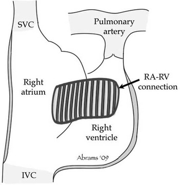
A young woman with a Bjork modification of the Fontan operation (Fig 1) was referred for pre-pregnancy counselling. Although reportedly New York Heart Association Class II, on objective exercise testing, she completed 6 minutes of the Bruce protocol, maximum workload eight metabolic equivalents (METs), stopping due to fatigue. Cardiac magnetic resonance imaging provided three-dimensional visualisation of the right atrium to right ventricular conduit, which measured 14 × 8 millimetres. It was freely regurgitant and moderately obstructed, mean pull-back gradient of 7 millimetres of mercury at cardiac catheterisation.
Line drawing showing a Bjork modification of the Fontan operation, in this patient for palliation of tricuspid atresia.

The Fontan circulation has a finely balanced physiology, which under normal circumstances, no arrhythmias, non-dilated right atrium, good ventricular function, low left ventricular end diastolic pressure, low pulmonary artery pressures, provides a good functional status. However, pregnancy is associated with a significant increase in cardiac workload and for good maternal outcome haemodynamics should be optimal.Reference Robson, Hunter, Boys and Dunlop1 It was therefore decided that the conduit obstruction and regurgitation should be alleviated before pregnancy, but because re-do surgery in the context of two prior sternotomies is associated with not insignificant risk, a percutaneous catheter intervention approach was chosen, with an attempt to implant a Melody valve within the conduit. Although never previously used for this indication, prior cardiac magnetic resonance imaging had defined the anatomy and morphology of the conduit, which looked suitable for Melody valve deployment.
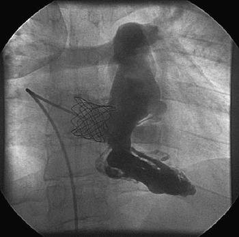
The procedure was performed under general anaesthesia without complication. The conduit was balloon-sized, pre-dilated (22 millimetres × 40 millimetres BIB; BVM Medical, NuMED Inc.) and pre-stented (EV3® MAX-LD IntraStent, Paris) to provide additional support and rigidity for the Melody valve. The Melody valve, 18 millimetres, was then implanted and post-dilated with a 20 millimetre high pressure balloon (EnsembleTM, Medtronic Inc.; MullinsTM, NuMed Inc). Post-implantation, there was no pull-back gradient across the conduit and right ventricular angiography demonstrated trivial regurgitation only (Fig 2).
Coronal view angiogram of right ventricle. Valved stent can be seen clearly in situ within the RA–RV conduit. The stenosis has been relieved across the conduit and there remains only trivial regurgitant flow.

The patient was discharged home the following day and at 6 months follow-up functional status had improved (New York Heart Assocation Class I) and echocardiography demonstrated a competent pulmonary stent valve within the conduit, with a peak velocity of 1.2 metres per second, trivial regurgitation and a mean gradient of 3.6 millimetres of mercury. On repeat exercise testing (Bruce protocol) she completed 7 minutes and 50 seconds, workload nine METS, and she is now planning a pregnancy.
Conclusion
We report the first case of Melody stent valve deployment within a Bjork Fontan connection, which successfully treated conduit obstruction and regurgitation. The improvement in haemodynamics resulted in a better exercise performance and functional class, which highlights its potential use for these patients both within and outside the context of pregnancy planning. Cardiac magnetic resonance imaging provided three-dimensional imaging, which enabled accurate definition of the right atrial to the right ventricular conduit anatomy for the Melody valve deployment, while the percutaneous catheter approach offered a low-risk treatment option for what was potentially a high-risk problem.Reference Khambadkone, Coats and Taylor2 This case also demonstrates that innovative thinking and teamwork are essential for the optimal management of adults with congenital heart disease.Reference Lutter, Ardehali, Cremer and Bonhoeffer3
Acknowledgements
The authors thank Professor P. Bonhoeffer, Great Ormond Street Hospital, London, Dr Marina Hughes, Consultant Cardiologist, Great Ormond Street Hospital, London, and Dr Dominic Abrams, Consultant Cardiologist, creator and designer of line image.