





A 28-year-old male with tricuspid atresia, normally related great vessels and ventricular septal defect status post staged surgical palliation with lateral tunnel Fontan at 3 years old was referred for catheterisation to treat Fontan stenosis and possibly embolise venous collaterals.
A chest/abdomen CT obtained for other clinical indications showed a dilated, tortuous venous vessel connecting the left renal vein to the hemiazygos system; the superior end of the connection was not identified (Figure 1).
Pre-procedure CT. ( a ), ( b ) 3D reconstruction of anterior view of large venous collateral from left renal vein (arrowheads). ( c ) Posterior view: the superior end of the collateral could not be defined (†).

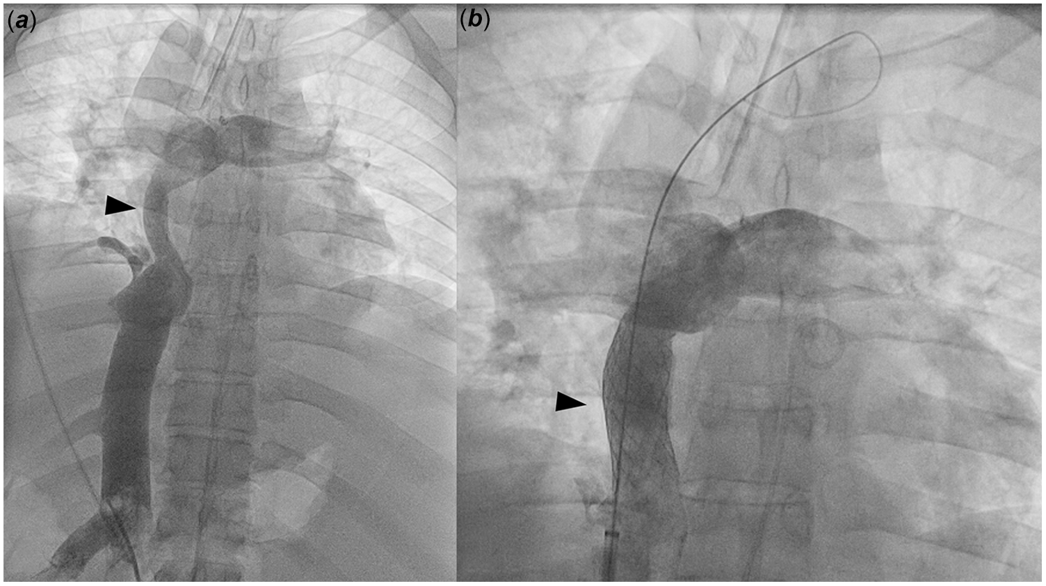
Haemodynamics showed top-normal Fontan pressure (14–15 mmHg) with a 2-mmHg gradient across the Fontan. Inferior caval vein angiography showed a dilated vessel with a severely stenotic Fontan conduit (narrowing from 18 mm to 9.8 mm); the left renal vein and collateral were not seen (Figure 2( a )). An 18 mm stent was placed in the Fontan with improved flow and gradient resolution (Figure 2( b )).
IVC angiography and first intervention. ( a ) Inferior caval vein angiogram showing dilated inferior caval vein and stenotic Fontan conduit (arrowhead); left renal vein not seen. ( b ) Improved patency after stenting.
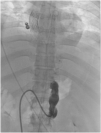
The left renal vein was directly engaged, and the venous collateral to the hemiazygos was visualised; it crossed midline and drained to the right pulmonary vein, decompressing the inferior caval vein from severe Fontan stenosis (Figure 3). To optimise antegrade flow through the freshly stented Fontan conduit and minimise the risk of thrombosis, the collateral was embolised with two 17 mm Amplatzer Septal Occluders (Abbott, Chicago, Illinois) with complete resolution of the shunt (Figure 4).
Collateral angiogram. ( a ) Angiogram of venous collateral from the left renal vein going cephalad and crossing the midline at the diaphragm (arrowheads). ( b ) Superior portion of the collateral (arrowheads) draining to right pulmonary veins. ( c ) Lateral imaging of the collateral vein (dashed lines) entering posteriorly into the pulmonary veins. Note the anterior systemic venous structures highlighted by the embolisation coils off the innominate vein and stent in the Fontan conduit.

Final intervention. Complete occlusion of venous collateral after placement of closure devices.

Systemic venous hypertension post-Fontan with decompression via systemic-pulmonary collaterals is common, but in this case, since the kidneys do not extract much oxygen, the patient did not experience systemic desaturation. This case highlights the importance of a high index of suspicion and the value of advanced imaging when intervening on patients with complex congenital heart disease.
Financial support
This research received no specific grant from any funding agency, commercial or not-for-profit sectors.
Competing interests
None.
Ethical standard
The authors assert that all procedures contributing to this work comply with the ethical standards of the relevant national guidelines on human experimentation and with the Helsinki Declaration of 1975, as revised in 2008; institutional review board oversight was not obtained for the report due to the de-identified nature.



 Open access

