Introduction
Successful cryopreservation of oocytes is important not only for human assisted reproductive technology, but also for the preservation of genetic resources in endangered species (Pukazhenthi & Wildt, Reference Pukazhenthi and Wildt2004). Cryopreservation technology also provides strong support for in vitro fertilization, in vitro culture systems and nuclear transplantation programmes (Dinnyes et al., Reference Dinnyes, Dai, Jiang and Yang2000). Cryopreservation of mammalian oocytes remains more difficult than cryopreservation of many other types of cells, despite much progress having been made since the technique was first reported by Whittingham (Reference Whittingham1977). Freezing and thawing procedures and cryoprotectants themselves can affect oocytes and related structures negatively, such as the zona pellucida (Larman et al., Reference Larman, Sheehan and Gardner2006; Meng et al., Reference Meng, Li, Wu, Dinnyes and Zhu2007; Tian et al., Reference Tian, Yan, Yang, Zhou, Yang and Zhu2007), spindle apparatus (Huang et al., Reference Huang, Chen, Tan and Chian2007), organelles (Valojerdi & Salehnia, Reference Valojerdi and Salehnia2005; Shi et al., Reference Shi, Jin, Kim, Kumar, Balasubramanian, Choe and Rho2007), cytoskeleton (Anon, 2006; Morato et al., Reference Morato, Izquierdo, Albarracin, Anguita, Palomo, Jimenez-Macedo, Paramio and Mogas2008) and chromosomes (Rojas et al., Reference Rojas, Palomo, Albarracin and Mogas2004; Peng et al., Reference Peng, Wang, Yin, Li, Li, Wang and Liu2008), resulting in impaired developmental potential of oocytes (Shaw et al., Reference Shaw, Oranratnachai and Trounson2000).
DNA methylation is an essential component of the epigenetic modification during development of the mammalian genome. Cytosine methylation at position 5 of the pyrimidine ring of CpG dinucleotides is a major site for epigenetic modification of DNA (Robertson & Wolffe, Reference Robertson and Wolffe2000). In general, DNA methylation plays a crucial role in transcriptional regulation such as X chromosome inactivation, genome imprinting and inactivation of transposable elements (Li, Reference Li2002; Santos & Dean, Reference Santos and Dean2004; Morgan et al., Reference Morgan, Santos, Green, Dean and Reik2005). Genome-wide demethylation is known to take place during preimplantation development in the mouse (Monk et al., Reference Monk, Boubelik and Lehnert1987; Howlett & Reik, Reference Howlett and Reik1991; Kafri et al., Reference Kafri, Ariel, Brandeis, Shemer, Urven, Mccarrey, Cedar and Razin1992). The global change in DNA methylation status is thought to be a key player in the nuclear reprogramming mechanism that erases epigenetic memories. Active demethylation of the paternal genome occurs in zygotes (Mayer et al., Reference Mayer, Niveleau, Walter, Fundele and Haaf2000; Oswald et al., Reference Oswald, Engemann, Lane, Mayer, Olek, Fundele, Dean, Reik and Walter2000), followed by passive demethylation of the maternal genome during cleavage stages (Howlett & Reik, Reference Howlett and Reik1991; Monk et al., Reference Monk, Adams and Rinaldi1991; Rougier et al., Reference Rougier, Bourc'his, Gomes, Niveleau, Plachot, Paldi and Viegas- Pequignot1998). After passive and active demethylation, the genome acquires a de novo methylation pattern from the activities of newly synthesized methyltransferases (Monk et al., Reference Monk, Boubelik and Lehnert1987). De novo methylation occurs specifically in the inner cell mass (ICM) but not in the trophectoderm of the blastocyst, establishing that cells can differentiate into various lineages (Santos et al., Reference Santos, Hendrich, Reik and Dean2002). Therefore, DNA methylation is crucial for the proper differentiation and development of early embryos, and an aberrant DNA methylation pattern can contribute to abnormal cleavage of blastomeres and interfere with normal development of embryos (Okano et al., Reference Okano, Bell, Haber and Li1999). Importantly, the DNA methylation patterns of vitrified–thawed oocytes and their IVF embryos are not known and need to be clarified based upon experimental results.
As immunoassay with an antibody 5-methylcytosine (5-MeC) was first used for the detection and quantitation of DNA methylation in the urine of cancer patients in 1991 (Reynauld et al., Reference Reynauld, Bruno, Boullanger, Grange, Barbesti and Niveleau1991), most analyses of global DNA methylation status in embryogenesis have been performed using immuno-staining for 5-MeC (Santos & Dean, Reference Santos and Dean2006). This method can quantify the extent of methylation in the nucleus at a single cell level, especially when there are very few cells, such as in early embryos (Santos & Dean, Reference Santos and Dean2006). Consequently, we analysed global DNA methylation using indirect immunocytochemical staining for 5-MeC.
In this study, we measured 5-MeC levels in the vitrified–thawed metaphase II (MII) mouse oocytes and their in vitro fertilized embryos, to determine whether potential changes in global genomic methylation due to oocyte vitrification compromise subsequent development when fertilized.
Materials and methods
Unless otherwise indicated, all chemicals and media were purchased from Sigma Chemical Co. (St. Louis, MO, USA).
Animals
Animals used in the study were 6–8-week-old Kunming white mice (Academy of Military Medical Sciences, Beijing, China), which were maintained at 20–22°C under a 14-h (6:00–20:00) light and 10-h (20:00–6:00) dark schedule. Experimental protocols for handing the mice were in accordance with the requirements of the Institutional Animal Care and Use Committee of the China Agricultural University.
Oocyte collection
One week after being housed in our animal facilities, mice were superovulated with 10 IU (intraperitoneal) equine chorionic gonadotropin (eCG; Ningbo Hormone Products Co., Ningbo, Zhejiang, People's Republic of China, L/N: 20100704), followed 48 h later by injection of 10 IU of human chorionic gonadotropin (hCG; Ningbo Hormone Products, L/N: 100801). Fourteen hours after hCG injection, the ampulla of the oviducts was opened with forceps and cumulus–oocyte complexes were recovered in M2 medium supplemented with 4 mg/ml bovine serum albumin (BSA), albumin fraction V powder; Roche Diagnostics GmbH, Mannheim, Germany). Cumulus cells were removed from oocytes with hyaluronidase (300 IU/ml) for 3 to 5 min in M2 medium. Only oocytes with normal morphology were used.
Vitrification and thawing of oocytes
Open-pulled straws (OPS) were made according to the method described by Vajta et al. (Reference Vajta, Booth, Holm, Greve and Callesen1997) with some modifications. Briefly, the straws (0.25 ml; IMV, L'Aigle, France) were heated to soften and pulled manually to get a straw of approximately 2 to 3 cm in length, 0.05 mm in inner diameter, and 0.10 mm in outer diameter.
Oocyte handling was performed at ambient temperature (25 ± 0.5°C); all vitrification media and oocytes were maintained at 37°C on a warming plate (Wenesco Inc., Chicago, USA). Oocytes were vitrified in EDFS30 (vitrification solution) that contained 15% (v/v) ethylene glycol (EG) and 15% (v/v) dimethylsulfoxide (DMSO) in Ficoll–sucrose (FS) medium (containing 300 g/L Ficoll, 171.2 g/L sucrose, and 3 mg/ml BSA in modified phosphate-buffered saline (mPBS) medium) by OPS method. First, oocytes were pretreated in 10% EG + 10% DMSO (pretreatment solution) for 30 s, then transferred to the EDFS30 in the narrow end of the pulled straw and held for 25 s, after which the straws were immediately plunged into liquid nitrogen (LN2). Ten oocytes were loaded into each OPS. Oocytes were stored for at least 24 h in LN2, then removed and thawed. The tip of each OPS was put into 0.5 mol/l sucrose, and released oocytes were kept in other fresh 0.5 M sucrose for 5 min to remove EDFS30. Finally, the oocytes were placed into 100 μl droplets of M2 medium in a Petri dish (35 mm × 10 mm, Corning Incorporated, Corning, New York, USA) for experimental use. The oocytes exposed to pretreatment solution and EDFS30 without being plunged into LN2 were designated as the toxicity group, and untreated oocytes were the controls.
IVF and embryo culture
Oocytes in each of the three groups were placed individually into 75 μl drops of human tubal fluid (HTF) medium under mineral oil, into which a suspension of 10 μl spermatozoa was added for fertilization, with a final concentration of 5 × 10⁶ sperm/ml. Five hours after IVF, eggs were removed from the fertilization drop, washed in HTF medium and cultured in 75 μl drops of HTF medium. Then 2-cell, 4-cell, 8-cell, morulae and blastocysts stages were collected individually about 24 h, 48 h, 60 h, 72 h and 96 h after fertilization.
Immunocytochemical staining of oocytes and early embryos
The MII oocytes and early embryos to be subjected to staining were washed with 0.1% Tween-20 in mPBS, fixed for 30 min in 4% paraformaldehyde in mPBS, permeabilized with 0.5% Triton X-100 in mPBS for 30 min and treated in 2 M HCl for 30 min at 25°C. After extensive washing with 0.1% Tween-20 in mPBS, cells were blocked for 2 h in 2% BSA in mPBS. Cells were incubated with anti-5-MeC antibodies (Epigentek Group Inc., USA) overnight at 4°C. After this step, oocytes and embryos were washed extensively and probed with fluorescein isothiocyanate (FITC)-conjugated anti-mouse IgG (Santa Cruz Biotech, USA) for 1.5 h at 37°C. The DNA was visualized by counterstaining the cells with 10 μg/ml propidium iodide (PI) for 10 min. After extensive washing, oocytes and embryos were treated in 10% triethylene diamine (DABCO) in mPBS, mounted on slides, and fluorescence detected using a Nikon spectral confocal scanning microscope (Nikon Corporation, Tokyo, Japan). System settings were constant for examination of all cells. Each treatment was repeated three times with a minimum of 15 oocytes or embryos for each treatment. Oocytes were treated with 2% BSA instead of monoclonal primary antibody as the negative control.
Fluorescence intensities were quantified by EZ-C1 Free Viewer software (Nikon), as described by Aoki et al. (Reference Aoki, Worrad and Schultz1997), with some modifications. In brief, the pixel value of fluorescence was measured within a constant area from 10 different regions of chromosomes and 10 different regions of cytoplasm, and the average cytoplasmic value was subtracted from the average chromosomal value.
Statistical analysis
Oocytes and embryos were distributed randomly in each experimental group, and at least three replicates were conducted for each experiment. Statistical analysis was performed using SPSS 17.0 software. In our experiment, percentage data such as rates of cleavage and blastocyst were arcsine-transformed before analysis to maintain homogeneity of variance, and the normal distribution of the data was checked using the Shapiro-Wilk test. Differences among the treatments in each experiment were evaluated by Duncan's test, and values P < 0.05 were considered as the criterion for statistical significance.
Results
Effect of oocyte vitrification on development of embryos derived by IVF
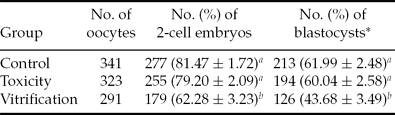
To evaluate the effect of OPS vitrification on oocyte development, the percentage of cultured OPS vitrified oocytes that reached advanced stages of development were compared with those of fresh and toxicity cultured oocytes (Table 1). Percentages of successful 2-cell embryos (62.28%) and blastocysts (43.68%) from vitrified–thawed oocytes were lower (P < 0.01) than those of fresh (81.47%, 61.99%) and toxicity (79.20%, 60.04%) oocytes. However, the toxicity group did not differ significantly from the control group for either trait (P > 0.05).
Effect of vitrification on developmental competence following in vitro fertilization

a ,b Different superscripts within the same column identify significant differences (P < 0.01). *Blastocyst rate is the percentage of total oocytes that reached the blastocyst stage.
Effect of vitrification on DNA methylation in oocytes and early embryos
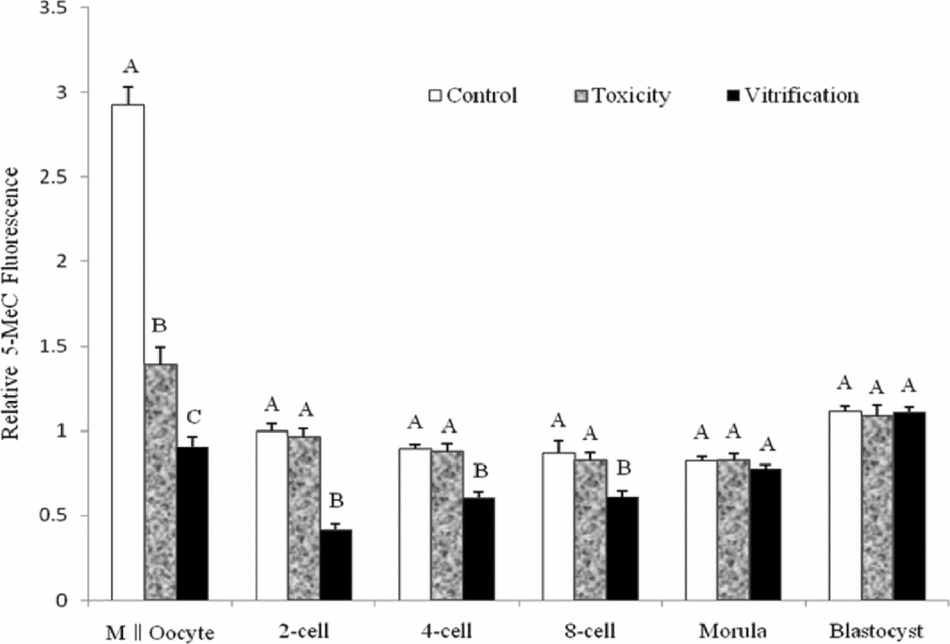
Trends across developmental stages in the pattern of DNA methylation of early embryos were similar in the three treatment groups (Fig. 1). Metaphase II oocytes were strongly stained, and after in vitro fertilization, substantial losses of 5-MeC fluorescence intensity were observed, reaching the lowest point at the morula stage. However, an intense signal was clearly visible by the blastocyst stage, representing the beginning of de novo methylation.
Effect of oocyte vitrification on relative intensity of anti-5-methylcytosine (anti-5-MeC) fluorescence of metaphase II (MII) oocytes and early embryos. Different letters signify treatments within an embryonic stage of development that differ at P < 0.01. The fluorescence intensity of a 2-cell embryo from the control group was set as 1.
As shown in Fig. 1, the 5-MeC fluorescence intensity declined (P < 0.01) significantly across developmental stage for oocytes in the toxicity group, and DNA methylation level of the vitrified–thawed oocytes was significantly lower (P < 0.01) than that of fresh or vitrification solution treated oocytes. During early embryo development, 5-MeC fluorescence intensity in the vitrification group was significantly decreased (P < 0.01) in 2-cell, 4-cell and 8-cell embryos compared with those in the control and toxicity groups, but the difference was not significant between the toxicity and control groups (P > 0.05).
Immunofluorescence results on 5-MeC of MII oocyte and early embryos from the control and vitrification groups are shown in Fig. 2.
Immunofluorescence results on anti-5-methylcytosine (anti-5-MeC) of metaphase II (MII) oocytes and early embryos from the control and vitrification groups. Immunofluorescence results on 5-MeC from MII oocytes (A); 2-cell embryos (B); 4-cell embryos (C); 8-cell embryos (D); morulae (E); and blastocysts (F) in the control group; and those from MII oocytes (G); 2-cell embryos (H); 4-cell embryos (I); 8-cell embryos (J); morulae (K); and blastocysts (L) in the vitrification group are shown. (a–l) Oocytes/embryos in panels (a–l) were counterstained with propidium iodide to identify the nuclear compartment. Bar = 20 μm.
Discussion
Effect of oocyte vitrification on development of embryos derived by IVF
Despite recent technological advances, cryopreservation of mammalian oocytes remains difficult, as reflected by reduced rates of fertilization and embryonic development compared with those of fresh oocytes and embryos (Chen et al., Reference Chen, Lien, Cheng, Chen, Ho and Yang2001; Larman et al., Reference Larman, Sheehan and Gardner2006; Tian et al., Reference Tian, Yan, Yang, Zhou, Yang and Zhu2007; Fan et al., Reference Fan, Li, Liu, Meng, Wang, Hou, Zhou and Zhu2008; Li et al., Reference Li, Pei, Zhou, Suo, Wang, Wu, Fu, Hou and Zhu2011). In order to improve the efficiency of oocyte cryopreservation, the OPS method for oocyte vitrification was developed subsequently (Vajta et al., Reference Vajta, Holm, Kuwayama, Booth, Jacobsen, Greve and Callesen1998). Due to the smaller inner diameter (0.05 mm) and thinner wall (0.025 mm) of the OPS in comparison with a standard straw, (0.25 ml, IMV, L'Aigle, France; Vajta, Reference Vajta2000), the OPS method is considered to have advantages of increased cooling and warming rates (20,000°C/min), reduced cryoprotectant concentration and toxicity, and decreased chilling injury.
Our study showed that cleavage and blastocyst formation rates both were decreased significantly in the vitrification group, but not in the toxicity group as we reported previously (Meng et al., Reference Meng, Li, Wu, Dinnyes and Zhu2007; Li et al., Reference Li, Pei, Zhou, Suo, Wang, Wu, Fu, Hou and Zhu2011). This finding suggests that the compromised developmental potential was mainly due to the process of vitrification of oocytes rather than to toxic effects of cryoprotectants.
Effect of vitrification on DNA methylation in oocytes and early embryos
Dynamic reprogramming of DNA methylation in the early mouse embryo has been suggested in a number of investigations (Monk et al., Reference Monk, Boubelik and Lehnert1987; Howlett & Reik, Reference Howlett and Reik1991; Kafri et al., Reference Kafri, Ariel, Brandeis, Shemer, Urven, Mccarrey, Cedar and Razin1992; Santos et al., Reference Santos, Hendrich, Reik and Dean2002). We observed substantial loss of 5-MeC fluorescence intensity during early embryonic development. The signal reached its lowest point during the morula stage, but it was restored by the blastocyst stage. These results are in close agreement with those reported by Santos et al. (Reference Santos, Hendrich, Reik and Dean2002).
In comparing the 5-MeC fluorescence intensity of oocytes among the three groups prior to in vitro fertilization, we found that the immunoreaction product was greatly reduced in the oocytes exposed to vitrification solution but that it was reduced to a lesser extent in the vitrified–thawed oocytes. Wang et al. (Reference Wang, Xu and He2010) also reported that cryopreservation led to alteration of DNA methylation of imprinted genes (H19, Igf2) in the mouse. Additionally, DMSO could affect epigenetic changes by acting on one or more DNA methyltransferases (Dnmts; Iwatani et al., Reference Iwatani, Ikegami, Kremenska, Hattori, Tanaka, Yagi and Shiota2006). Thus, on the one hand, the change of 5-MeC level in oocytes due to OPS vitrification in our study may have resulted partly from DMSO in the vitrification solution. On the other hand, the observation that DNA methylation of the vitrified–thawed oocytes was significantly lower than that of the toxicity oocytes suggests that the chilling stress after vitrification solution treatment might have aggravated the aberrant expression of 5-MeC.
By comparing the 5-MeC fluorescence intensity of embryos among the three groups after in vitro fertilization, we observed significant loss of immunoreaction product in 2-cell, 4-cell and 8-cell embryos in the vitrification group but not in the toxicity group. These observations suggest that the process of oocyte vitrification might result not only in abnormal DNA methylation in the mouse oocytes themselves, but also in their IVF early embryos.
Given the fact that DNA methylation participates in induced chromatin deacetylation (Eden et al., Reference Eden, Hashimshony, Keshet, Cedar and Thorne1998), the reduction in global genomic methylation in the present study supports our previous results that cryopreservation leads to histone acetylation by decreasing HDAC1 (histone deacetyltransferase1) expression (Li et al., Reference Li, Pei, Zhou, Suo, Wang, Wu, Fu, Hou and Zhu2011). Global hypomethylation is one of the common mechanisms responsible for aberrant expression of genes, including cell aging (Issa, Reference Issa2000) and tumorogenesis (Baylin, Reference Baylin1997; Soares et al., Reference Soares, Pinto, Cunha, Andre, Barao, Sousa and Cravo1999), as DNA methylation establishes a silent chromatin state by collaborating with histone modifications (Bird & Wolffe, Reference Bird and Wolffe1999; Urnov & Wolffe, Reference Urnov and Wolffe2001; Wolffe & Matzke, Reference Wolffe and Matzke1999). Furthermore, it was proposed in 1975 that DNA methylation could be responsible for the stable maintenance of a particular gene expression pattern through mitotic cell division (Holliday & Pugh, Reference Holliday and Pugh1975; Riggs, Reference Riggs1975). Since then, ample evidence has been obtained to support this concept, and DNA methylation as part of a genome-wide defense system is now recognized to be a chief contributor to chromosome stability and structural integrity (Yoder et al., Reference Yoder, Walsh and Bestor1997; Jaenisch & Bird, Reference Jaenisch and Bird2003), indicating that an appropriate level of global genomic methylation is essential for early embryonic development (Rogers et al., Reference Rogers, Francis, Sulik, Alles, Massaro, Zucker, Elstein, Rosen and Chernoff1994; Carambula et al., Reference Carambula, Oliveira and Hansen2009). Accordingly, we hypothesize in our study that inadequate DNA methylation due to oocyte vitrification decreases genomic stability and leads to aberrant expression of genes, which consequently blocks early embryo development and increases apoptosis in embryos.
Although cryoprotectants appeared to affect DNA methylation in oocytes, the extent of reduction was relatively small; and DNA methylation level could basically return to normal promptly after fertilization. Thus, the developmental potential of the oocytes exposed to vitrification solution remained similar to the fresh oocytes and apparently was unaffected. DNA methylation level was decreased to a lesser extent in the oocyte vitrification group than in the toxicity group, and its restorative speed in the early embryos was also lower. These findings lead to the deduction why oocyte vitrification had an overall negative impact on early embryo development.
In conclusion, a reduction in global genomic methylation due to MII oocyte vitrification may result in compromised in vitro developmental potential in early mouse embryos.
Acknowledgements
This work was supported by grants from the National 863 project (No. 2008AA101007) and the National Natural Science Foundation of the People's Republic of China (No. 30972102). The authors thank Professor William D. Hohenboken for proofreading this manuscript.