Introduction
Sialolithiasis is one of the most common diseases of the salivary glands, with an incidence of 2.9–5.5 per 100 000 per year.Reference Sherman and McGurk 1 – Reference Schroder, Homoe, Wagner, Vataire, Lundager Madsen and Bardow 4 It affects men and women equally, and typically presents in patients aged 30–60 years.Reference Schroder, Homoe, Wagner, Vataire, Lundager Madsen and Bardow 4 – Reference Sigismund, Zenk, Koch, Schapher, Rudes and Iro 6 Sialolithiasis may obstruct the duct and prevent the physiological flow of saliva. Symptoms are recurrent and painful, and often present as meal-related (chewing and taste related) swelling of the involved gland.Reference Sigismund, Zenk, Koch, Schapher, Rudes and Iro 6 , Reference Lustmann, Regev and Melamed 7 Treatment depends on the size of the sialolith, and can range from gentle massage of the gland and duct to endoscopic or surgical removal of the stone, or, in recurrent and severe cases, gland resection.Reference Wilson, Meier and Ward 8 , Reference Witt, Iro, Koch, McGurk, Nahlieli and Zenk 9
The aetiology of sialolithiasis is debated; theories include retrograde formation of stones, the existence of intracellular micro calculi or intraductal mucoplugs.Reference Harrison 10 , Reference Marchal, Kurt, Dulguerov and Lehmann 11 Furthermore, tobacco smoking, diuretic treatments,Reference Huoh and Eisele 12 – Reference Yiu, Kalejaiye, Amdur, Todd Hesham and Bandyopadhyay 14 or systemic factors such as elevated levels of serum potassium, sodium and parathyroid hormone (PTH) have been suggested to influence sialolithiasis formation.Reference Lustmann, Regev and Melamed 7 , Reference Harrison 10 , Reference Yiu, Kalejaiye, Amdur, Todd Hesham and Bandyopadhyay 14 Thus, while the aetiology remains elusive, the observation that decreased salivary flow increases risk of stone formationReference Triantafyllou, Harrison and Garrett 15 has led to various speculations that local factors play a role in sialolithiasis.
Sialolithiasis occurs mainly in the submandibular gland,Reference Sigismund, Zenk, Koch, Schapher, Rudes and Iro 6 , Reference Lustmann, Regev and Melamed 7 , Reference Carter 16 potentially influenced by a combination of the anatomy of the gland with a long ascending duct, and the higher viscosity of the mucous submandibular gland secretion compared to the strictly serous secretion from the parotid gland where sialolithiasis is less frequent.Reference Delli, Spijkervet and Vissink 3 , Reference Carter 16 Sialolithiasis is also rarely bilateral, which argues against a systemic aetiology.Reference Blatt 17 Apart from local factors, exogenous factors such as drinking water concentration of calcium and magnesium may also play a role in sialolithiasis formation, and constitute a pre-condition for stone formation.Reference Schroder, Homoe, Wagner, Vataire, Lundager Madsen and Bardow 4
The ultra-structural composition and the structure of sialoliths are well described; sialoliths are made of a calcified solid mass mainly consisting of a core of organic material surrounded by inorganic layers, either arranged concentrically or irregularly, and with variable degrees of mineralisation.Reference Harrison 10 , Reference Grases, Santiago, Simonet and Costa-Bauza 18 – Reference Mimura, Tanaka, Ichinose, Kimijima and Amagasa 21 The inorganic components are primarily calcium and phosphate in the form of hydroxyapatite (Ca10(PO4)6(OH)2), magnesium whitlockite (Ca9MgH(PO4)7), and other calcium phosphate forms such as brushite (CaHPO4.2H2O).Reference Teymoortash, Buck, Jepsen and Werner 22 , Reference Gopal, Calvo, Ito and Sabine 23 Therefore, it is not surprising that chemical analyses of saliva from sialolithiasis patients confirm an increased concentration of calciumReference Grases, Santiago, Simonet and Costa-Bauza 18 , Reference Su, Zhang, Ke, Zheng, Chu and Liao 24 together with decreased concentrations of crystallisation inhibitors (inositol phosphate), which may promote precipitation.Reference Grases, Santiago, Simonet and Costa-Bauza 18 However, it is not known whether this difference is found when both the sialolith is present and when it has been surgically removed.
This study aimed to examine whether the salivary inorganic composition in patients with sialolithiasis differs from that of healthy controls, and whether the salivary inorganic composition changes after the sialolith has been surgically removed. We hypothesised that specific inorganic components found in relation to sialoliths are increased in sialolithiasis patients, both before and after surgery. Finally, we expected the salivary flow to be low before surgery and to normalise after surgical removal of the sialolith.
Materials and methods
Study population
Within a one-year period (2012–2013), we prospectively and consecutively recruited patients (n = 40) who were undergoing surgery for sialolithiasis at Nordsjaellands University Hospital, by sialendoscopy or sialodochotomy, or by a combination of these procedures. The inclusion criterion was a known diagnosis of sialolithiasis, confirmed by ultrasound, by direct visualisation during diagnostic sialendoscopy, or by typical symptomatology such as swelling and pain of the gland. The exclusion criteria were: previous sialendoscopy, known stenosis of the duct, pregnancy, Sjögren's disease, age of less than 18 years, pre-operative antibiotic treatment or use of mouth washes.
The healthy controls (n = 40) were identified through advertising. These individuals were matched to the patients by gender, age and geography. Saliva was collected once, following the same procedure as for the patient group.
The required sample size was derived from a power calculation and based on previous data on the total calcium concentration in saliva (in terms of delta values, reflecting differences in calcium concentration between cases and controls, and standard deviations (SDs) for calcium concentrationsReference Grases, Santiago, Simonet and Costa-Bauza 18 ). The input parameters were as follows: delta = 18.21 mg/l, SD = 19.92 mg/l, significance level = 0.01, power = 0.80, with a two-sided hypothesis. The level of significance in the power calculation was set to 1 per cent, corresponding to the maximal Bonferroni correction (κ > 5), because of multiple analyses on related data. This revealed a necessary sample size of 30 patients. We aimed to include at least 40 patients and a similar number of healthy controls to avoid dropout problems.
The study was approved by the Ethical Committees of the Capital Region, Denmark (H-4-2011-022).
Saliva sample collection
Patients were examined twice: before surgery (range, 1–28 days before) and again 3–6 months after surgery. At both consultations, unstimulated whole saliva was collected for 10 minutes using the spitting method.Reference Navazesh and Kumar 25 Samples were kept frozen at −80 °C until all chemical determinations could be performed on all samples simultaneously. The container utilised for the collection of saliva was weighed before and after collection, and the flow rate was calculated as millilitres per minute, assuming a saliva density of 1 g/ml. Saliva collection was standardised: it was carried out during the daytime, and eating, oral hygiene methods, smoking and chewing of gum were prohibited 1 hour prior to examination. One physician who was trained in the sampling technique collected all saliva samples. Blood samples were obtained for each patient before surgery, and included evaluation of PTH and free ionised calcium (Ca++).
Saliva analyses
Salivary concentrations of sodium and potassium were determined by atomic absorption spectroscopy (Perkin Elmer, Norwalk, Connecticut, USA), in the emission mode, at lines 589.0 nm and 766.5 nm respectively, keeping spectral overlap at a minimum. Calcium and magnesium concentrations in saliva were also determined by atomic absorption spectroscopy, but in the absorption mode, at lines 422.7 nm and 285.2 nm respectively. For all determinations conducted by atomic absorption spectroscopy, the samples were diluted with a solution of 99 per cent Millipore™ water and 1 per cent concentrated nitric acid. Sodium and potassium samples were diluted 1000 fold, calcium samples were diluted 100 fold and magnesium samples were diluted 50 fold. For calcium and magnesium, strontium chloride and potassium chloride were added to the matrix to reduce complexation and oxysalt formation. Colorimetric methods were used to determine phosphorous and chloride concentrations in saliva: phosphorous by the molybdenum reaction at 700 nm and chloride by the mercury-chloride/iron-TPTZ reaction at 610 nm.Reference Fried, Hoeflmayr and Velosy 26 All measurements were performed twice; in cases of discrepancies, a third determination was performed. The pH of saturation with respect to hydroxyapatite (that is, ionic product equal to the solubility product of hydroxyapatite, i.e. 10−117. 3 M18) was determined by iterative search and testing,Reference Bardow, Lykkeaa, Qvist, Ekstrand, Twetman and Fiehn 27 and iterations were repeated until the pH used for determination of phosphate differed less than 1 per cent from the estimated pH of saturation.
Statistical analyses
Statistical analyses were conducted using SAS® version 9.4 software. Because of considerable variations in results, the non-parametric Wilcoxon signed rank tests (paired and unpaired) were used to compare the groups (pre-operative, post-operative, controls). The significance level was set to p < 0.05.
Results
Of the 40 patients enrolled, 20 were men and 20 women, with a median age of 45 years (range, 21–76 years). The 40 matched healthy controls also consisted of 20 men and 20 women, with a median age of 43 years (range, 18–73 years). Among the patients, 36 had sialoliths removed from the duct of the submandibular gland and 4 had sialoliths removed from the duct of the parotid gland.
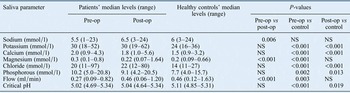
Prior to surgery, the patient group had a median unstimulated salivary flow of 0.27 ml/minute (range, 0.09–0.82 ml/min), which was significantly lower than that of the healthy controls (Figure 1a; p < 0.01). However, after surgery, the flow among patients increased to 0.46 ml/minute, and thereby matched the flow level of the healthy controls (p = 0.60).
Boxplots showing (a) unstimulated whole saliva flow rate, (b) sodium concentration, (c) potassium concentration and (d) chloride concentration, in 40 patients (before and after salivary stone removal) and in 40 matched healthy controls (asterisks indicate outliers). Pre-op = pre-operative; post-op = post-operative

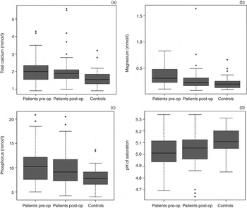
Pre-operative sialolithiasis patients had higher concentrations of potassium (Δ6 mmol/l), chloride (Δ6 mmol/l), calcium (Δ0.5 mmol/l), magnesium (Δ0.1 mmol/l) and phosphorous (Δ2.5 mmol/l) compared to the healthy controls (Table I, Figure 1c and d, and Figure 2a–c; p < 0.01 or less). The salivary concentrations of potassium, chloride, calcium and phosphorous remained higher in the sialolithiasis patient group compared to the healthy controls, even after surgical removal of the sialoliths (p ≤ 0.01). Magnesium concentration in the sialolithiasis patients decreased significantly after surgery (p < 0.001) to a level that did not differ from that of the healthy controls (p = 0.07) (Table I, Figure 2b). The sodium concentration increased in the sialolithiasis patients after surgery (p = 0.006), but neither the pre-operative concentration nor the post-operative concentration differed from that of the healthy controls (p = 0.23 and p = 0.09, respectively). The pH of saturation with respect to hydroxyapatite was lower for the sialolithiasis patients, both before and after surgery, compared to that of the healthy controls (Figure 1d; p = 0.02 or less). Sialolithiasis patients had mean PTH and mean ionised calcium levels of 4.4 ± 2.1 pmol/l and 1.24 ± 0.04 mmol/l, respectively.
Boxplots showing (a) total calcium concentration, (b) magnesium concentration, (c) phosphorous concentration and (d) unstimulated whole saliva pH of saturation with respect to hydroxyapatite, in 40 patients (before and after salivary stone removal) and in 40 matched healthy controls (asterisks indicate outliers). Pre-op = pre-operative; post-op = post-operative

Inorganic composition of unstimulated whole saliva*

Comparisons among the groups were made using Wilcoxon signed rank tests (paired and unpaired). *Obtained from 40 patients (before and after surgical removal of sialoliths) and from 40 healthy controls. Pre-op = pre-operative; post-op = post-operative; NS = non-significant
Discussion
In this series, 4 out of 40 patients (10 per cent) had stones in the parotid gland, compared to 36 (90 per cent) with stones in the submandibular gland. This distribution corresponds to previous findings.Reference Sigismund, Zenk, Koch, Schapher, Rudes and Iro 6 , Reference Kraaij, Karagozoglu, Kenter, Pijpe, Gilijamse and Brand 13 , Reference Lustmann and Shteyer 28 The salivary obstruction was cured by the sialolithiasis surgery, and the patients achieved a normal salivary flow rate after surgery. Because the flow rate normalised after surgery, there is no apparent physiological explanation for the remaining differences in the saliva composition between sialolithiasis patients and healthy controls.
In general, the patients had higher saliva concentrations of calcium, magnesium and phosphorous compared to the healthy controls. The higher concentrations of calcium and phosphorous are in accordance with findings of previous studies.Reference Grases, Santiago, Simonet and Costa-Bauza 18 , Reference Su, Zhang, Ke, Zheng, Chu and Liao 24 However, in contrast to earlier studies, we observed a significantly higher concentration of magnesium in the patients before sialolith surgery, although this concentration decreased to a level similar to that of the healthy controls after surgery.Reference Grases, Santiago, Simonet and Costa-Bauza 18 , Reference Nolasco, Anjos, Marques, Cabrita, da Costa and Mauricio 19 , Reference Su, Zhang, Ke, Zheng, Chu and Liao 24 We speculate that the magnesium concentration decreases after surgery may have been caused by a small signal (Mg++ ions detaching from the stone surface) arising via the saliva from the stone. Nonetheless, as magnesium is a main part of some sialoliths, high levels of this ion will increase the pressure for precipitation of magnesium whitlockite, which coincides well with the fact that this mineral phase is abundant in some sialoliths. Thus, as calcium, magnesium and phosphate comprise the main components of most sialoliths in the form of hydroxyapatite, magnesium whitlockite and brushite,Reference Grases, Santiago, Simonet and Costa-Bauza 18 , Reference Teymoortash, Buck, Jepsen and Werner 22 high concentrations of these ions will make the saliva substantially oversaturated with respect to these calcium phosphate salts, and therefore will likely cause precipitation and stone formation. This correlates with the theory that sialoliths form as calcium precipitates on the phospholipids of degenerated mucosal membranes.Reference Harrison 10 This also supports the fact that sialolithiasis occurs primarily in the submandibular gland, as this gland has a high content of calcium compared to the parotid gland.Reference Harrison, Triantafyllou, Baldwin, Garrett and Schafer 29
Given the saliva collection method utilised (i.e. whole saliva), we were not able to measure the saliva pH. This can only be done in a closed system because of extensive carbon dioxide evaporation and thereby a misleading pH.Reference Bardow, Moe, Nyvad and Nauntofte 30 Therefore, the degree of saturation with respect to hydroxyapatite could not be calculated. Instead, the pH value of saturation with respect to this mineral form was calculated, as this measure is independent of the actual pH. The lower the pH value of saturation becomes, the greater the likelihood of mineral precipitation. As the sialolithiasis patients had a significantly lower pH value of saturation with respect to hydroxyapatite, both before and after surgery, compared to the healthy controls, we assume that the low pH value of saturation together with the highest content of ions involved in calculus formation, especially calcium and phosphorous, could be important factors for an increased risk of sialolithiasis formation in sialolith patients compared to the healthy controls.
Sodium and chloride are extensively reabsorbed in the striated ducts of the major salivary glands at low salivary flow rates. As sialoliths obstructing the duct can cause stagnation of saliva in the duct, this may allow more time for re-absorption and may explain the lower concentration in the sialolithiasis patients before surgery compared to that of the healthy controls, although this theory does not apply for chloride that is also reabsorbed in the salivary duct. Thus, the high chloride concentration cannot be explained by common physiological mechanisms. However, it could be speculated that tissue damage due to stone formation and surgery may have influenced these values, in addition to attempts by the gland to re-establish electroneutrality (cation and anions) given the larger amount of cations in the saliva.
-
• Sialoliths consist mainly of calcium and phosphate salts
-
• Sialolithiasis patients had higher salivary concentrations of the ions that constitute the main inorganic phase of most sialoliths
-
• The pH of saturation with respect to hydroxyapatite was lower for sialolithiasis patients than healthy controls, before and after surgery
-
• Sialolithiasis patients had reduced salivary flow prior to surgery, but flow normalised after sialolith removal
Apart from the saliva composition, exogenous factors may also play a role, or function as a pre-condition for sialolithiasis,Reference Schroder, Homoe, Wagner, Vataire, Lundager Madsen and Bardow 4 but the mechanism remains unknown. It is interesting to speculate how this can influence sialolith formation, as the calcium metabolism is strictly regulated in humans. For all patients, we measured PTH and ionised calcium to find evidence of a poorly regulated calcium metabolism. However, all blood test results fell within the normal range (PTH = 4.4 ± 2.1 pmol/l, ionised calcium = 1.24 ± 0.04 mmol/l).
The strength of this study is that all patients attended a follow-up appointment. Furthermore, comprehensive measurements of inorganic components were conducted; such detailed analysis is likely to indicate the contribution of these interrelated factors. The study limitations include the fact that additional data collection could have further elucidated the importance of organic salivary composition, as this may also play a role in precipitation. The study did not allow for the characterisation of patient duct anatomy, including length and diameter, which also constitute risk factors for sialolithiasis.Reference Delli, Spijkervet and Vissink 3 Finally, although the study had a prospective case–control design, selection bias cannot be ruled out; the patients were from only one regional hospital, and the findings may therefore not be transferable to other areas because of different drinking water compositions between regions.Reference Schroder, Homoe, Wagner, Vataire, Lundager Madsen and Bardow 4
In conclusion, the present study adds to the knowledge concerning the pathophysiology of sialolithiasis. Sialolithiasis patients had reduced salivary flow prior to surgery, but salivary flow normalised after the sialoliths were surgically removed. Sialolithiasis patients had a different inorganic salivary composition, with a higher concentration of the ions that constitute the main inorganic phase of most sialoliths. For calcium and phosphorous, this was the case both before and after surgery, and for magnesium this was the case prior to surgery. We conclude that these variations in saliva composition, with higher concentrations especially for calcium, magnesium and phosphorous, seem to be associated with an increased risk of sialolithiasis.
Acknowledgements
The authors wish to thank the nurses and staff at Nordsjaellands University Hospital for their assistance during the study. This work was supported by: the Research Foundation at Nordsjaellands University Hospital (grant number E2012), the A P Møller Foundation for the Advancement of Medical Science (grant number 15-355), and Inge and Per Refshall's Research Grant (number 2016-1840/68KBN).