Introduction
Schizophrenia and bipolar disorder are common, disabling psychiatric illnesses that together affect one in 50 people at some stage in their lifetime. There are no diagnostic tests or specific symptoms that uniquely distinguish the two disorders, which are instead separated on the basis of characteristic clusters of symptoms and their longitudinal course. Evidence from imaging studies of each disorder suggests that there may be differences between schizophrenia and bipolar disorder in the structure and function of the hippocampal formation, a region of the brain known to play a prominent role in memory formation (Heckers, Reference Heckers2001; Frey et al. Reference Frey, Andreazza, Nery, Martins, Quevedo, Soares and Kapczinski2007). However, to date no studies have directly compared hippocampal function in schizophrenia and bipolar disorder patients using non-invasive neuroimaging techniques.
There is extensive evidence of abnormalities in both the structure and the function of the hippocampus in schizophrenia (Heckers, Reference Heckers2001). Structural neuroimaging studies have shown reduced hippocampal volumes in the disorder, a finding that has been confirmed by meta-analysis (Wright et al. Reference Wright, Rabe-Hesketh, Woodruff, David, Murray and Bullmore2000). Hippocampal volume reductions are also seen in the unaffected and at-risk relatives of individuals with schizophrenia, suggesting that such changes are in part heritable and are not simply secondary to the consequences of illness (Boos et al. Reference Boos, Aleman, Cahn, Pol and Kahn2007). Behavioural studies have shown memory deficits in schizophrenia in tasks known to depend on the hippocampus, such as tests of episodic and associative memory (Aleman et al. Reference Aleman, Hijman, de Haan and Kahn1999; Danion et al. Reference Danion, Rizzo and Bruant1999; Achim & Lepage, Reference Achim and Lepage2003; Toulopoulou et al. Reference Toulopoulou, Rabe-Hesketh, King, Murray and Morris2003). Functional neuroimaging studies have confirmed decreased recruitment of the hippocampus in subjects with schizophrenia, which is particularly pronounced in hippocampal memory tasks (Heckers et al. Reference Heckers, Rauch, Goff, Savage, Schacter, Fischman and Alpert1998; Heckers, Reference Heckers2001; Achim & Lepage, Reference Achim and Lepage2005; Ongur et al. Reference Ongur, Cullen, Wolf, Rohan, Barreira, Zalesak and Heckers2006).
Studies of hippocampal structure and function in bipolar disorder have in general revealed less evidence for hippocampal pathology. Structural imaging studies of bipolar disorder have failed to find consistent evidence of alterations in hippocampal volume, although there is some evidence of volume reduction in paediatric onset cases (Haldane & Frangou, Reference Haldane and Frangou2004; Strakowski et al. Reference Strakowski, DelBello and Adler2004; Frey et al. Reference Frey, Andreazza, Nery, Martins, Quevedo, Soares and Kapczinski2007). Behavioural studies have shown some evidence of memory impairment in bipolar disorder (Frey et al. Reference Frey, Andreazza, Nery, Martins, Quevedo, Soares and Kapczinski2007), but it has not been possible to determine whether these results derive from deficits in hippocampal function or from impairments in other brain regions involved in memory encoding and retrieval such as the prefrontal cortex (PFC). Functional neuroimaging studies have shown some evidence of increased activation of the hippocampus in bipolar disorder in affect processing tasks (Lawrence et al. Reference Lawrence, Williams, Surguladze, Giampietro, Brammer, Andrew, Frangou, Ecker and Phillips2004; Chen et al. Reference Chen, Lennox, Jacob, Calder, Lupson, Bisbrown-Chippendale, Suckling and Bullmore2006), but no studies have investigated hippocampal function in bipolar disorder in memory tasks known to depend upon this brain region.
In the present study we compared hippocampal activation in patients with schizophrenia and bipolar disorder and also in unaffected comparison subjects during the learning of novel face–name pair associations. The formation of complex cross-modal associations, such as face–name pairs, is known to depend on the hippocampus (Vargha-Khadem et al. Reference Vargha-Khadem, Gadian, Watkins, Connelly, Van Paesschen and Mishkin1997). Functional imaging studies have shown that activation of the anterior hippocampus during face–name pair learning correlates with successful memory encoding (Sperling et al. Reference Sperling, Chua, Cocchiarella, Rand-Giovannetti, Poldrack, Schacter and Albert2003; Zeineh et al. Reference Zeineh, Engel, Thompson and Bookheimer2003; Kirwan & Stark, Reference Kirwan and Stark2004). Here we show that patients with schizophrenia and bipolar disorder show distinct patterns of hippocampal and PFC activation during the encoding and retrieval of face–name pairs despite closely matched behavioural performance. These results suggest a differential dysregulation of fronto-temporal neural systems in the two disorders.
Method
Participants
Patients meeting DSM-IV diagnostic criteria for schizophrenia or bipolar disorder (type I) were recruited from out-patient and clinically stable in-patient populations in Edinburgh. Diagnoses were confirmed using the Structured Clinical Interview for DSM-IV (SCID). Individuals meeting diagnostic criteria for schizo-affective disorder were excluded from the study. Further exclusion criteria were age under 18 or over 65 years, neurological disease, current dependence on alcohol or non-prescribed drugs, and other concomitant axis I disorders. Clinical symptoms were assessed on the day of scanning using the Positive and Negative Syndrome Scale (PANSS; Kay et al. Reference Kay, Fiszbein and Opler1987), the Hamilton Depression Rating Scale (HAMD; Hamilton, Reference Hamilton1960) and the Young Mania Rating Scale (YMRS; Young et al. Reference Young, Biggs, Ziegler and Meyer1978). Healthy control volunteers were recruited from the same regions and communities as the patients. The diagnostic status of the control subjects was also confirmed by the SCID. Control participants were screened for any family history of psychiatric illness and were also subject to the same exclusion criteria as the patients.
A total of 48 subjects were recruited (16 in each group); however, two subjects were excluded from further analysis because of a failure to make behavioural responses during scanning (one each from the control and bipolar groups) and three subjects were excluded because of scanner and movement artefacts. The final groups consisted of 15 patients with schizophrenia, 14 patients with bipolar disorder and 14 controls.
The study was approved by the local ethics committee, and after complete description of the study, written informed consent was obtained from all participants.
Experimental paradigm
A repeated face–name pair encoding and retrieval task was used to investigate the acquisition of associative memory over time, based on previous studies (Zeineh et al. Reference Zeineh, Engel, Thompson and Bookheimer2003). Subjects were required to learn a series of six face–name pairs in each of three task runs. Runs consisted of four blocks of encoding (40 s per block), four blocks of retrieval (40 s per block) and interspersed baseline blocks of fixation (14 s per block). During each of the encoding blocks subjects viewed a 2.5-s instruction screen followed by six face–name pairings in random order with each pair shown for 5 s and with a 1.25-s break between pairs. The pairs remained the same within a given run, but were changed between runs. During retrieval blocks subject were shown the six faces in random order in addition to all six names and were required to press a one of six buttons (three with each hand) to indicate which name had previously been paired with the face. Timings during the retrieval blocks were the same as for the encoding blocks. During fixation blocks subjects viewed a fixation cross. For functional magnetic resonance imaging (fMRI) analysis, sessions were divided into early (blocks 1 and 2) and late (blocks 3 and 4) encoding and retrieval periods based on prior evidence that the hippocampus is recruited preferentially during early encoding (Zeineh et al. Reference Zeineh, Engel, Thompson and Bookheimer2003). Supplementary analyses confirmed that hippocampal activation did not differ between blocks within the early and late training periods in control subjects or patient groups. The number of successfully retrieved face–name pairings was recorded during each retrieval block, providing a within-scanner measure of behavioural performance.
Scanning procedure
Imaging was carried out at the Brain Imaging Research Centre (BIRC) for Scotland on a GE 1.5-T Signa scanner (GE Medical, USA). The imaging protocol consisted of a localizer scan, followed by a T2-weighted fast spin–echo sequence, two functional imaging paradigms (only one of which is described here), and finally a structural T1-weighted sequence. For the face–name pair task, axial gradient-echo planar images (EPIs) [repetition time (TR)=2000 ms, echo time (TE)=40 ms, matrix=64×64, field of view (FOV)=24 cm] were acquired continually over two runs. Twenty-eight contiguous interleaved 5-mm slices aligned to the anterior and posterior commissure were acquired within each TR period. Each acquisition consisted of 227 volumes per run, of which the first four volumes were discarded. The T1 sequence yielded 128 contiguous 1.7-mm coronal slices of 256×192 voxels [acquisition parameters: TR=8.1 ms, TE=3.3 ms, inversion time (TI)=600 ms, number of excitations (NEX)=1, flip angle=15°, FOV=220 mm].
Scan processing and analysis
EPIs were reconstructed offline into analyze format (Mayo Foundation, USA) using dicom convert functions available in SPM (Statistical Parametric Mapping; The Wellcome Department of Cognitive Neurology and collaborators, Institute of Neurology, UK) running in Matlab (MathWorks, USA). T1 structural images were reconstructed using MRIcro (University of South Carolina, USA). To assess data quality, reconstructed images were examined using ‘Art Repair’ software (Centre for Interdisciplinary Brain Sciences Research, Stanford University, USA). Images were corrected for differences in image acquisition time between slices (slice timing) and then realigned to the mean functional image using a two-pass procedure to correct for movement artefact throughout the period of image acquisition. The structural (source) and functional (reference) images were then co-registered and the anatomical image was then segmented, creating grey- and white-matter images. The spatial normalization parameters generated from the previous step were then used to normalize the realigned functional EPI data. Finally, the slice timed, realigned and normalized images were smoothed with a 6-mm full-width half-maximum (FWHM) Gaussian filter to meet assumptions for statistical analysis.
Statistical analysis was performed using the general linear model approach as implemented in SPM5. At the individual participant level, the data were modelled with each of the task conditions (encoding blocks, retrieval blocks and fixation blocks) modelled by a boxcar function convolved with a synthetic haemodynamic response function. Estimates of head movement from the realignment stage of pre-processing were included as additional regressors in the first-level model. Before fitting the model, the participants' data were filtered in the time domain using a high-pass filter (128 s cut-off) and serial correlations were accounted for by using the autoregressive [AR(1)] model. All pre-processing and analyses were conducted using default settings unless stated otherwise. Contrast images for each participant were then constructed representing a subject-specific summary of brain responses to the different conditions for (1) early encoding versus fixation, (2) late encoding versus fixation, (3) early retrieval versus fixation and (4) late retrieval versus fixation.
Contrast images were entered into a second-level random effects analysis to examine areas of activation within each of the three groups (one-sample t test) and differences in activation between the groups (ANOVA) for the main contrasts of interest. Pairwise comparisons between groups were conducted within a design matrix incorporating all three groups. Between-group statistical maps were thresholded at a level of p=0.001 uncorrected, and regions were considered significant at the p<0.05 cluster level corrected for multiple comparisons across the whole brain volume. Based on our prior hypothesis, small volume corrections (SVCs) were applied for the bilateral hippocampal formation derived from the WFU PickAtlas (Tzourio-Mazoyer et al. Reference Tzourio-Mazoyer, Landeau, Papathanassiou, Crivello, Etard, Delcroix, Mazoyer and Joliot2002; Maldjian et al. Reference Maldjian, Laurienti, Kraft and Burdette2003).
Correlation analysis
Correlation analyses were performed to investigate the relationship of the between-group differences to clinical variables. Data were extracted using the SPM VOI function from spheres of 4 mm radius around the peak voxel from clusters showing between-group differences in the encoding and retrieval sessions. These data were then correlated within each of the patient groups with symptom scores on the PANSS (positive, negative, general and total), HAMD, YMRS and medication variables. Antipsychotic medication doses were converted into chlorpromazine equivalents (Woods, Reference Woods2003) for correlation analysis.
Discriminant function analysis
To assess whether between-group differences in activation separated the patient groups with potentially clinically useful precision, a discriminant function analysis was conducted using the extracted data from the clusters showing between-group differences. We assumed that the groups could be separated by a straight line on a scatter plot of their activation scores; in other words, that the discriminant function was linear. The usefulness of fMRI activations as discriminators between the groups was assessed by comparing a subject's known diagnosis with that predicted from their pattern of fMRI activation (McIntosh et al. Reference McIntosh, Whalley, McKirdy, Hall, Sussmann, Shankar, Johnstone and Lawrie2008).
Results
Participant characteristics and behaviour
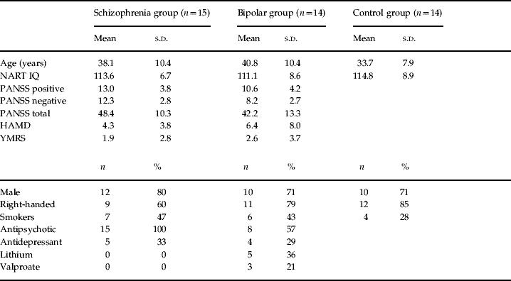
The demographic and clinical characteristics of the participants are presented in Table 1 and full details of medication doses are shown in Supplementary Table 1 (available online). There were no significant differences between the groups in terms of age, IQ measured by the National Adult Reading Test (NART; Nelson & Willison, Reference Nelson and Willison1991), gender, handedness or number of smokers. Control subjects smoked on average a lower total number of cigarettes per day than the patient groups, but there was no difference between the schizophrenia and bipolar groups in the mean number of cigarettes smoked (10 cigarettes/day on average in the schizophrenia group and 11 cigarettes/day on average in the bipolar group). There were no significant difference between the groups in terms of PANSS positive, PANSS general or PANSS total scores, but patients with schizophrenia had higher PANSS negative syndrome scores than patients with bipolar disorder (p<0.001). Depression and mania rating scores did not differ significantly between the patient groups. Four patients with bipolar disorder and two patients with schizophrenia had HAMD scores >8 on the day of scanning. Two patients with bipolar disorder and no patients with schizophrenia had YMRS scores >8 on the day of scanning. Twelve out of 14 patients with bipolar disorder had a lifetime history of psychotic symptoms during mood episodes.
Demographic and clinical characteristics

NART, National Adult Reading Test; PANSS, Positive and Negative Syndrome Scale; HAMD, Hamilton Depression Rating Scale; YMRS, Young Mania Rating Scale; s.d., standard deviation.
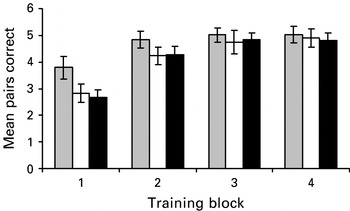
The groups were well matched in terms of in-scanner behavioural performance (Fig. 1). All subject groups showed learning of the face–name pair association across training, with the majority of new learning occurring during the early encoding sessions. Repeated-measures ANOVA confirmed a main effect of session (F=60, df=3, 120, p<0.001) but no main effect of group (F=1.0, df=2, 40, p=0.4) or group by session interaction (F=1.5, df=6,120, p=0.2). Notably, the schizophrenia and bipolar groups had almost identical performance across all training sessions.
Number of face–name pairs correctly remembered at each retrieval block by group. Error bars show standard error of the mean.  , Control subjects; □, bipolar disorder subjects; ▪, schizophrenia subjects.

Task-related brain activation
A full list of areas activated by the task in control subjects is shown in Supplementary Table 2 (available online). Activation was seen in the anterior hippocampus during early encoding but not during late encoding. This pattern of results is highly consistent with previous studies of face–name pair learning (Zeineh et al. Reference Zeineh, Engel, Thompson and Bookheimer2003; Kirwan & Stark, Reference Kirwan and Stark2004) and with more general evidence supporting a selective role for the anterior hippocampus in encoding (Lepage et al. Reference Lepage, Habib and Tulving1998; Dolan & Fletcher, Reference Dolan and Fletcher1999). Pronounced activation of the posterior, but not the anterior, hippocampus was seen during both retrieval periods. Activation of frontal cortical areas, including dorsal, inferior and superior frontal cortices, was also seen during both encoding and retrieval (Supplementary Table 2). Full details of the within-group activation patterns for the patient groups are shown in Supplementary Tables 3 and 4 (available online).
Between-group activation differences

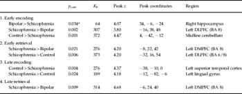
DLPFC, Dorsolateral prefrontal cortex; DMPFC, dorsomedial prefrontal cortex; BA, Brodmann area.
a Within a hippocampal small volume correction (SVC). Only contrasts with significant between-group differences are shown.
Between-group comparisons
Regions showing significant between-group differences during the encoding and retrieval of face–name pairs are presented in Table 2.
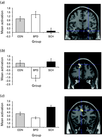
During early encoding, patients with schizophrenia showed significantly less relative activation of the anterior right hippocampus than patients with bipolar disorder [corrected p=0.034 within bilateral hippocampal SVC, z=4.07, total cluster size=64 voxels, peak Montreal Neurological Institute (MNI) coordinates 34, −6, −24 (Fig. 2)]. No other brain regions showed this pattern of activation differences between the groups. Inspection of the parameter estimates for this cluster revealed that this effect derived from lower hippocampal activation in the schizophrenia group than in the bipolar and control groups (Fig. 2). By contrast, bipolar subjects showed significantly lower activation of the left dorsolateral prefrontal cortex (DLPFC) during early encoding relative to subjects with schizophrenia [Brodmann area (BA) 8, corrected p=0.002, z=3.80, cluster=307 voxels, peak MNI coordinates −16, 38, 48 (Fig. 2)]. Inspection of the parameter estimates confirmed that bipolar subjects had lower activation of this brain region than either patients with schizophrenia or control subjects (Fig. 2). Unaffected comparison subjects showed greater activation of the midline cerebellum than patients with schizophrenia during early encoding (corrected p=0.001, z=4.47, cluster=372 voxels, peak MNI coordinates 4, −42, −12).
Clusters showing discrimination between the patient groups. (a) Decreased anterior hippocampal activation during early encoding in patients with schizophrenia compared to patients with bipolar disorder. (b) Decreased dorsolateral prefrontal cortex (DLPFC) activation during early encoding in patients with bipolar disorder compared to patients with schizophrenia. (c) Decreased dorsomedial prefrontal cortex (DMPFC) activation in subjects with bipolar disorder compared to subjects with schizophrenia during early retrieval. SPMs thresholded at p=0.001. Blue cross-hairs show voxel of peak difference. Error bars show standard error of the mean. CON, control subjects; BPD, bipolar disorder subjects; SCH, schizophrenia subjects.

During early retrieval, patients with schizophrenia showed greater activation than bipolar subjects of the dorsomedial prefrontal cortex (DMPFC), extending to the dorsal cingulate cortex [BA 8, corrected p=0.021, z=4.20, cluster=276 voxels, peak MNI coordinates −8, 22, 42 (Fig. 2)]. Patients with schizophrenia also showed greater activation of the dorsal PFC than control subjects during this task phase across a larger cluster that extended to include the region discriminating the patient groups and also to an additional area of DLPFC (BA 6/8, corrected p=0.006, z=4.20, cluster=373 voxels, peak MNI coordinates −32, 16, 54).
There were no clusters showing discrimination between the patient groups during late encoding. Control subjects showed greater activation of the left superior temporal cortex than patients with schizophrenia in late encoding (corrected p=0.004, z=4.37, cluster=276 voxels, peak MNI coordinates −38, −10, 0) whereas patients with schizophrenia showed greater activation of the lingual gyrus than controls during this task period (corrected p=0.024, z=4.18, cluster=189 voxels, peak MNI coordinates −12, −82, −6).
During late retrieval, patients with schizophrenia continued to show greater activation of the DMPFC than patients with bipolar disorder (BA 8, corrected p=0.009, z=4.68, cluster=314 voxels, peak MNI coordinates −6, 24, 40).
Symptom and medication correlations
We investigated whether the differences between the schizophrenia and bipolar groups related to mood state, psychotic symptoms or medication dose. Data were extracted from brain regions showing group differences and correlated with symptom scores (HAMD, YMRS, PANNS positive, PANSS negative, PANSS general and PANSS total) and medication dose (chlorpromazine equivalents, lithium dose, valproate dose) within each group. There was a positive correlation between YMRS score and hippocampal activation during early encoding in the bipolar group (p=0.01) and a negative correlation between YMRS and hippocampal activation during early encoding in the schizophrenia group (p<0.05). The latter effect was accounted for by patients with schizophrenia scoring on the items in the YMRS rating abnormal thought content and lack of insight (correlation p<0.05 for these items only). No significant correlations were found with any of the other symptom measures. There was no evidence of any effect of medication on brain activation in the regions of interest except for a positive correlation between antipsychotic dose and dorsal prefrontal activation during late retrieval in the bipolar group (p<0.05). Notably, this effect of medication operated to reduce, rather than increase, the difference between patient groups. Comparison of bipolar subjects with (n=8) and without (n=6) mood stabilizer medication also did not reveal any differences in blood oxygen level-dependent (BOLD) signal due to medication.
Discriminant function analysis
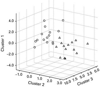
Data were extracted from the four clusters showing differences in activation between the schizophrenia and bipolar groups and entered into a discriminant function analysis. The model that best differentiated the patient groups included the hippocampal and dorsal prefrontal clusters from the early encoding period and the dorsal prefrontal cluster from the early retrieval period. This model classified 96% of patients correctly (Fig. 3). Notably, this analysis was performed only to explore the degree to which these activation differences discriminated the groups within the current study, and further prospective investigation would be required to investigate the predictive and diagnostic value of these findings.
Three-dimensional scatter plot showing separation of the schizophrenia and bipolar groups based on functional magnetic resonance imaging (fMRI) measures. Cluster 1, anterior hippocampal cluster, early encoding; cluster 2, dorsolateral prefrontal cluster, early encoding; cluster 3, dorsomedial prefrontal cluster, early retrieval; circles, schizophrenia subjects; triangles, bipolar subjects.

Discussion
Very few studies have compared the neural basis of schizophrenia and bipolar disorder using functional neuroimaging (Curtis et al. Reference Curtis, Dixon, Morris, Bullmore, Brammer, Williams, Sharma, Murray and McGuire2001; McIntosh et al. Reference McIntosh, Whalley, McKirdy, Hall, Sussmann, Shankar, Johnstone and Lawrie2008). The few studies that have made such a direct comparison have focused on tests of frontal lobe function (Curtis et al. Reference Curtis, Dixon, Morris, Bullmore, Brammer, Williams, Sharma, Murray and McGuire2001; McIntosh et al. Reference McIntosh, Whalley, McKirdy, Hall, Sussmann, Shankar, Johnstone and Lawrie2008). The degree to which abnormalities in brain function in other regions, such as the temporal lobes, separate the two disorders therefore remains largely unclear. Previous evidence from studies focusing on each disorder separately suggests that they may differ in terms of hippocampal function. We therefore investigated whether hippocampal activation could distinguish schizophrenia from bipolar disorder using fMRI in participants performing an associative memory task known to depend on this brain region. Our results demonstrate differential activation of the hippocampus in the two disorders, which was evident despite closely matched behavioural performance measured during scanning.
Patients with schizophrenia showed decreased anterior hippocampal activation during memory encoding, which distinguished them from subjects with bipolar disorder. Examination of parameter estimates demonstrated lower hippocampal activation in the schizophrenia group than in either the control group or the bipolar disorder group, although the difference between the schizophrenia group and the control subjects failed to reach cluster-level significance (Fig. 2). The anterior hippocampal formation is known to play a central role in the memory encoding, and specifically in the encoding of cross-domain associations such as face–name pairs (Lepage et al. Reference Lepage, Habib and Tulving1998; Zeineh et al. Reference Zeineh, Engel, Thompson and Bookheimer2003; Kirwan & Stark, Reference Kirwan and Stark2004). The formation of integrative, cross-domain associations is thought to be central to the ability to encode and recall episodic memories (Mayes et al. Reference Mayes, Montaldi and Migo2007). Patients with schizophrenia have memory impairments that are particularly marked for tasks that depend on the hippocampus such as tests of associative and episodic memory (Huron et al. Reference Huron, Danion, Giacomoni, Grange, Robert and Rizzo1995; Danion et al. Reference Danion, Rizzo and Bruant1999; Achim & Lepage, Reference Achim and Lepage2003; Toulopoulou et al. Reference Toulopoulou, Rabe-Hesketh, King, Murray and Morris2003). Similar deficits in memory function are also seen in the relatives of patients with schizophrenia, suggesting that they represent a heritable vulnerability for the disorder (Toulopoulou et al. Reference Toulopoulou, Rabe-Hesketh, King, Murray and Morris2003). Functional imaging studies have demonstrated impairments in hippocampal activation in schizophrenia that are particularly prominent in memory tasks (Heckers et al. Reference Heckers, Rauch, Goff, Savage, Schacter, Fischman and Alpert1998; Heckers, Reference Heckers2001; Achim & Lepage, Reference Achim and Lepage2005; Ongur et al. Reference Ongur, Cullen, Wolf, Rohan, Barreira, Zalesak and Heckers2006). This failure of activation may derive in part from tonic hyperactivation of this brain region, which has been associated with abnormal regulation of ascending dopaminergic function in models of schizophrenia (Lodge & Grace, Reference Lodge and Grace2007; Lisman et al. Reference Lisman, Coyle, Green, Javitt, Benes, Heckers and Grace2008). By contrast, there is little consistent evidence of either structural or functional abnormalities in the hippocampus in bipolar disorder, other than increased activation in response to emotional stimuli, which may relate to the close functional integration of the hippocampus with the adjacent amygdala (Haldane & Frangou, Reference Haldane and Frangou2004; Strakowski et al. Reference Strakowski, DelBello and Adler2004; Frey et al. Reference Frey, Andreazza, Nery, Martins, Quevedo, Soares and Kapczinski2007). In the present study we found a positive correlation between mania symptoms and hippocampal activation in the bipolar group, which may similarly reflect an influence of affective state on hippocampal activation in bipolar subjects.
Subjects with bipolar disorder showed decreased activation of the DLPFC during the encoding of face–name pairs compared to patients with schizophrenia, who did not differ significantly from control subjects in activation of this region during encoding (Fig. 2). Frontal lobe regions, including the DLPFC, play a central role in organizational processes related to memory encoding (Blumenfeld & Ranganath, Reference Blumenfeld and Ranganath2007) and have been shown to be particularly important for the encoding of associative memories (Summerfield et al. Reference Summerfield, Greene, Wager, Egner, Hirsch and Mangels2006). There is evidence of impaired function of both dorsal and ventral prefrontal cortical regions in bipolar disorder, which may account for both the neuropsychological deficits and dysregulation of emotional control seen in patients with this disorder (Blumberg et al. Reference Blumberg, Leung, Skudlarski, Lacadie, Fredericks, Harris, Charney, Gore, Krystal and Peterson2003; Strakowski et al. Reference Strakowski, DelBello and Adler2004; Phillips & Vieta, Reference Phillips and Vieta2007). Decreased dorsal PFC activation has previously been reported in euthymic bipolar patients during a verbal encoding task (Deckersbach et al. Reference Deckersbach, Dougherty, Savage, McMurrich, Fischman, Nierenberg, Sachs and Rauch2006) and in fMRI studies of working memory (Monks et al. Reference Monks, Thompson, Bullmore, Suckling, Brammer, Williams, Simmons, Giles, Lloyd, Harrison, Seal, Murray, Ferrier, Young and Curtis2004; Lagopoulos et al. Reference Lagopoulos, Ivanovski and Malhi2007). The present results support the view that dorsal PFC activation is abnormal during memory encoding in bipolar disorder, a finding that may reflect more general impairments in executive control in the disorder (Phillips & Vieta, Reference Phillips and Vieta2007). Impairments in frontal lobe function are therefore more likely to underlie the reported deficits in memory function in bipolar disorder than deficits in hippocampal function.
Participants with schizophrenia showed increased activation of left DMPFC during recall periods compared to patients with bipolar disorder and increased activation of the DMPFC and the DLPFC during retrieval relative to control participants. The PFC is strongly implicated in memory retrieval, both for the adoption and maintenance of retrieval strategies and for the monitoring of retrieval success (Fletcher et al. Reference Fletcher, Frith and Rugg1997). Left dorsal PFC activation has been shown to be greater for items that are consciously recalled rather than being simply familiar (Henson et al. Reference Henson, Rugg, Shallice, Josephs and Dolan1999) and to correlate with successful recall of face–name pair memories (Zeineh et al. Reference Zeineh, Engel, Thompson and Bookheimer2003). Increased activation of the DMPFC and additional recruitment of DLPFC in patients with schizophrenia may reflect the higher demand placed on this cortical region in schizophrenia subjects to maintain task performance. This increase in demand could reflect either the relatively inefficient prefrontal activation in schizophrenia or a compensation for impaired hippocampal activation (Heckers et al. Reference Heckers, Rauch, Goff, Savage, Schacter, Fischman and Alpert1998; Callicott et al. Reference Callicott, Mattay, Verchinski, Marenco, Egan and Weinberger2003).
The current study is the first to directly compare brain activations in schizophrenia and bipolar disorder using a memory task designed to recruit the hippocampus. A strength of the study was the balanced task performance across subject groups, which meant that the between-group differences in brain activation could not be accounted for by differences in performance. Future studies, however, could also use event-related fMRI and subsequent memory analysis to isolate only those events that were successfully remembered by a given subject. A limitation of the current study is that all the patients were, of necessity, on medication. There was, however, substantial overlap in medication between the patient groups, and medication effects did not explain the observed group differences in hippocampal and prefrontal activation, with the only significant effect of medication tending to decrease, rather than increase, between-group differences in the PFC. In addition, the current study had relatively small group numbers, and studies with larger sample sizes would be required to confirm the reliability and replicability of the current findings. Although discriminant function analysis suggested that this task produced a significant separation between the schizophrenia and bipolar groups, this represented a post-hoc investigation of the data, which would require confirmation in an independent sample. Furthermore, it should be noted that the discriminant function analysis compared the two patient groups, and did not examine the separation of patients from controls.
Overall, the present results show that schizophrenia and bipolar disorder can be distinguished in terms of activation of the hippocampus and PFC during memory encoding and retrieval. The ability to separate the two disorders using non-invasive imaging thus supports the view that there are separable components to the pathology underlying the two disorders and is of direct relevance to efforts to construct valid classifications of psychiatric illness.
Acknowledgements
J.H. was supported by a Medical Research Council (MRC) Clinical Research Training Fellowship. A.M. was supported by the Health Foundation. The work was supported by an award (EU_036) from the Translational Medicine Research Collaboration, a consortium made up of the Universities of Aberdeen, Dundee, Edinburgh and Glasgow, the four associated National Health Service (NHS) Health Boards (Grampian, Tayside, Lothian, and Greater Glasgow and Clyde), Scottish Enterprise, and Wyeth Pharmaceuticals. The study was carried out at the SFC Brain Imaging Research Centre (www.sbirc.ed.ac.uk) and the Wellcome Trust Clinical Research Facility, University of Edinburgh, Western General Hospital, Edinburgh. The work was carried out on a 1.5-T Signa Horizon MR/I 1.5T HDX operating under a research collaboration with GE Medical Systems (USA), operating as IGE in the UK.
Declaration of Interest
J.H., A.McI., H.C.W. and S.M.L. have received grants from the Translational Medicine Research Collaboration, a consortium made up of the Universities of Aberdeen, Dundee, Edinburgh and Glasgow, the four associated NHS Health Boards (Grampian, Tayside, Lothian, and Greater Glasgow and Clyde), Scottish Enterprise, and Wyeth Pharmaceuticals. H.I.W. is an employee of Wyeth Pharmaceuticals.
Note
Supplementary material accompanies this paper on the Journal's website (http://journals.cambridge.org/psm).