Introduction
Toxoplasma gondii is an intestinal coccidium in the cat family as the definitive hosts and other warm-blooded vertebrates including humans and most species of domestic animals and birds as intermediate hosts (Dubey, Reference Dubey2010; Rostami et al., Reference Rostami, Riahi, Fakhri, Saber, Hanifehpour, Valizadeh, Gholizadeh, Pouya and Gamble2017). This protozoon is considered medically important due to its congenital transmission and ability to cause opportunistic infections in immunocompromised patients (Rostami et al., Reference Rostami, Keshavarz, Shojaee, Mohebali and Meamar2014; Shiadeh et al., Reference Shiadeh, Niyyati, Fallahi and Rostami2016a, Reference Shiadeh, Rostami, Pearce, Gholipourmalekabadi, Newport, Danesh, Mehravar and Seyyedtabaei2016b). According to different studies, the prevalence of toxoplasmosis in Iran ranges between 50 and 75% (Daryani et al., Reference Daryani, Sarvi, Aarabi, Mizani, Ahmadpour, Shokri, Rahimi and Sharif2014; Rostami et al., Reference Rostami, Seyyedtabaei, Aghamolaie, Behniafar, Lasjerdi, Abdolrasouli, Mehravar and Alvarado-Esquivel2016). The clinical signs and symptoms of toxoplasmosis depend on the host immune status. In people with strong immune systems, self-limited infections usually occur. In those with impaired immune systems, particularly patients with impaired cellular immune systems, there is a risk of infection recurrence or spread (Yazar et al., Reference Yazar, Demirtas, Yalcin, Yaman, Tokgoz, Utas and Sahin2003a; Macpherson, Reference Macpherson2005; Arab-Mazar et al., Reference Arab-Mazar, Fallahi, Koochaki, Haghighi and Tabaei2016a, Reference Arab-Mazar, Zamanian and Yadegarynia2016b). Patients with malignancy, those undergoing kidney transplantation, patients treated with immunosuppressive drugs and haemodialysis patients with chronic renal failure are susceptible to Toxoplasma infection (Sibley et al., Reference Sibley, Weidner and Krahenbuhl1985; Howe and Sibley, Reference Howe and Sibley1995; Yazar et al., Reference Yazar, Demirtas, Yalcin, Yaman, Tokgoz, Utas and Sahin2003a; Omrani et al., Reference Omrani, Fallahi, Rostami, Siyadatpanah, Barzgarpour, Mehravar, Memari, Hajialiani and Joneidi2015). Thus, the identification of toxoplasmosis in these patients is important.
In general, because of the high prevalence of this parasite in Iran and due to the involvement of the nervous system in immunocompromised patients, this infection is a crucial health point. However, physicians may consider the diagnosis of toxoplasmosis that causes importance health care and social problems. On the other hand, clinical findings alone cannot solely diagnose toxoplasmosis, accordingly, parasitological, serological and molecular evaluations are needed for confirmation (Petersen et al., Reference Petersen, Borobio, Guy, Liesenfeld, Meroni, Naessens, Spranzi and Thulliez2005). Toxoplasmosis patients may be at risk for reactivation of the infection due to defect in cell-mediated immunity (Athlin and DomellÖF, Reference Athlin and DomellÖF1986; Ferreira and Borges, Reference Ferreira and Borges2002). Studies have shown that diagnosis of toxoplasmosis as an opportunistic infection in renal failure patients, such as those under haemodialysis, is of great significance; therefore, diagnosis at an earlier stage with definitive methods can be highly desirable in these patients (Omrani et al., Reference Omrani, Fallahi, Rostami, Siyadatpanah, Barzgarpour, Mehravar, Memari, Hajialiani and Joneidi2015). Thus, we sought to investigate immunodiagnosis, molecular validation, disease awareness and risk factors for T. gondii infection among patients with end-stage renal disease (ESRD) undergoing haemodialysis.
Materials and methods
Study design and population
In this case-control study, the whole blood samples of 260 haemodialysis patients from five main dialysis centres in Tehran, the capital of Iran, and 259 healthy individuals (control group) were collected during 2016 (ethics committee code: 9127, 18.10.2016). Based on physician consultant consideration, laboratory examination and documents, the control group subjects did not have any kidney problems. A previously published questionnaire was used to assess the risk factor with some modification (Rostami et al., Reference Rostami, Seyyedtabaei, Aghamolaie, Behniafar, Lasjerdi, Abdolrasouli, Mehravar and Alvarado-Esquivel2016). Briefly, the following variables were included to measure demographics and potential risk factors: age, sex, place of residence, educational level, exposure to cat and contaminated soil as shown in Tables. It was tried that the two cases and control groups to be matched for different variables such as age, gender and place of residence.
Serological assessment of anti-Toxoplasma antibodies
Venous blood samples (5 mL) were taken from each subject and sera were obtained from the collected blood using centrifugation. Moreover, for molecular evaluations, a buffy coat was obtained from each blood sample. Sera and buffy coat were kept at −20 °C until use. All the sera samples were tested for immunoglobulin G (IgG) and immunoglobulin M (IgM) anti-Toxoplasma antibodies using a commercial enzyme-linked immunosorbent assay kits (ACON, San Diego, USA) as instructed by the manufacturers. According to the manufacturer's data sheet, the sensitivity and specificity of kits were 99.9 and 99%, respectively. On the basis of the kit protocol, the results were reported in International Units (IU). Sera with values of <9.0 IU mL−1 were considered negative; those with values between 9.0–11.0 IU mL−1 were considered suspicious (grey zone), and those with values >11.0 IU mL−1 were considered positive, for both T. gondii IgG and IgM antibodies.
GRA6-PCR
DNA was extracted from the buffy coat in the IgM-positive samples by using a DNG plus kit (Sinaclon, Tehran, Iran) according to the manufacture's protocol. Positive DNA as a control was extracted from the RH strain of T. gondii which was prepared from the Department of Parasitology at Shahid Beheshti Medical University. The extracted DNA transferred to −80 °C until use.
Polymerase chain reaction (PCR) was performed to amplify a GRA6 gene of T. gondii by using following primers: F:5′-GTAGCGTGCTT-GTTGGCGAC-3′ and R: 5′-TACAAGACATAGAGTGCCCC-3′ (Norouzi et al., Reference Norouzi, Seyyed Tabaei, Niyyati, Saber and Behniafar2016). PCR assay was carried out in a 15 µL reaction mixture containing 7.5 µL master mix (Bie & Berntsen A/S), 1 µL of equally-mixed primers, 1.5 µL DNA and 5 µL distilled water. The following thermal cycling condition was used for the PCR: a 5 min hold at 96 °C, then followed by 33 three-step cycles of denaturation at 95 °C for 30 s, annealing at 56 °C for 30 s, and extension at 72 °C for 45 s. The reaction was ended with a final 10 min extension at 72 °C. The amplified DNA was run on 1.5% agarose gel containing ethidium bromide and was then visualized under an ultraviolet transilluminator. The molecular weight of the PCR product was determined and verified using a 100 bp standard ladder (Fermentas, Berlin, Germany).
SAG1-LAMP
The LAMP reaction was carried out using the specific primers for the SAG1 gene of T. gondii (GenBank accession No. X14080) presented by Lau and colleagues [19]. The final volume of 25 µL reaction mixture was containing 1.4 mm dNTP, 8 mm MgSO4, 0.8 M betaine, 8 U Bst DNA polymerase (New England Biolabs, UK), 1× buffer enzyme [KCL (10 mm), Tris-HCL (20 mm), (NH4)2SO4 (10 mm), MgSO4 (9 mm)], 40 pmol each of FIP and BIP primers, 5 pmol each of F3 and B3 primers, 6.5 µL distilled water and 1 µL of template DNA. For DNA amplification, the microtubes were placed in a water bath at 63 °C for 60 min. Subsequently, 1 µL of 0.1% SYBR Green I (Invitrogen, USA) was added to each tube and then the tubes were evaluated under natural light and UV light with a wavelength of 312 nm.
Statistical analysis
The collected data were analysed using χ 2 by using statistical package for the social sciences (SPSS) software Ver. 21 (SPSS Inc., Chicago, USA). The probability level of 0.05 was accepted as statistically significant.
Results
Serological assessment of anti-T. gondii antibodies
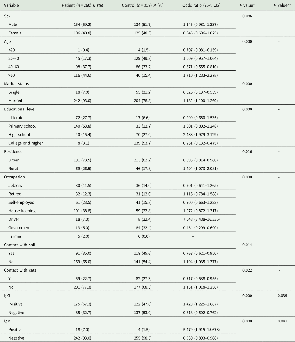
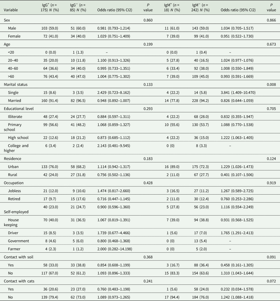
Among 519 participants of the study, the overall seroprevalence of toxoplasmosis was 57.2% (297/519). The frequency of T. gondii infection was 67.3% (175/260) in haemodialysis patients and 47% (122/259) in the control group which shows a statistically significant association between T. gondii seropositivity and haemodialysis (OR = 2.312; 95% CI = 1.620–3.300; P = 0.00). This significant difference remained unchanged even after adjustment of confounding variables including, age, marital status, education, residence, occupation, contact with soil and cats. In haemodialysis patients, 18 cases were positive for both anti-Toxoplasma IgG and IgM antibodies while only four people of the control group were positive for both IgG and IgM antibodies. Our results also showed that the percentage of haemodialysis patients which were positive for anti-T. gondii IgM antibodies was found to be greater than that in the control group, as well the difference between the groups was statistically significant (P = 0.05). Seroprevalence of toxoplasmosis according to the demographic characteristic in haemodialysis patients and the control group is summarized in Table 1. The T. gondii IgM antibodies indicating acute infection were detected in 7 women and 11 men among patients. Comparison of the demographic characteristic of haemodialysis patients, according to anti-Toxoplasma IgG and IgM antibodies is summarized in Table 2.
Sociodemographic characteristics and T. gondii serostatus of haemodialysis patients and controls

*Raw P value; **P value after adjustment of age, marital status, education, residence, occupation, contact with soil and cats.
Demographic characteristic of haemodialysis patients, according to Toxoplasma IgG and IgM antibodies

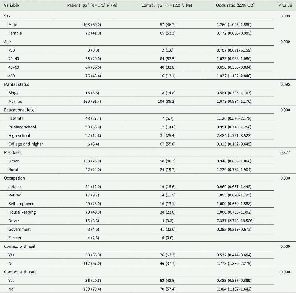
As shown in Table 3, in the patients group, the frequency of anti-T. gondii IgG antibodies was higher in men (59%) but in the control group the frequency of seropositivity in women was higher (53.3%) that this difference was statistically significant (OR = 1.631; 95% CI = 1.024–2.600; P = 0.040). In addition, seropositivity is elevated with an increase of age in patients group, which showed significantly different between the patient and control group (P = 0.00). Of the 175 seropositive haemodialysis patients, 133 (76%) patients were residing in the urban area and the seroprevalence rate for the control group was 98/122 (80.3%). Statistical analysis showed no significant between seropositivity of T. gondii, marital status and residency. The prevalence of IgG antibodies in the primary school category was higher than other categories, although this difference was not statistically significant. The majority of haemodialysis patients were housewives (101/38.8%).
Comparison of demographic characteristic between haemodialysis patients and controls, according to the Toxoplasma IgG antibody

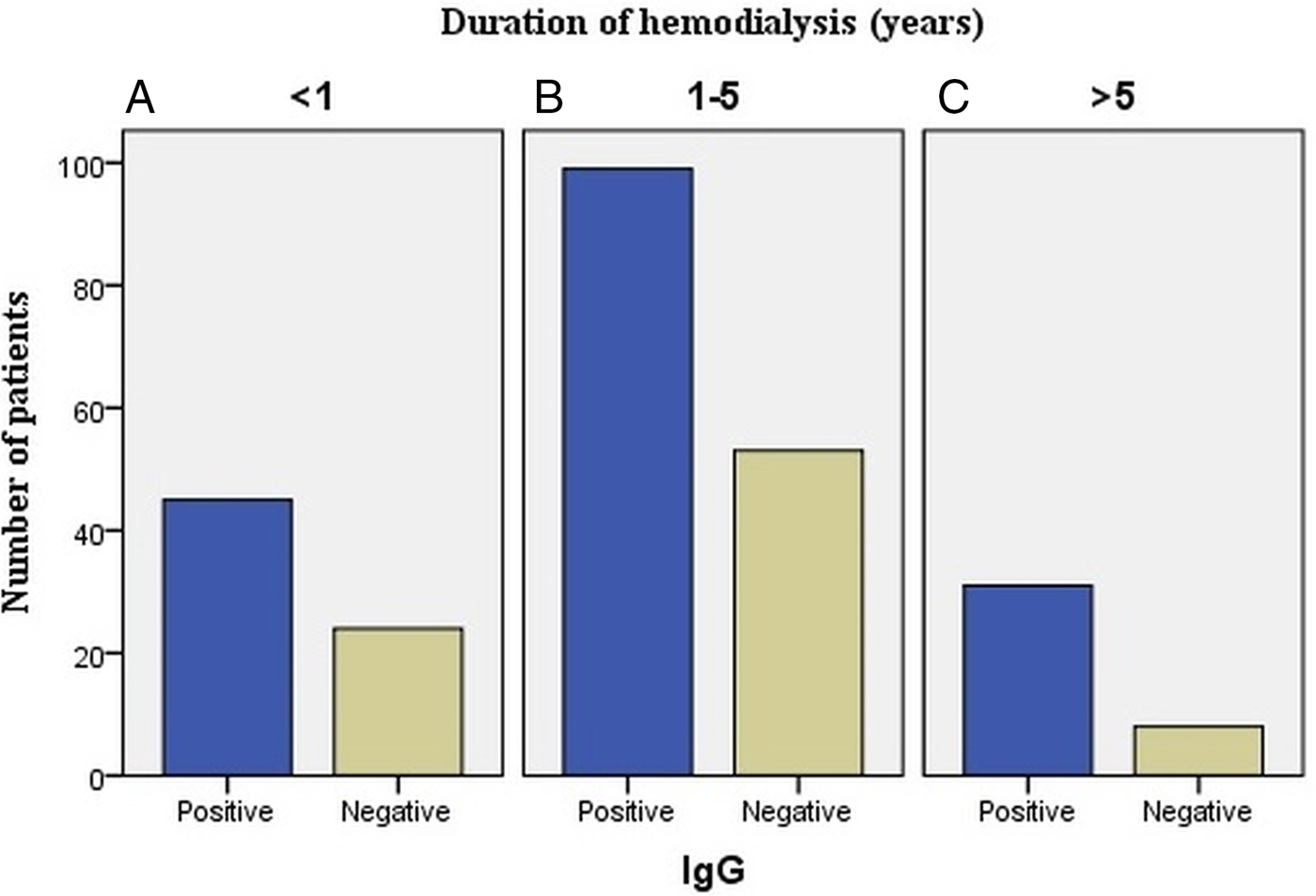
The relativity between the haemodialysis duration and seropositivity of T. gondii antibody was also evaluated in this study. Our study indicates that there is no significant association between anti-T. gondii due to the IgG antibody frequency and the duration of haemodialysis (Fig. 1). We observed an increase in the seropositivity rate with increasing length of time on dialysis treatment.
Relationship between the haemodialysis duration and seropositivity of the T. gondii antibody. The number of patients with positive and negative T. gondii IgG antibodies with the duration of haemodialysis: (A) less than 1 year, (B) between 1 to 5 years (C) and over 5 years.

GRA6-PCR
The GRA6-based PCR gave a positive result with 2 of the 18 anti-Toxoplasma IgM positive blood samples in the haemodialysis patients group and in the IgM positive control subjects there was no positive PCR result. The size of the PCR product was about 791 bp.
SAG1-LAMP
Upon addition of SYBR Green I to microtubes, the positive tubes were green while the negative tubes remained to be pale orange. SYBR Green I is an asymmetrical cyanine dye that preferentially binds to double-stranded DNA. The resulting DNA-dye-complex absorbs blue light and emits green light. The results of SAG1-LAMP showed that nine samples (50%) in the IgM-positive haemodialysis patients and two samples (50%) in the IgM-positive control group were positive for T. gondii DNA. This means that in the present study half of IgM-seropositive people have had Toxoplasma DNA in their blood sample.
Discussion
T. gondii is the most frequent protozoan causing opportunistic infections in immunocompromised patients (Yazar et al., Reference Yazar, Demirtas, Yalcin, Yaman, Tokgoz, Utas and Sahin2003b; Arab-Mazar et al., Reference Arab-Mazar, Fallahi, Koochaki, Haghighi and Tabaei2016a). Considering that current therapies are not fully efficient and there is a lack of safe and effective vaccines, it seems that it is vital to prevent or reduce toxoplasmosis transmission (Arab-Mazar et al., Reference Arab-Mazar, Seyyed-Tabaei and Mirahmadi2014). Haemodialysis is currently used as a life-sustaining therapy for patients with ESRD. These patients face severe complications such as congestive heart failure, psychotic disorders, various cutaneous abnormalities and weakness of the immune system (House, Reference House1987; Szepietowski et al., Reference Szepietowski, Reich and Pacan2004; Vonesh et al., Reference Vonesh, Snyder, Foley and Collins2004). There are about 1 million ESRD patients under haemodialysis worldwide (Grassmann et al., Reference Grassmann, Gioberge, Moeller and Brown2005).
Elevated urea can cause mortality in patients with chronic kidney disease if they do not undergo dialysis, because uremia impairs the function of lymphocytes, leukocytes such as neutrophils and monocytes, nitric oxide (NO) and platelets (Kato et al., Reference Kato, Chmielewski, Honda, Pecoits-Filho, Matsuo, Yuzawa, Tranaeus, Stenvinkel and Lindholm2008). Several studies have supported the high incidence of opportunistic infections among haemodialysis patients (Tonelli et al., Reference Tonelli, Wiebe, Culleton, House, Rabbat, Fok, McAlister and Garg2006; Omrani et al., Reference Omrani, Fallahi, Rostami, Siyadatpanah, Barzgarpour, Mehravar, Memari, Hajialiani and Joneidi2015). Our study showed a significantly high percentage of positivity for T. gondii IgG and IgM antibodies in patients compared to controls, which indicates the role of kidney and the haemodialysis procedure in patients with T. gondii infection. The present findings are comparable with those of previous studies, which found that anti-Toxoplasma IgG and IgM antibodies were higher in haemodialysis patients than controls (Ocak et al., Reference Ocak, Duran, Eskiocak and Aytac2005; Solhjoo et al., Reference Solhjoo, Jahromi and Parnian-Rad2010). Despite the direct association between the prevalence of T. gondii antibodies and the geographical region, studies in different countries have supported the high prevalence of toxoplasmosis among ESRD and dialysis patients. In a serological study performed in Egypt on 60 patients, the prevalence of toxoplasmosis was reported to be 38.3%. The present study found that housewives and self-employed people have high T. gondii seropositivity. This finding is in agreement with those of previous studies conducted in Iran and other countries (Al-Qurashi, Reference Al-Qurashi2004; Jones et al., Reference Jones, Dargelas, Roberts, Press, Remington and Montoya2009; Daryani et al., Reference Daryani, Sarvi, Aarabi, Mizani, Ahmadpour, Shokri, Rahimi and Sharif2014).
Further analysis revealed that T. gondii seropositivity in the older age group (>60 years) was two times higher than in a younger age group (20–40 years). This finding is agreement with the reports from the majority of international studies (Bahia-Oliveira et al., Reference Bahia-Oliveira, Jones, Azevedo-Silva, Alves, Oréfice and Addiss2003; Al-Qurashi, Reference Al-Qurashi2004). This result may be due to the higher consumption of raw or undercooked infected meat and higher chance of contact with cats or contaminated soil. In addition, our results revealed that higher level of education is associated with the reduced rate of T. gondii seropositivity. The lower level of education is associated with the lack of knowledge regarding this disease, especially in haemodialysis patients, and lower hygiene status (Jones et al., Reference Jones, Dargelas, Roberts, Press, Remington and Montoya2009). In our study, a contact with cats was associated with T. gondii seropositivity. Similar results were observed in a study conducted by Mahmoudvand et al., on blood donors in southeast of Iran (Mahmoudvand et al., Reference Mahmoudvand, Saedi Dezaki, Soleimani, Baneshi, Kheirandish, Ezatpour and Zia-ali2015; Fallahi et al., Reference Fallahi, Rostami, Birjandi, Zebardast, Kheirandish and Spotin2017).
In the present study, GRA6-PCR and SAG1-LAMP were used to detect T. gondii DNA in the buffy coat of IgM-positive patients and controls. PCR only detected two patients and there was no positive PCR result in the IgM-positive control subjects, while the LAMP technique was able to detect nine patients and two cases of controls. These findings may indicate the higher sensitivity of the LAMP technique compared to the PCR. Due to the use of 4–6 primers, which target 6–8 distinct regions on the target DNA (Fallahi et al., Reference Fallahi, Tabaei, Pournia, Zebardast and Kazemi2014), and the low likelihood of non-specific binding of primers, the specificity of the LAMP technique has been reported 100% in all studies. Usability in the field, cost-effectiveness, promptness, and no need for expensive equipment are the other advantages of the LAMP technique (Notomi et al., Reference Notomi, Okayama, Masubuchi, Yonekawa, Watanabe, Amino and Hase2000; Mori et al., Reference Mori, Nagamine, Tomita and Notomi2001; Fallahi et al., Reference Fallahi, Mazar, Ghasemian and Haghighi2015).
In conclusion, the results of this study indicated that haemodialysis patients are considered as a high-risk group for Toxoplasma infection, and it is assumed that patients with chronic renal failure who undergo long-term haemodialysis are candidates for kidney transplantation. Kidney recipients should be screened for toxoplasmosis to prevent the dissemination of infection. Our results demonstrated the high prevalence of Toxoplasma infection in haemodialysis patients. Thus, health programs should be implemented in these patients to increase their knowledge regarding toxoplasmosis. Also, parasitological investigation of haemodialysis patients should be performed regularly in endemic areas to prevent its severe complications.
Acknowledgements
The authors would like to thank the administrator and staff of participate haemodialysis centers and Mrs. Naderi and Mr. Nazarimanesh for their kind cooperation during sample collection. Most importantly, the authors would like to thank all participates for giving their permission to collect samples.
Financial support
This study was financially supported by Infectious Diseases and Tropical Medicine Research Center, Shahid Beheshti University of Medical Sciences (SBMU) Tehran, Iran.
Conflict of interest
The authors have no conflict of interest.
Ethical standards
This study was approved by the Ethical Committee of the Shahid Beheshti University of Medical Science, Tehran, Iran (ethics committee code: 9127, 18.10.2016). All the enrolled participants were informed about the study and written informed consent was obtained.